<code lang="o6XJPD0E"></code> Skip to main page content
U.S. flag

An official website of the United States government

Dot gov

The . gov means it’s official. Federal government websites often end in . gov or VSports app下载. mil. Before sharing sensitive information, make sure you’re on a federal government site. .

Https

The site is secure V体育官网. The https:// ensures that you are connecting to the official website and that any information you provide is encrypted and transmitted securely. .

. 2021 Mar 3;12(3):230.
doi: 10.1038/s41419-021-03520-2.

Sodium iodate induces ferroptosis in human retinal pigment epithelium ARPE-19 cells

Affiliations

Sodium iodate induces ferroptosis in human retinal pigment epithelium ARPE-19 cells

Binghua Liu et al. Cell Death Dis. .

"V体育2025版" Abstract

Sodium iodate (SI) is a widely used oxidant for generating retinal degeneration models by inducing the death of retinal pigment epithelium (RPE) cells VSports手机版. However, the mechanism of RPE cell death induced by SI remains unclear. In this study, we investigated the necrotic features of cultured human retinal pigment epithelium (ARPE-19) cells treated with SI and found that apoptosis or necroptosis was not the major death pathway. Instead, the death process was accompanied by significant elevation of intracellular labile iron level, ROS, and lipid peroxides which recapitulated the key features of ferroptosis. Ferroptosis inhibitors deferoxamine mesylate (DFO) and ferrostatin-1(Fer-1) partially prevented SI-induced cell death. Further studies revealed that SI treatment did not alter GPX4 (glutathione peroxidase 4) expression, but led to the depletion of reduced thiol groups, mainly intracellular GSH (reduced glutathione) and cysteine. The study on iron trafficking demonstrated that iron influx was not altered by SI treatment but iron efflux increased, indicating that the increase in labile iron was likely due to the release of sequestered iron. This hypothesis was verified by showing that SI directly promoted the release of labile iron from a cell-free lysate. We propose that SI depletes GSH, increases ROS, releases labile iron, and boosts lipid damage, which in turn results in ferroptosis in ARPE-19 cells. .


Conflict of interest statement

The authors declare no competing interests.

Figures
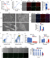
Fig. 1
Fig. 1. ARPE-19 cells treated with SI manifest necrotic phenotype.
A Cell viabilities determined by CCK assays after NaIO3 treatment for 12 h or 24 h at indicated concentrations (n = 4). B ARPE-19 cells were cultured with NaIO3 at indicated concentrations for 24 h. After the treatment, the LDH levels were determined by the LDH assays (n = 4). C Left: Fluorescence images of cells stained with JC-1, an indicator of mitochondrial membrane potential (MMP). ARPE-19 cells were treated with 10 mM NaIO3 for 24 h. Scale bar: 100 μm. Right: The ratio of red/green fluorescence intensity shows the change of MMP (n = 3). D ARPE-19 cells treated with 10 mM NaIO3 for 24 h were observed by scanning electron microscope. Scale bar: 20 μm. E Upper: Fluorescence images of cells treated with 10 mM NaIO3 for 24 h followed by Hoechst 33342 and PI double staining. Scale bar: 100 μm. Lower: Cells positively stained with Hoechst and PI were counted using Image J software. Graphs represent the percentage of Hoechst and PI stained cells against total cells (n = 3). F Fluorescence-activated cell sorting (FACS) histograms of Annexin V-FITC/PI stained cells with (right) and without (left) the treatment of 10 mM NaIO3 for 24 h (Q1: cellular debris or necrotic cells; Q2: necrotic or late apoptotic cells; Q3: early apoptotic cells; Q4: viable cells). G Left: Effect of the caspase inhibitor Z-VAD on ARPE-19 cell death. Cells were pretreated with Z-VAD or solvent alone (DMSO) at indicated concentrations for 3 h and followed by co-treatment with or without 20 mM NaIO3 for 24 h (n = 4). Right: A positive control for apoptosis inhibition by Z-VAD. Jurkat cells were pretreated with 100 μM Z-VAD or solvent alone (DMSO) for 3 h followed by co-treatment with 1.5 μM doxorubicin (DOX) for 18 h (n = 4). H Left: TUNEL staining of apoptotic ARPE-19 cells. Cells were exposed to 10 mM NaIO3 for the indicated time. Scale bar: 100 μm. Right: numbers of apoptotic cells and normal cells in the microscope field of view were counted by image J software (n = 3).
Fig. 2
Fig. 2. Necroptosis is not the main pathway related to the death of ARPE-19 cells induced by SI.
A Cell viabilities of ARPE-19 cells treated with 200 μM Nec-1, 2 μM NSA or 10 μM GSK′872 for 24 h before 20 mM NaIO3 treatment for 24 h (n = 4). B HT-29 cells with or without necrosulfonamide (NSA) pretreatment was treated with the indicated necrosis-inducing agents for 16 h. T + Z + S: TNF-α (200 ng/ml), Z-VAD (20 μM), and SM-164 (10 μM) (n = 4). C Phospho-RIPK1 (S166), total RIPK1, total RIPK3, Phospho-MLKL (S358), and total MLKL were detected in ARPE-19 or HT-29 cells by western blotting. SI: 10 mM NaIO3 treatment for 24 h. T + Z + S: TNF-α (100 ng/ml), Z-VAD (20 μM), and SM-164 (10 μM) treatment for 3 h. D Cell viabilities of SH-SY5Y cells treated with the indicated necrosis-inducer agent(s) for 3 h. T: 100 ng/ml TNF-α; S: 10 μM SM-164; Z: 20 μM Z-VAD (n = 4). E Cell viabilities of ARPE-19 cells treated with the indicated necrosis-inducer agent(s) for 48 h. T1: 50 ng/ml TNF-α; T2: 100 ng/ml TNF-α; S: 10 μM SM-164; Z: 20 μM Z-VAD (n = 4).
Fig. 3
Fig. 3. Death of ARPE-19 cells induced by SI is related to the intracellular labile iron.
A, B Alteration of labile iron levels in ARPE-19 cells with NaIO3 treatment in concentration (A) or time (B) dependent manner. The labile iron levels were assessed as the increase in fluorescence (ΔF) 30 min after PIH addition (n = 4). C Viabilities of ARPE-19 cells treated with indicated concentrations of FAC (ammonium ferric citrate) for 24 h followed by additional 24 h treatment with or without 20 mM NaIO3 (n = 5). D Viabilities of ARPE-19 cells treated with indicated concentrations of Heme or vehicle alone (DMSO) for 24 h followed by additional 24 h treatment with or without 20 mM NaIO3 (n = 5). E The protective role of DFO for SI-induced cell death. ARPE-19 cells were pretreated with DFO at indicated concentrations or solvent alone (PBS) for 24 h prior to co-treatment with 20 mM NaIO3 for an additional 24 h (n = 4). F The protective role of PIH for SI-induced cell death. ARPE-19 cells were pretreated with PIH at indicated concentrations or solvent alone (DMSO) for 48 h prior to co-treatment with 20 mM NaIO3 for an additional 24 h (n = 4).
Fig. 4
Fig. 4. SI promotes ROS generation and oxidative damage of lipids in ARPE-19 cells.
A Concentration dependent induction of ROS by SI in ARPE-19 cells. Cells were stained with the green fluorescent ROS probe DCFH-DA for 30 min and then exposed to 10, 20, or 30 mM NaIO3 or 50 μM tBH as a positive control for 30 min. Scale bar: 100 μm. B Detection of MDA content by TBA test in cells treated for 24 h with 10 mM NaIO3 or 150 μM tBH. MDA level was expressed in μmol/g protein (n = 3). C In vitro reaction between SI and MDA. The residual MDA level was detected by the TBA test after 50 μM MDA was incubated with indicated concentrations of NaIO3 for 30 min at 37 °C (n = 4). D Detection of MDA content by MDA-6. ARPE-19 cells seeded in a 96-well plate were treated with or without 5 mM or 10 mM NaIO3 for 2 h followed by washing with PBS and staining with 10 μM MDA-6. Left: Fluorescence images of cells stained with MDA-6. Scale bar: 100 μm. Right: Quantification of fluorescence intensity of fluorescence images. E Peroxidation of lipids analyzed by BODIPY 581/591 C11. APRE-19 cells were preloaded with the probe BODIPY 581/591 C11 for 30 min followed by treatment with SI or tBH and heme at indicated concentrations for 0.5 or 6 h. Fluorescence of probe in oxidized (488/520 nm) and non-oxidized (575/600 nm) forms were monitored. The ratio of the oxidized BODIPY 581/591 C11 to the total BODIPY 581/591 C11 (oxidized plus non-oxidized) was calculated to represent the extent of lipid peroxidation (n = 6). F The protective role of Fer-1 for SI-induced cell death. ARPE-19 cells pretreated with or without 100 μM Fer-1 for 24 h were incubated with indicated concentrations of SI for an additional 24 h. The survival rate of cells was measured by the CCK-8 method (n = 4).
Fig. 5
Fig. 5. SI reacts directly with cysteine and GSH in ARPE-19 cells.
A Upper panel: Western blotting detection of GPX4 expression in cells treated with 10 mM NaIO3 for the indicated time. Lower panel: the quantifications of three repeats from the upper panel. BE The glutathione levels in ARPE-19 cells treated for 24 h with or without 10 mM NaIO3. B Total glutathione levels; C reductive glutathione levels; D oxidative glutathione levels; and E ratios of reductive to oxidative glutathione levels (n = 3). F The concentration of cysteine in ARPE-19 cells with or without 10 mM NaIO3 treatment for 24 h (n = 3). G, H In vitro reaction between SI and cysteine or GSH. 10 mM cysteine (G) or 10 mM GSH (H) was incubated with NaIO3 at indicated concentrations for 10 min at 37 °C. The residual sulfhydryl was assayed using a cysteine assay kit (n = 4).
Fig. 6
Fig. 6. SI induces the increase of labile iron by promoting the release of sequestered iron in ARPE-19 cells.
A, B Ferrous iron influx (A) or efflux (B) in SI-treated ARPE-19 cells was indicated by the quenching and reversing of calcein fluorescence, a marker of the intracellular labile iron level. The relative fluorescence intensity represented the ratios of the fluorescence intensity of the samples at the indicated time relative to its initial fluorescence intensity. F and P values were calculated by ordinary two-way ANOVA (n = 4). C Quantitative real-time PCR analysis of ferritin (ferritin light chain, FTL) mRNA levels in ARPE-19 cells treated with or without 10 mM NaIO3 for 24 h (n = 3). D Upper panel: Western blots of ferritin in ARPE-19 cells incubated with 10 mM NaIO3 for the indicated time. Lower panel: the quantifications of three repeats from the upper panel (n = 3). E SI did not promote the release of free iron in ferritin from the horse spleen. Totally, 0.16 mg/ml ferritin from the horse spleen was treated with NaIO3 at the indicated concentrations for 30 min or 24 h and the level of free iron in ferritin solution was determined by calcein fluorescence quenching (n = 5). F SI promoted the release of free iron in the ARPE-19 cell lysate. ARPE-19 cell lysate was treated with 20 mM NaIO3 for 30 min, 24 h, or 48 h and the level of free iron in ferritin solution was determined by calcein fluorescence quenching (n = 4). G The activity of aconitase (ACO) in the ARPE-19 cell-free lysate treated with or without 10 mM NaIO3 for 10 min (n = 4).

"V体育ios版" References

    1. Datta S, Cano M, Ebrahimi K, Wang L, Handa JT. The impact of oxidative stress and inflammation on RPE degeneration in non-neovascular AMD. Prog. Retin. Eye Res. 2017;60:201–218. doi: 10.1016/j.preteyeres.2017.03.002. - DOI (VSports最新版本) - PMC - PubMed
    1. Hellinen L, Pirskanen L, Tengvall-Unadike U, Urtti A, Reinisalo M. Retinal pigment epithelial cell line with fast differentiation and improved barrier properties. Pharmaceutics. 2019;11:412. doi: 10.3390/pharmaceutics11080412. - DOI (V体育平台登录) - PMC - PubMed
    1. Wang W, et al. Metabolic deregulation of the blood-outer retinal barrier in retinitis pigmentosa. Cell Rep. 2019;28:1323–34 e4. doi: 10.1016/j.celrep.2019.06.093. - DOI - PMC - PubMed
    1. Zhang Q, et al. Highly differentiated human fetal RPE cultures are resistant to the accumulation and toxicity of lipofuscin-like material. Investig. Ophthalmol. Vis. Sci. 2019;60:3468–3479. doi: 10.1167/iovs.19-26690. - DOI - PMC - PubMed
    1. Saint-Geniez M, Kurihara T, Sekiyama E, Maldonado AE, D’Amore PA. An essential role for RPE-derived soluble VEGF in the maintenance of the choriocapillaris. Proc. Natl Acad. Sci. USA. 2009;106:18751–18756. doi: 10.1073/pnas.0905010106. - DOI - PMC - PubMed

"V体育ios版" Publication types

MeSH terms

LinkOut - more resources (VSports app下载)