Skip to main page content
U.S. flag

An official website of the United States government

Dot gov

The . gov means it’s official. Federal government websites often end in . gov or VSports app下载. mil. Before sharing sensitive information, make sure you’re on a federal government site. .

Https

The site is secure. The https:// ensures that you are connecting to the official website and that any information you provide is encrypted and transmitted securely V体育官网. .

. 2021 Jan 29;135(2):347-365.
doi: 10.1042/CS20200573.

Sepsis plasma-derived exosomal miR-1-3p induces endothelial cell dysfunction by targeting SERP1

Affiliations

Sepsis plasma-derived exosomal miR-1-3p induces endothelial cell dysfunction by targeting SERP1

Min Gao et al. Clin Sci (Lond). .

Abstract

Acute lung injury (ALI) is the leading cause of death in sepsis patients. Exosomes participate in the occurrence and development of ALI by regulating endothelial cell inflammatory response, oxidative stress and apoptosis, causing serious pulmonary vascular leakage and interstitial edema. The current study investigated the effect of exosomal miRNAs on endothelial cells during sepsis. We found a significant increase in miR-1-3p expression in cecal ligation and puncture (CLP) rats exosomes sequencing and sepsis patients' exosomes, and lipopolysaccharide (LPS)-stimulated human umbilical vein endothelial cells (HUVECs) in vitro. However, the specific biological function of miR-1-3p in ALI remains unknown. Therefore, mimics or inhibitors of miR-1-3p were transfected to modulate its expression in HUVECs. Cell proliferation, apoptosis, contraction, permeability, and membrane injury were examined via cell counting kit-8 (CCK-8), flow cytometry, phalloidin staining, Transwell assay, lactate dehydrogenase (LDH) activity, and Western blotting. The miR-1-3p target gene was predicted with miRNA-related databases and validated by luciferase reporter. Target gene expression was blocked by siRNA to explore the underlying mechanisms. The results illustrated increased miR-1-3p and decreased stress-associated endoplasmic reticulum protein 1 (SERP1) expression both in vivo and in vitro. SERP1 was a direct target gene of miR-1-3p. Up-regulated miR-1-3p inhibits cell proliferation, promotes apoptosis and cytoskeleton contraction, increases monolayer endothelial cell permeability and membrane injury by targeting SERP1, which leads to dysfunction of endothelial cells and weakens vascular barrier function involved in the development of ALI. MiR-1-3p and SERP1 may be promising therapeutic candidates for sepsis-induced lung injury VSports手机版. .

Keywords: SERP1; endothelial cell dysfunction; exosomal miR-1-3p; sepsis-induced lung injury V体育安卓版. .


Conflict of interest statement

The authors declare that there are no competing interests associated with the manuscript.

"V体育2025版" Figures

Figure 1
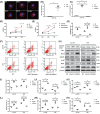
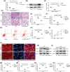
Figure 1. Exosomal miR-1-3p overexpressed in vivo and in vitro
(A) Endotoxin level of rat blood was assessed using an endotoxin detection kit 12 h after the CLP model was established (n=4, from four individuals). (B) The wet and dry weight (24 h) of the left lower lung was recorded and calculated for the wet/dry ratio (n=4, from four individuals). (C) Representative H&E staining of lung tissue under microscopy observation. The images are representative of four individuals. (D) Observation of CLP rat lung by electron microscopy. The images are representative of four individuals. (E–G) Western blotting analysis of active-casp3, Bcl-2, and Bax expression in the lung tissue of the CLP model, and β-actin served as an internal reference. The blots are representative of at least three independent experiments with similar results. (H) Exosomes were extracted from the plasma of sham and CLP rats by precipitation and then subjected to miRNA sequencing. KEGG analysis of the top 20 most significantly different miRNAs’ pathways. (I) MiR-1-3p was significantly expressed in the sequencing of CLP plasma-derived exosomes (CLP-Exo) (n=3, from three independent experiments). (J,K) RT-PCR to confirm the expression of miR-1-3p in CLP rats and sepsis patients’ (n=3, from three independent experiments) plasma-derived exosomes. (L) RT-PCR test of miR-1-3p expression in lung tissue of CLP rat (n=4, from four independent experiments). (M) RT-PCR test of miR-1-3p expression in the lung tissue of LPS administrated rat (n=4, from four independent experiments). (N) RT-PCR test of miR-1-3p expression in the intestine of CLP rat (n=4, from four independent experiments). (O) MiR-1-3p expression in Sham-Exo and CLP-Exo (precipitated from 50 μl of plasma) treated HUVECs for 24 h by RT-PCR (n=4, from four independent experiments). Data are presented as mean ± SE, *P<0.05. Abbreviation: RT-PCR, real-time polymerase chain reaction.
Figure 2
Figure 2. MiR-1-3p regulates proliferation, apoptosis, and related protein expression of HUVECs in vitro
(A) HUVECs were treated with LPS at different concentrations (0.1, 1, 5, 10, and 100 μg/ml). Cytoskeleton F-actin was stained with phalloidin and observed under a fluorescence microscope (400×). (B) MiR-1-3p expression analysis of LPS (10 μg/ml) stimulated HUVECs for 24 h by RT-PCR (n=4, from four independent experiments). (C) MiR-1-3p expression of HUVECs transfected with miR-1-3p mimics, inhibitor, and NC by RT-PCR detection (n=4, from four independent experiments). (D,E) CCK-8 for cell proliferation tests with or without LPS stimulation (n=6, from three independent experiments with duplicate, *P<0.05 in NC vs mimics, **P<0.05 in NC vs inhibitor, ***P<0.05 in mimics vs inhibitor). (F,G) Cell apoptosis assay by flow cytometry (n=3, from three independent experiments). (HN) Western blotting evaluated the expression of apoptosis-related proteins active-casp3, Bcl-2, Bax, ER stress protein GRP78, VEGF, and proinflammatory cytokines IL-1β and iNOS. β-actin served as an internal reference. The blots are representative of at least three independent experiments with similar results. Data are presented as mean ± SE, *P<0.05.
Figure 3
Figure 3. MiR-1-3p contribute to enhanced permeability of HUVECs
(A) HUVECs cytoskeleton F-actin was stained with phalloidin after miR-1-3p NC/mimics/inhibitor transfection with or without LPS treatment. The images are representative of three experiments with similar results. (BE) Western blotting analysis of p-MLC, p-FAK397, and Cx43 expression. β-actin served as an internal reference. The blots are representative of at least three independent experiments with similar results. (F) Transwell assays measured the effect of miR-1-3p on monolayer cell permeability by detecting the absorbance of BSA-Evans Blue (n=3, from three independent experiments). (G) LDH activity detection in the supernatant of transfected cells by an LDH detection kit (n=3, from three independent experiments). Data are presented as mean ± SE, *P<0.05.
Figure 4
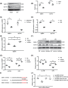
Figure 4. MiR-1-3p directly targets the 3′UTR of SERP1 and inhibits its expression
(A,B) Western blotting analysis of SERP1 and GLP1R expression in the lung tissue of CLP rats. The blots are representative of at least three independent experiments with similar results. (C,D) MRNA expression of SERP1 and GLP1R in CLP-exo and LPS (10 μg/ml) treated HUVECs for 24 h by RT-PCR (n=3, from three independent experiments). (E) SERP1 mRNA expression under the treatment of miR-1-3p mimics, inhibitor, and NC (n=3, from three independent experiments). (F–H) Western blotting detection of GLP1R and SERP1 protein levels in HUVECs transfected with miR-1-3p mimics, inhibitor, and NC. β-actin served as an internal reference. The blots are representative of at least three independent experiments with similar results. (I) Binding site of SERP1 mRNA 3′-UTR (SERP1-WT) with miR-1-3p and designed SERP1 3′-UTR mutant (SERP1-Mut) sequence. (J) Dual-luciferase reporter assay of miR-1-3p and SERP1 mRNA 3′-UTR binding sites (n=3, from three independent experiments). Data are presented as mean ± SE, *P<0.05.
Figure 5
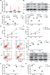
Figure 5. Down-regulated SERP1 inhibits proliferation and promotes apoptosis of HUVECs
(A) HUVECs were transfected with SERP1 siRNA (si-001, si-002, and si-003), and mRNA expression of SERP1 was assessed by RT-PCR to select the most efficient siRNA (n=4, from four independent experiments). (BD) Western blotting for SERP1 and GLP1R protein levels under siRNA transfection. Si-003 was the most efficient siRNA. The blots are representative of at least three independent experiments with similar results. (E,F) Effect of decreased SERP1 on HUVECs proliferation with or without LPS stimulation (n=6, from three independent experiments with duplicate). (G,H) Effect of decreased SERP1 on HUVEC apoptosis with or without LPS stimulation (n=3, from three independent experiments). (I–K) Western blotting analysis of apoptosis-related proteins active-casp3, Bcl-2, and Bax expression. The blots are representative of at least three independent experiments with similar results. β-actin served as an internal reference. Data are presented as mean ± SE, *P<0.05.
Figure 6
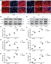
Figure 6. SERP1 induces HUVECs dysfunction by prompting permeability, membrane injury, and inflammatory response
(A) HUVECs cytoskeleton F-actin was stained with phalloidin after transfection with si-SERP1 with or without LPS treatment (400×). The images are representative of three experiments with similar results. (B–D) Western blotting detection of HUVECs permeability related p-MLC and Cx43 expression after transfection with si-SERP1. The blots are representative of at least three independent experiments with similar results. (EG) Western blotting detection of inflammatory cytokine IL-1β and iNOS expression after transfection with si-SERP1 in HUVECs. The blots are representative of at least three independent experiments with similar results. (H) Transwell assays measured the effect of SERP1 on monolayer cell permeability by detecting the absorbance of BSA-Evans Blue (n=3, from three independent experiments). (I) LDH activity analysis in the supernatant of si-SERP1 transfected HUVECs by an LDH detection kit (n=3, from three independent experiments). β-actin served as an internal reference. Data are presented as mean ± SE, *P<0.05.
Figure 7
Figure 7. SERP1 overexpression protects lung tissue and HUVECs from deleterious effects of miR-1-3p
(A) SERP1 mRNA expression in the lung tissue after miR-1-3p antagomir was locally trachea-administrated to the CLP rat model (n=4, from four individuals). (B,C) Western blotting analysis of SERP1 expression in the lung tissue, with β-actin served as an internal reference. The blots are representative of at least three independent experiments with similar results. (D) H&E staining of lung tissue under microscopic observation. The images are representative of three experiments with similar results. (E) The wet/dry ratio of the left lower lung in each group (n=4, from four individuals). (F) HUVECs were transfected with SERP1 plasmid (SERP1−over) and/or mimics, and mRNA expression of SERP1 was assessed by RT-PCR to verify the transfection efficiency (n=3, from three independent experiments). (G,H) Flow cytometry analysis of apoptosis after SERP1 overexpression (n=3, from three independent experiments). (I) Effect of SERP1 overexpression on HUVEC proliferation was assessed by CCK8 (n=6, from three independent experiments with duplicate). *P<0.05 in LPS+NC vs LPS+mimics, **P<0.05 in LPS+NC vs LPS+mimics+SERP1−over. (J) HUVECs cytoskeleton F-actin was stained with phalloidin after transfecting with SERP1 plasmid (SERP1−over) and/or mimics under LPS treatment (fluorescence microscope, 400×). The images are representative of three experiments with similar results. (K,M,N) Western blotting analysis of permeability related p-MLC and Cx43 expression after transfecting with SERP1 plasmid (SERP1−over) and/or mimics under LPS treatment. β-actin served as an internal reference. The blots are representative of at least three independent experiments with similar results. (L,O,P) Western blotting detection of inflammatory cytokine IL-1β and iNOS expression after transfection with SERP1 plasmid (SERP1 over) and/or mimics under LPS situation. The blots are representative of at least three independent experiments with similar results. β-actin served as an internal reference.

References

    1. Rhodes A., Evans L.E., Alhazzani W., Levy M.M., Antonelli M., Ferrer R. et al. . (2017) Surviving Sepsis Campaign: International Guidelines for Management of Sepsis and Septic Shock: 2016. Intensive Care Med. 43, 304–377 10.1007/s00134-017-4683-6 - DOI - PubMed
    1. Miyashita T., Ahmed A.K., Nakanuma S., Okamoto K., Sakai S., Kinoshita J. et al. . (2016) A three-phase approach for the early identification of acute lung injury induced by severe sepsis. In Vivo (Brooklyn) 30, 341–349, http://www.ncbi.nlm.nih.gov/pubmed/27381595 - PubMed
    1. Sevransky J.E., Martin G.S., Shanholtz C., Mendez-Tellez P.A., Pronovost P., Brower R. et al. . (2009) Mortality in sepsis versus non-sepsis induced acute lung injury. Crit. Care 13, R150 10.1186/cc8048 - DOI - PMC - PubMed
    1. Deutschman C.S. and Tracey K.J. (2014) Sepsis: current dogma and new perspectives. Immunity 40, 463–475 10.1016/j.immuni.2014.04.001 - DOI - PubMed
    1. Park E.J., Appiah M.G., Myint P.K., Gaowa A., Kawamoto E. and Shimaoka M. (2019) Exosomes in sepsis and inflammatory tissue injury. Curr. Pharm. Des. 25,4486–4495 - PubMed

Publication types

MeSH terms (VSports最新版本)

"VSports注册入口" Substances