Skip to main page content
U.S. flag

An official website of the United States government

Dot gov

The . gov means it’s official. Federal government websites often end in . gov or . mil VSports app下载. Before sharing sensitive information, make sure you’re on a federal government site. .

Https

The site is secure. The https:// ensures that you are connecting to the official website and that any information you provide is encrypted and transmitted securely. V体育官网.

. 2021 Jun;28(6):1773-1789.
doi: 10.1038/s41418-020-00700-z. Epub 2020 Dec 16.

Deubiquitinating enzyme OTUB1 promotes cancer cell immunosuppression via preventing ER-associated degradation of immune checkpoint protein PD-L1

Affiliations

Deubiquitinating enzyme OTUB1 promotes cancer cell immunosuppression via preventing ER-associated degradation of immune checkpoint protein PD-L1

Dan Zhu et al. Cell Death Differ. 2021 Jun.

Abstract

Upregulation of programmed death ligand 1 (PD-L1) helps tumor cells escape from immune surveillance, and therapeutic antibodies targeting PD-1/PD-L1 have shown better patient outcomes only in several types of malignancies. Recent studies suggest that the clinical efficacy of anti-PD-1/PD-L1 treatments is associated with PD-L1 levels; however, the underlying mechanism of high PD-L1 protein levels in cancers is not well defined. Here, we report that the deubiquitinase OTUB1 positively regulates PD-L1 stability and mediates cancer immune responses through the PD-1/PD-L1 axis VSports手机版. Mechanistically, we demonstrate that OTUB1 interacts with and removes K48-linked ubiquitin chains from the PD-L1 intracellular domain in a manner dependent on its deubiquitinase activity to hinder the degradation of PD-L1 through the ERAD pathway. Functionally, depletion of OTUB1 markedly decreases PD-L1 abundance, reduces PD-1 protein binding to the tumor cell surface, and causes increased tumor cell sensitivity to human peripheral blood mononuclear cells (PBMCs)-mediated cytotoxicity. Meanwhile, OTUB1 ablation-induced PD-L1 destabilization facilitates more CD8+ T cells infiltration and increases the level of IFN-γ in serum to enhance antitumor immunity in mice, and the tumor growth suppression by OTUB1 silencing could be reversed by PD-L1 overexpression. Furthermore, we observe a significant correlation between PD-L1 abundance and OTUB1 expression in human breast carcinoma. Our study reveals OTUB1 as a deubiquitinating enzyme that influences cancer immunosuppression via regulation of PD-L1 stability and may be a potential therapeutic target for cancer immunotherapy. .


Conflict of interest statement

The authors declare that they have no conflict of interest.

V体育ios版 - Figures

Fig. 1
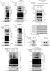
Fig. 1. OTUB1 maintains PD-L1 protein stability.
a Screening of OTU family members that regulate PD-L1 protein level. MDA-MB-231 cells individually were infected with shRNAs targeting different OTU subfamily DUBs, and the expression of PD-L1 was detected. b Immunoblotting of PD-L1 expression in HEK293T cells transfected with increasing amounts of Flag-OTUB1 together with a constant amount of HA-PD-L1 plasmid in each group. GFP expression was used to confirm the constant transfection efficiency across the four experimental groups. c Immunoblot analysis of PD-L1 in MDA-MB-231 and BT-549 cells infected with shOTUB1- or shControl-encoding lentivirus, respectively. d The qRT-PCR analyses of PD-L1 mRNA expression in MB-MDA-231 and BT-549 cells stably expressing shControl or shOTUB1. Data are shown as the mean ± SEM (n = 3). Statistical analysis was performed using Student’s t test. n.s., not significant. e Immunoblotting of PD-L1 expression in RKO, HepG2, and A549 cells after introducing shControl or shOTUB1 lentiviruses. f Half-life analysis of PD-L1 in shControl or shOTUB1 MDA-MB-231 cells treated with 50 µg/mL cycloheximide for the indicated times before harvesting. g Semi-quantification of PD-L1 levels, with β-tubulin used as a loading control. Relative PD-L1 levels at time 0 were set as 1.
Fig. 2
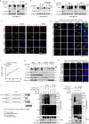
Fig. 2. OTUB1 specifically interacts with PD-L1 in vivo and in vitro.
a HEK293T cells were transfected with HA-PD-L1 or HA vector, and immunoprecipitation was performed with anti-HA antibody to examine the interaction between HA-PD-L1 and endogenous OTUB1. b Association between endogenous OTUB1 and PD-L1 in MDA-MB-231 cell lines was detected by Co-IP assays using IgG or PD-L1 antibodies. c In situ interaction between OTUB1 and PD-L1. Cells were fixed with 4% paraformaldehyde, immunostained with OTUB1 and PD-L1 antibodies and then assessed using the Duolink PLA assay. Scale bar, 10 µm. Quantification of the PLA dots indicating PD-L1-OTUB1 interactions was shown as mean ± SEM. d The direct interaction between recombinant Flag-PD-L1 19-290 aa proteins and His-OTUB1 proteins examined by in vitro pull-down assays. e Schematic representation of various OTUB1 truncations. f Mapping of OTUB1 domains critical for PD-L1 binding. HEK293T cells were transfected with different OTUB1 truncations, and cell lysates were immunoprecipitated with anti-Flag antibody to detect their PD-L1 binding ability. g Schematic representation of PD-L1 full-length and ΔC-tail constructs. h The interaction between Flag-OTUB1 and PD-L1 full-length or ΔC-tail detected by Co-IP assays.
Fig. 3
Fig. 3. OTUB1 stabilizes PD-L1 through cleaving K48-linked poly-ubiquitin chains of PD-L1.
a HA-PD-L1 ubiquitination levels in HEK293T cells transfected with Flag-vector or Flag-OTUB1 were analyzed by His pull-down assay. b The effect of PD-L1 ubiquitination by depleted OTUB1 was examined in OTUB1WT, OTUB1-/- and re-expressed OTUB1 HEK293T cells by His pull-down assay. Two OTUB1-/- strains were obtained through CRISPR/Cas9 genome editing. c The shControl- and shOTUB1- MDA-MB-231 cells were subjected to TUBE pull-down assays to examine the effect of depleted OTUB1 on endogenous ubiquitination levels of PD-L1. d The influence of OTUB1 on specific ubiquitination types of endogenous PD-L1 was determined by immunoblotting analysis using the K48- or K63- ubiquitin linkage-specific antibodies. MDA-MB-231 cells infected with shControl- or shOTUB1-encoding lentiviruses were treated with MG132 overnight and then subjected to immunoprecipitation. e In vitro deubiquitination assays of recombinant OTUB1 proteins and enriched K48-linked or K63-linked ubiquitinated PD-L1 from cell extracts. The mixture was incubated at 30 °C for 4 h and then analyzed by immunoblotting. f Schematic representation of the OTUB1 C91S, D88A, ASA (D88A/C91S/H265A) mutants and ΔN truncation. Stars represent mutated amino acids in the OTU domain. g Examination of the effect of wild-type OTUB1 or ASA mutant on PD-L1 abundance. GFP expression was used to confirm constant transfection efficiency across the three experimental groups. h Determination of PD-L1 levels after re-expression of the indicated OTUB1 mutants. The shOTUB1 MDA-MB-231 cells were infected with lentiviruses stably expressing shRNA-resistant wild-type OTUB1 or ASA mutant and then subjected to immunoblotting of PD-L1. i Immunoblot analysis of PD-L1 ubiquitination affected by wild-type OTUB1, ASA, C91S, D88A, or ΔN truncation mutants. HEK293T cells were transfected with HA-tagged PD-L1 and His-ubiquitin along with different OTUB1 mutants. Following MG132 treatment, cell extracts were enriched by Ni+-NTA agarose and then prepared for immunoblotting with the indicated antibodies. j Detection of endogenous K48-linked polyubiquitination of PD-L1 affected by OTUB1. MDA-MB-231 cells with shControl or shOTUB1 and OTUB1-depleted cells with re-overexpressed wild-type or mutation constructs of Flag-OTUB1 were subjected to K48-TUBE pull-down analyses.
Fig. 4
Fig. 4. OTUB1 regulates PD-L1 abundance through ERAD pathway.
a The MDA-MB-231 cells harboring shControl or shOTUB1 treated with or without 10 µM MG132 for 4 h were collected to analyze PD-L1 expression. Red stars represent incomplete glycosylated PD-L1. b Immunoblot analysis of PD-L1 in shControl or shOTUB1 MDA-MB-231 cells treated with or without the lysosomal inhibitor chloroquine (20 µM). c MDA-MB-231 cells with shControl and shOTUB1 treated with MG132 were immunostained with a PD-L1 antibody (red), a calnexin antibody (green), and a DAPI antibody (blue). Scale bar, 50 µm. d The shOTUB1 MDA-MB-231 cells were treated with the ERAD pathway inhibitor Eer I (10 µM) at the indicated times for immunoblot detection of PD-L1 expression. Red stars represent incomplete glycosylated PD-L1. e MDA-MB-231 cells were subjected to immunofluorescence assays with calnexin (ER), TGN46 (Golgi), Rab5 (endosome), Rab11 (recycling endosome), and Na+/K+ ATPase (plasma membrane) antibodies following Duolink PLA to identify the location of OTUB1 interaction with PD-L1. f The positive correlation between PD-L1-OTUB1 PLA dots and the fluorescence intensity of ER marker calnexin was shown in scatterplot. Pearson’s coefficient tests were performed to assess statistical significance (Pearson r2 = 07379, p < 0.001, n = 20). g The presence of PD-L1 and OTUB1 in different fractions was analyzed by immunoblotting, using antibodies against PD-L1 and OTUB1, the ER protein calnexin, cytosolic α-Tubulin, as well as the nuclear protein histone H3. h The in situ binding between PD-L1 and OTUB1 was detected by Duolink PLA immunofluorescence using two antibodies specific for the PD-L1 intracellular domain (ICD) (EPR19759 and 13684 S) and extracellular domain (ECD) (17952-1-AP). i Schematic representation of HA-PD-L1 ΔC-tail and 5KR constructs. OTUB1WT and OTUB1-/- HEK293T cells transfected with PD-L1 full-length or the 5KR mutant were subjected to His pull-down and SDS-PAGE analyses. j The effect of OTUB1 on the ubiquitination of full-length PD-L1 or ΔC-tail truncation was monitored by His-ubiquitin pull-down assay.
Fig. 5
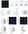
Fig. 5. Depletion of OTUB1 impairs PD-L1 function in T cell-mediated cancer cell killing.
a Immunofluorescence assays to detect PD-1 binding intensity. MDA-MB-231 cells stably expressing shControl or shOTUB1 were incubated with recombinant human PD-1 Fc protein and then anti-human Alexa Fluor 488 dye. Scale bar, 50 µm. b Normalized intensity of PD-1 binding and immunoblotting of OTUB1 expression to show knockdown efficiency. c FACS analysis of PBMCs-mediated killing of tumor cells using Annexin V and propidium iodide (PI) double staining. Healthy cells are negative for both stains in the Q4 quadrant. The Q3 quadrant shows Annexin V-positive cells, which are in the early stage of apoptosis. The Q2 quadrant shows cells that are both Annexin V- and PI-positive, which are in the late stage of apoptosis. d Quantitative apoptotic measurement of Q2 and Q3 quadrants. Greater than 104 cells were counted for each group. e Immunoblotting of cleaved caspase-3 levels in shControl or shOTUB1 MDA-MB-231 cells after incubation with activated PBMCs. f The qRT-PCR analyses of IFN-γ and TNF-α mRNA expressions in PBMCs after co-culture with MDA-MB-231 shControl or shOTUB1 cells. g, h The Ki67 or TUNEL staining of PBMCs after co-culture with MDA-MB-231 shControl or shOTUB1 cells. The percentage of Ki67 or TUNEL positive staining cells were analyzed using the ImageJ software. The results of b, d, f, g and h are shown as the mean ± SEM (n = 3). Statistical analysis was performed using Student’s t test. **p < 0.01, ***p < 0.001.
Fig. 6
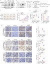
Fig. 6. OTUB1 silencing-mediated downregulation of PD-L1 promotes antitumor immunity.
a Immunoblot analysis of PD-L1 in murine 4T1, B16-F10, and CT26 tumor cells with overexpressed OTUB1. b Experimental design for the in vivo tumorigenesis assay and the expression levels of OTUB1 and PD-L1 in four different 4T1 cell types (shOTUB1, PD-L1, shOTUB1/PD-L1, and control) were confirmed by immunoblot analysis. c Photographs of mice tumors of each group (n = 8) at the end of the experiment. d The curve graph exhibited the tumor growth measured at different time-points after inoculation (n = 8). e The image shows the tumor weight for each group (n = 8). f The abundance of IFN-γ (pg/mL) in mice serum (n = 5) detected by ELISA assays. g IHC of PD-L1 expression (Scale bar, 50 µm), Ki67 staining (Scale bar, 20 µm), and CD8+ cell infiltration (Scale bar, 20 µm) in tumor sections (n = 6 for each group). Statistical results are quantified by ImageJ software. Data in d, e, f, and g are presented as the mean ± SEM in each group, and statistical analysis was performed using Student’s t test. n.s., not significant, *p < 0.05, **p < 0.01, ***p < 0.001.
Fig. 7
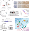
Fig. 7. OTUB1 correlates with PD-L1 abundance in breast carcinoma.
a Bioinformatic analyses of PD-L1 and OTUB1 expression in breast invasive carcinoma (BRCA) patients (n = 1085) relative to those of healthy controls (n = 291). b Representative images of IHC staining of PD-L1 and OTUB1 in human BRCA specimens. Scale bars, 50 µm. c Pearson correlation analysis to determine the degree of association between OTUB1 and PD-L1 by IHC staining (n = 86). P-value was obtained by Student’s t test. d Immunoblotting of PD-L1 and OTUB1 expression in the indicated human breast cancer cell lines. e Kaplan–Meier curve of overall survival of patients in the TCGA BRCA database. Patients were divided into the high OTUB1 group (n = 399) and the low OTUB1 group (n = 398). The difference in the overall survival between these two groups was determined using a log-rank test. f Crystal violet staining and CCK8 analysis to detect tumor cell viability after incubation with activated PBMCs. The results of CCK8 assay are shown as the mean ± SEM (n = 3) and statistical analysis was performed using Student’s t test (**p < 0.01). The expression levels of OTUB1 and PD-L1 in different MDA-MB-231 cell types were confirmed by immunoblot analysis. g Working model for regulation of PD-L1 stability and cancer cell immunosuppression by OTUB1.

References

    1. Zou W, Wolchok JD, Chen L. PD-L1 (B7-H1) and PD-1 pathway blockade for cancer therapy: Mechanisms, response biomarkers, and combinations. Sci Transl Med. 2016;8:328rv324. - "VSports注册入口" PMC - PubMed
    1. Chen L, Han X. Anti-PD-1/PD-L1 therapy of human cancer: past, present, and future. J Clin Invest. 2015;125:3384–91. - PMC (VSports最新版本) - PubMed
    1. Bardhan K, Anagnostou T, Boussiotis VA. The PD1:PD-L1/2 pathway from discovery to clinical implementation. Front Immunol. 2016;7:550. - PMC - PubMed
    1. Freeman GJ, Long AJ, Iwai Y, Bourque K, Chernova T, Nishimura H, et al. Engagement of the PD-1 immunoinhibitory receptor by a novel B7 family member leads to negative regulation of lymphocyte activation. J Exp Med. 2000;192:1027–34. - PMC - PubMed
    1. Iwai Y, Hamanishi J, Chamoto K, Honjo T. Cancer immunotherapies targeting the PD-1 signaling pathway. J Biomed Sci. 2017;24:26. - PMC - PubMed

Publication types

MeSH terms

Substances