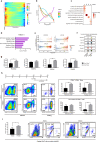
<b draggable="Hxk8V6p"><bdo draggable="UoUbw"></bdo></b><area dropzone="x1ZBBP"></area> Skip to main page content
U.S. flag

An official website of the United States government

Dot gov

The . gov means it’s official. Federal government websites often end in . gov or . mil. Before sharing sensitive information, make sure you’re on a federal government site. VSports app下载.

Https

The site is secure. The https:// ensures that you are connecting to the official website and that any information you provide is encrypted and transmitted securely. V体育官网.

. 2020 Dec 1;6(1):89.
doi: 10.1038/s41421-020-00213-6.

Characterization and generation of human definitive multipotent hematopoietic stem/progenitor cells

Affiliations

"VSports最新版本" Characterization and generation of human definitive multipotent hematopoietic stem/progenitor cells

V体育官网 - Yanling Zhu et al. Cell Discov. .

VSports手机版 - Abstract

Definitive hematopoiesis generates hematopoietic stem/progenitor cells (HSPCs) that give rise to all mature blood and immune cells, but remains poorly defined in human. Here, we resolve human hematopoietic populations at the earliest hematopoiesis stage by single-cell RNA-seq. We characterize the distinct molecular profiling between early primitive and definitive hematopoiesis in both human embryonic stem cell (hESC) differentiation and early embryonic development VSports手机版. We identify CD44 to specifically discriminate definitive hematopoiesis and generate definitive HSPCs from hESCs. The multipotency of hESCs-derived HSPCs for various blood and immune cells is validated by single-cell clonal assay. Strikingly, these hESCs-derived HSPCs give rise to blood and lymphoid lineages in vivo. Lastly, we characterize gene-expression dynamics in definitive and primitive hematopoiesis and reveal an unreported role of ROCK-inhibition in enhancing human definitive hematopoiesis. Our study provides a prospect for understanding human early hematopoiesis and a firm basis for generating blood and immune cells for clinical purposes. .


Conflict of interest statement

The authors declare that they have no conflict of interest.

Figures
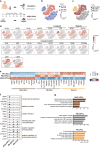
Fig. 1
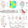
Fig. 1. Single-cell RNA sequencing of hPSCs-derived HPCs and adult peripheral blood HSPCs.
a Scheme of the experiment design. Fresh LinCD34+CD38 HSPCs mobilized in human peripheral blood (PB-HSPCs) were sorted by FACS. Human ESCs were differentiated into blood lineages in a monolayer, defined condition. The floating blood cells at differentiation day 8 were sorted by CD43. The sorted cells were analyzed by 10× genomics for single-cell RNA sequencing (scRNA-seq). b t-SNE projection of PB-HSPC and hESC-HPC sample cells assigned based on samples. Each dot represents one cell and colors represent cell samples. c t-SNE projection of hESC-HPC, PB-HSPC, and PB-LPC cluster cells assigned based on clusters. Each dot represents one cell and cells are colored according to their assigned clusters. d Expressions of known hematopoietic and blood lineage marker genes at single-cell resolution. Color displays expression level (TPM, Log-scaled). e Expression profiles of top highly expressed genes in hESC-HPC, PB-HSPC and PB-LPC clusters. Color displays the scaled expression level (TPM, z-normalized) and diameter denotes fractional expression. f Violin plots show expression levels (TPM, Log-scaled) of selected hematopoietic and blood lineage marker genes with GO terms enriched in hESC-HPC, PB-HSPC and PB-LPC clusters. Colors represent clusters. g Top Gene Ontology (GO) terms enriched in hESC-HPC, PB-HSPC, and PB-LPC clusters. Colors represent clusters. The length of bar represents P value.
Fig. 2
Fig. 2. hPSCs-derived HPCs contain cells with different haematopoiesis states.
a t-SNE projection of pseudotime order of hESC-HPC sample cells. Each dot represents one cell and cells are colored based on their putative pseudotime values. b Expression profiles of selected hematopoietic, myeloid and lymphoid marker genes along with the pseudotime progression of hESC-HPC sample. Color displays the scaled expression level (TPM, z-normalized). c t-SNE projection of hESC-HPC sample cells assigned to different stages based on pseudotime progression. Each dot represents one cell and cells are colored according to their assigned stages. d Expression profiles of selected hematopoietic, myeloid, and lymphoid marker genes in hESC-HPC sample at different stages of pseudotime progression. Color displays the scaled expression level (TPM, z-normalized) and diameter denotes fractional expression. e Pearson correlation between PB-HSPC sample cells and hESC-HPC sample cells at different pseudotime stages. Color represents coefficient of correlation computed based on bulk profiles merged from single cells. f Expression profiles of top highly expressed genes in hESC-HPC sample cells at different pseudotime stages. Color displays expression level (TPM, Log-scaled) and diameter denote fractional expression. g Expression profiles of selected blood marker genes in hESC-HPC sample. Each dot represents one cell and color displays expression level (TPM, Log-scaled). h Expression profiles of selected blood marker genes in hESC-HPC sample along the pseudotime. Each dot represents one cell and color represents pseudotime value. Line shows mean expression level (TPM, Log-scaled).
Fig. 3
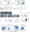
Fig. 3. Identification of definitive multi-potent HSPCs in hESC differentiation.
a Digital cytometry analysis on hESC-HPC and PB-HSPC sample cells based on CD44 and GATA1 expressions. Gate shows the fraction of CD44+GATA1 cells in PB-HSPC and hESC-HPC samples. Colors represent density of cells. b Frequencies of cells of hESC-HPCs or PB-HSPCs based on expressions of CD44 and/or GATA1. c FACS analysis of CD44 expression in PB-HSPCs and hESC-HPCs. d Experimental strategy for functional characterization of hESC-HPCs. e Representative pictures of CFUs formed by hESC-HPCs sorted by CD43+CD44+ or CD43+CD44. Scale bar, 100 μm; ND, not detected; E, erythroid; G, granulocytes; M, macrophages; GM, granulocyte and monocyte-macrophage; Mix, mixed erythro-myeloid. f Quantitative analysis of various blood CFUs formed by the CD43+CD44+ or CD43+CD44 hESC-HPCs sorted at differentiation day 8. g May–Grunwald–Giemsa staining of different blood cells isolated from CFUs derived from CD43+CD44+ hESC-HPCs. Scale bar, 10 μm. h: Flow cytometry analysis on lineage output (indicated in quadrants) of CD44-sorted hESC-HPCs at differentiation day 8 co-cultured with MS5 or OP9-DL4 (OL4). The human hematopoietic cells were gated 2 weeks (MS5) or 4 weeks (OL4) after differentiation and analyzed for known lineage markers as indicated. G, granulocytes; M, macrophages; Mk, megakaryocytes; E, erythroid; NK, nature killer cells; T, T cells.
Fig. 4
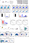
Fig. 4. Analysis of multi-potency of hESC-HSPCs by single-cell clonal assay and in vivo transplantation.
a Experimental design of clonal assays of lineage potential of hESC-HSPCs co-culturing with MS-5. CD43+CD44+ hESC-HSPCs were sorted at differentiation day 8 and seeded on MS-5 cells in 10 96-wells plates at single-cell level. Myeloid and/or lymphoid lineage outputs were examined 4 weeks after co-culturing. b Morphology of single hESC-HSPCs co-culturing with MS-5 for 4 weeks. Scale bar: 100 μm. c FACS analysis of myeloid and/or lymphoid lineage outputs of single hESC-HSPCs co-culturing with MS-5. The CD45+ human hematopoietic cells were gated 4 weeks after differentiation and analyzed for known lineage markers as indicated. CD33 for myeloid and CD56 for lymphoid/NK. Left lower panel: quantitative data of total cloning efficiency of single hESC-HSPCs that can form blood clones. Right lower panel: percentage of myeloid and/or lymphoid clones of total formed blood clones by single hESC-HSPCs. d FACS analysis of erythroid, megakaryocyte and/or myeloid lineage outputs of single hESC-HSPCs co-culturing with MS-5. The cells were gated 4 weeks after differentiation and erythroid clones were defined as hCD235a+ only (Er only). Megakaryocyte clones were defined as hCD41+ only (Mk only). Erythroid/megakaryocyte clones were defined as hCD235a+ and hCD41+ but negative for hCD11b (Er/Mk). Erythroid/myeloid clones were defined as hCD235a+ and hCD11b+ but negative for hCD41 (Er/My). Erythroid/megakaryocyte/ myeloid clones were defined as hCD235a+, hCD41+ and hCD11b+ (Er/Mk/My). Left lower panel: quantitative data of total cloning efficiency of single hESC-HSPCs that can form blood clones. Right lower panel: percentage of erythroid, megakaryocyte, and/or myeloid clones of total formed blood clones by single hESC-HSPCs. e Lineage cloning efficiency of single hESC-HSPCs shown in c and d, presented as the proportion of cells of various lineages in positive wells. Er, Erythroid potential (erythroid plus mixed); MK, Megakaryocyte potential (megakaryocyte plus mixed); My, Myeloid potential (myeloid plus mixed); Ly/NK, Lymphoid potential (lymphoid plus mixed). f Limiting-dilution assays for myeloid and/or lymphoid frequencies of hESC-HSPCs. LDA plots show the frequency (f: 1 in X cells can give rise to) of CD44+ hESC-HSPCs in myeloid, lymphoid and myeloid-lymphoid potential. Plots are generated by ELDA software. g Experimental design of in vivo transplantation of hESC-HSPCs. h Human CD45+ cell engraftment in the injected tibias of NSI mice 2 weeks after transplantation of hESC-HSPCs. i, j In vivo lineage potential of hESC-HSPCs. CD43+CD44+ hESC-HSPCs were sorted at differentiation day 8 and transplanted into the tibia of NSI mice. Multilineage outputs were examined 2 weeks after transplantation. Myeloids (My), Nature killer cells (NK), B cells, Granulocytes (G) and macrophages (M) were gated for human CD45+ events. Megakaryocytes and erythrocytes were defined as hCD41+ only (Mk) or hCD235a+ (E) only.
Fig. 5
Fig. 5. CD44 discriminates primitive and definitive hematopoiesis in hESC differentiation.
a Immunostaining analysis of nascent HPCs derived from hESCs by anti-CD31, anti-CD43, anti-CD44 at day 8 of hematopoietic differentiation. White arrow, CD43+CD44 cells; Yellow arrow, CD43+CD44+ cells. Scale bar, 20 μm. b FACS analysis of the adherent hESC-HPCs differentiated at day 8 by anti-CD31, anti-CD43, anti-CD44. c qRT-PCR analysis of the indicated primitive, definitive and endothelial genes in each subpopulation sorted by anti-CD31, anti-CD43, and anti-CD44 at differentiation day 8. The significance level was determined using unpaired two-tailed Student’s t-tests, ***P < 0.001. The data represent mean ± SD from three independent replicates (n = 3). d The expanding of CD43+CD44+ and CD43+CD44- HPCs. The cells were sorted at differentiation day 8 and cultured in human HSC medium. The total cell numbers of the indicated population were counted at indicated time points. Significance level was determined using unpaired two-tailed Student’s t-tests, **P < 0.01, ***P < 0.001. The data represent mean ± SD from three independent replicates (n = 3). e Morphology of CFU-E derived from CD43+CD44+ or CD43+CD44 HPCs. Scale bar, 100 μm. f qRT-PCR analysis of indicated globin genes in erythrocytes derived from CD43+CD44+ or CD43+CD44 HPCs. The significance level was determined using unpaired two-tailed Student’s t-tests, ***P < 0.001. The data represent mean ± SD from three independent replicates (n = 3).
Fig. 6
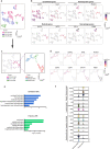
Fig. 6. Definitive human hematopoiesis in early embryonic development.
a t-SNE projection of endothelial and hematopoietic cells, resulting from sub-dividing the cells in Supplementary Fig. S1a as indicated in Supplementary Fig. S1c, assigned based on samples. Each dot represents one cell and colors represent cell samples. The legend shows the number of cells in each sample. b Expressions of known blood and endothelial marker genes at single-cell resolution. Color displays expression level (TPM, Log-scaled). c t-SNE projection of erythroid, definitive HSPC and primitive HPC cluster cells, resulting from sub-dividing the cells in Fig. 6a as indicated in the left panel, according to blood and endothelial marker gene expression as indicated in Fig. 6b. d Expression profiles of selected feature genes in erythroid, definitive HSPC and primitive HPC cluster cells. Each dot represents one cell and color displays expression level (TPM, Log-scaled). e Top Gene Ontology (GO) terms enriched in definitive HSPC and primitive HPC clusters. Colors represent cell clusters. f Violin plots show expression levels (TPM, Log-scaled) of selected myeloid/erythroid, lymphoid and hematopoietic stem/progenitor cell marker genes in definitive HSPC and primitive HPC clusters.
Fig. 7
Fig. 7. ROCK inhibition promotes the generation of definitive HSPCs from hESCs.
a, b Expression profiles of genes significantly upregulated or downregulated along the pseudotime progression. Based on changing profile, genes were clustered hierarchically into four different patterns (b). Lines show the mean values of scaled expression levels (TPM, z-normalized) in each pattern. c Featured GO terms enriched in genes of each pattern. Color displays P-value (Log-scaled) of GO terms. d KEGG enriched genes in pattern 1 indicated in b. e Expression profiles of ROCK pathway-related genes along the pseudotime. Each dot represents one cell and color represents pseudotime value. Line shows the mean expression level (TPM, Log-scaled). f Violin plots show expression levels (TPM, Log-scaled) of these ROCK signaling genes in early definitive and primitive HSPCs identified in human embryo. g qRT-PCR analysis of the ROCK pathway-related genes in CD43+CD44+ or CD43+CD44 HPCs sorted at differentiation day 8. The significance level was determined using unpaired two-tailed Student’s t-tests, *P < 0.05, **P < 0.01, ***P < 0.001. The data represent mean ± SD from three independent replicates (n = 3). h Analysis of ROCK inhibition in hESC differentiation. The chemical inhibitor, Y-27632, was added in EHT medium at differentiation day 4 as indicated. The adherent or floating cells at differentiation day 8 were analyzed by FACS for the expression of CD31, CD43, and CD44. Right panel: quantitative data of each indicated population at differentiation day 8 of hESCs. The significance level was determined using unpaired two-tailed Student’s t-tests, ***P < 0.001. The data represent the mean ± SD from three independent replicates (n = 3). i Function Analysis of CD43+ HSPCs generated under ROCKi treatment. Y-27632 was added in EHT medium at differentiation day 4 as indicated. The floating CD43+ cells at differentiation day 8 were counted and co-cultured with MS5 with 10,000 cells per well. Human CD45+ cells were gated 2 weeks after differentiation and analyzed for the known lineage markers as indicated. My, myeloid; Ly, lymphoid. The significance level was determined using unpaired two-tailed Student’s t-tests, *P < 0.05. The data represent mean ± SD from three independent replicates (n = 3).

References

    1. Laurenti E, Gottgens B. From haematopoietic stem cells to complex differentiation landscapes. Nature. 2018;553:418–426. doi: 10.1038/nature25022. - DOI - PMC - PubMed
    1. Haas S, Trumpp A, Milsom MD. Causes and consequences of hematopoietic stem cell heterogeneity. Cell Stem Cell. 2018;22:627–638. doi: 10.1016/j.stem.2018.04.003. - DOI - PubMed
    1. Cvejic A. Mechanisms of fate decision and lineage commitment during haematopoiesis. Immunol. Cell Biol. 2015;94:230–235. doi: 10.1038/icb.2015.96. - DOI - PubMed
    1. Manz MG, Miyamoto T, Akashi K, Weissman IL. Prospective isolation of human clonogenic common myeloid progenitors. Proc. Natl Acad. Sci. USA. 2002;99:11872–11877. doi: 10.1073/pnas.172384399. - DOI - PMC - PubMed
    1. Galy A, Travis M, Cen D, Chen B. Human T, B, natural killer, and dendritic cells arise from a common bone marrow progenitor cell subset. Immunity. 1995;3:459–473. doi: 10.1016/1074-7613(95)90175-2. - DOI - PubMed