VSports最新版本 - Skip to main page content
U.S. flag

An official website of the United States government

Dot gov

The . gov means it’s official. Federal government websites often end in . gov or VSports app下载. mil. Before sharing sensitive information, make sure you’re on a federal government site. .

Https

The site is secure. The https:// ensures that you are connecting to the official website and that any information you provide is encrypted and transmitted securely V体育官网. .

. 2020 Dec 1;12(23):24394-24423.
doi: 10.18632/aging.202293. Epub 2020 Dec 1.

E3 ligase FBXW7 restricts M2-like tumor-associated macrophage polarization by targeting c-Myc

Affiliations

E3 ligase FBXW7 restricts M2-like tumor-associated macrophage polarization by targeting c-Myc (V体育安卓版)

Lijia Zhong (V体育2025版) et al. Aging (Albany NY). .

Abstract

FBXW7 functions as an E3 ubiquitin ligase to mediate oncoprotein degradation via the ubiquitin-proteasome system in cancer cells, effectively inhibiting the growth and survival of tumor cells. However, little is known about the functions of FBXW7 in macrophages and the tumor immune microenvironment. In this study, we find that FBXW7 suppresses M2-like tumor-associated macrophage (TAM) polarization to limit tumor progression. We identified a significant increase in the proportion of M2-like TAMs and aggravated tumor growth in mice with myeloid FBXW7 deficiency by subcutaneous inoculation with Lewis lung carcinoma cells (LLCs). When stimulated with LLCs supernatant in vitro, FBXW7-knockout macrophages displayed increased M2 macrophage polarization and enhanced ability of supporting cancer cells growth. In mechanism, we confirmed that FBXW7 inhibited M2-like TAM polarization by mediating c-Myc degradation via the ubiquitin-proteasome system VSports手机版. These findings highlight the role of FBXW7 in M2-like TAM polarization and provide new insights into the potential targets for cancer immunotherapies. .

Keywords: FBXW7; c-Myc; macrophage polarization; tumor-associated macrophages; ubiquitination. V体育安卓版.


Conflict of interest statement

CONFLICTS OF INTEREST: The authors have declared that no conflicts of interest exist.

"VSports" Figures

Figure 1
Figure 1
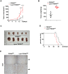
Lysm+FBXW7f/f mice show aggravated tumor progression in an LLC-inoculated model. (A) LLCs were subcutaneously injected into the mice. The volume of tumors from FBXW7f/f and Lysm+FBXW7f/f mice were measured and recorded in 16 days (n = 5 per group). (B) The weight of tumors at 16 days from both groups (n = 5 per group). (C) The appearance of tumors dissected 16 days after inoculation in FBXW7f/f and Lysm+FBXW7f/f mice. (D) The survival curve of tumor-bearing FBXW7f/f and Lysm+FBXW7f/f mice (n = 10 per group). (E) Representative images of tumors from two groups immunohistochemically stained with an antibody against Ki67. Scale bars: 10× was 200 μm, 40× was 50 μm. Data are expressed as the mean ± SD and are representative of three independent experiments. *P < 0.05; **P < 0.01; ****P < 0.0001 (two-way ANOVA (A), Student’s t test (B) and log rank test (D)).
Figure 2
Figure 2
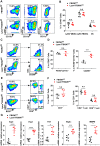
FBXW7 knockout in myeloid cells remodels the tumor immune microenvironment by promoting M2-like TAM polarization. (A) Tumors were digested to obtain single-cell suspensions. Ly6G+CD11b+ myeloid-derived suppressor cells (MDSCs), Ly6C+CD11b+ MDSCs, and MHCII+CD11c+ dendritic cells in the tumors were analyzed by flow cytometry. (B) Statistical analysis of the results in (A) (n = 5 per group). (C, D) Flow cytometry analysis (C) and statistical analysis (D) of F4/80+CD11b+ macrophages and CD206+ macrophages in tumors (n = 5 per group). (E, F) Flow cytometry analysis (E) and statistical analysis (F) of CD3+ T cells infiltrating the tumor, CD4+ and CD8+ T cells in CD3+ T cells (n = 5 per group). (G) Relative mRNA expression of Arg1, Fizz1, Ym1, VEGFα, and MMP9 in tumors was measured by qRT-PCR (n = 6 per group). Data are shown as the mean ± SD and are representative of three independent experiments. *P < 0.05; ***P < 0.001; ****P < 0.0001; n.s, no significance (Student’s t test (B, D, F, G)).
Figure 3
Figure 3
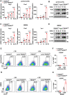
FBXW7 expression decreases in M2-like TAMs. (A, B) Peritoneal macrophages were stimulated with conditioned medium containing LLC supernatant for the indicated time periods. The mRNA (A) and protein (B) expression levels of Arg1, Ym1, and FBXW7 were measured by qRT-PCR and immunoblotting, respectively. (C, D) BMDMs were incubated in conditioned medium, and the mRNA (C) and protein (D) expression levels of Arg1, Ym1, and FBXW7 were measured by qRT-PCR and immunoblotting. Data are shown as the mean ± SD and are representative of three independent experiments. n=3. **P < 0.01; ***P < 0.001; ****P < 0.0001 (one-way ANOVA (A, C)).
Figure 4
Figure 4
FBXW7 knockout facilitates M2 macrophage polarization in a tumor microenvironment-mimicking condition. (A) Peritoneal macrophages extracted from FBXW7f/f and Lysm+FBXW7f/f mice were treated with conditioned medium containing LLC supernatant. The mRNA expression of Arg1, Fizz1, and Ym1 were analyzed by qRT-PCR. (B) The protein levels of Arg1 and Ym1 in wild-type and FBXW7-knockout peritoneal macrophages were detected by immunoblotting after conditioned medium stimulation. (C, D) The mRNA (C) and protein (D) expression of Arg1, Fizz1, and Ym1 were analyzed by qRT-PCR and western blotting, respectively, in BMDMs incubated with conditioned medium. (E, F) Flow cytometry analysis (E) and statistical analysis (F) of the percentage of M2 macrophages (CD206+) in wild-type and FBXW7-knockout peritoneal macrophages stimulated with conditioned medium (n = 3 per group). (G, H) Flow cytometry analysis (G) and statistical analysis (H) of the percentage of M2 macrophages (CD206+) in wild-type and FBXW7-knockout BMDMs stimulated with conditioned medium (n = 3 per group). Data are shown as the mean ± SD and are representative of three independent experiments. *P < 0.05; **P < 0.01; ***P < 0.001; ****P < 0.0001 (two-way ANOVA (A, C, F, H)).
Figure 5
Figure 5
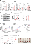
FBXW7 knockout promotes macrophage expressing pro-tumoral factors. (A) BMDMs from FBXW7f/f and Lysm+FBXW7f/f mice were stimulated with conditioned medium, and the mRNA expression of MMP9, IL-10, TGFβ, and VEGFα was examined by qRT-PCR. (B) The protein expression of MMP9 and VEGFα in BMDMs incubated with conditioned medium were detected by immunoblotting. (C) The protein levels of IL10 and TGFβ in the supernatant of BMDMs which co-cultured with LLCs for indicated time were measured by ELISA kit. (D) LLCs were cultured in serum-free RPMI-1640, supernatant from IL-4-induced wild-type or FBXW7-knockout macrophages. The proliferation of LLCs in three groups was measured by MTT assay. (E) Schematic representation of the co-injection experimental approach. IL4-induced BMDMs derived from FBXW7f/f and Lysm+FBXW7f/f mice were mixed with LLCs at a ratio of 1:1 and injected subcutaneously into healthy wild-type C57BL/6 mice. (F, G) The volume (F) and weight (G) of tumors in co-injection mice. (H) The appearance of tumors in two groups inoculated with a mixture of M2 macrophages and LLCs. Data are shown as the mean ± SD and are representative of three independent experiments. *P < 0.05; **P < 0.01; ***P < 0.001; ****P < 0.0001 (two-way ANOVA (A, C, D, F) and Student’s t test (G)).
Figure 6
Figure 6
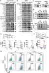
The role of FBXW7 in regulating M2 macrophage polarization is dependent on c-Myc. (A, B) Peritoneal macrophages (A) and BMDMs (B) derived from FBXW7f/f and Lysm+FBXW7f/f mice were stimulated with conditioned medium for the indicated time periods. The phosphorylated or total proteins related to M2 macrophage polarization were analyzed by immunoblotting. (C) Immunoblotting was used to analyze the interference efficiency of c-Myc siRNA in primary macrophages following stimulation with conditioned medium for 12 hours. (D, E) Immunoblotting (D) and qRT-PCR analysis (E) of Arg1 and Ym1 expression in primary macrophages from FBXW7f/f and Lysm+FBXW7f/f mice transfected with or without c-Myc siRNA and stimulated with conditioned medium. (F, G) Statistical analysis (F) and flow cytometry analysis (G) of the percentage of M2 macrophages (CD206+) in wild-type and FBXW7-knockout macrophages transfected with or without c-Myc siRNA and stimulated with conditioned medium. Data are shown as the mean ± SD and are representative of three independent experiments. ****P < 0.0001; n.s, no significance (two-way ANOVA (E, F)).
Figure 7
Figure 7
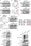
FBXW7 mediates degradation and ubiquitination of c-Myc in M2-like TAMs. (A) The protein expression of c-Myc and FBXW7 in wild-type and FBXW7-knockout primary macrophages stimulated with conditioned medium were measured by immunoblotting. (B) qRT-PCR analysis of c-Myc mRNA expression in wild-type and FBXW7-knockout primary macrophages after conditioned medium treatment. (C) Immunoblot analysis of c-Myc and FBXW7 in wild-type and FBXW7-knockout primary macrophages treated with CHX (20 μg/mL) for the indicated time period after conditioned medium pre-treatment. (D) The quantification of relative c-Myc levels in (C). (E) The protein expression of c-Myc and FBXW7 in wild-type and FBXW7-knockout primary macrophages after treatment with MG132 (10 μM) and conditioned medium. (F) The phosphodegron sequence alignment of c-Myc recognized by FBXW7 with MCL1, cyclin E, Notch1, and c-Jun. The FBXW7 phosphodegron sequence present in c-Myc is conserved across different species. Conserved residues within the degron sequences are shown in red. (G) Immunoblot analysis of BMDMs stimulated with conditioned medium for the indicated time periods and treated with MG132, followed by immunoprecipitation with specific antibody-conjugated agarose or immunoglobulin G (IgG)-conjugated agarose. (H) Immunoblot analysis of the K48 ubiquitination of c-Myc in wild-type and FBXW7-knockout BMDMs stimulated with conditioned medium. Data are shown as the mean ± SD and are representative of three independent experiments. *P < 0.05; **P < 0.01; ***P < 0.001; ****P < 0.0001; n.s, no significance (two-way ANOVA (B, D)).

References

    1. Biswas SK, Mantovani A. Macrophage plasticity and interaction with lymphocyte subsets: cancer as a paradigm. Nat Immunol. 2010; 11:889–96. 10.1038/ni.1937 - DOI (V体育官网入口) - PubMed
    1. Di Caro G, Cortese N, Castino GF, Grizzi F, Gavazzi F, Ridolfi C, Capretti G, Mineri R, Todoric J, Zerbi A, Allavena P, Mantovani A, Marchesi F. Dual prognostic significance of tumour-associated macrophages in human pancreatic adenocarcinoma treated or untreated with chemotherapy. Gut. 2016; 65:1710–20. 10.1136/gutjnl-2015-309193 - DOI - PubMed
    1. Murray PJ. Macrophage polarization. Annu Rev Physiol. 2017; 79:541–66. 10.1146/annurev-physiol-022516-034339 - DOI - PubMed
    1. Colegio OR, Chu NQ, Szabo AL, Chu T, Rhebergen AM, Jairam V, Cyrus N, Brokowski CE, Eisenbarth SC, Phillips GM, Cline GW, Phillips AJ, Medzhitov R. Functional polarization of tumour-associated macrophages by tumour-derived lactic acid. Nature. 2014; 513:559–63. 10.1038/nature13490 - "V体育官网" DOI - PMC - PubMed
    1. Casazza A, Laoui D, Wenes M, Rizzolio S, Bassani N, Mambretti M, Deschoemaeker S, Van Ginderachter JA, Tamagnone L, Mazzone M. Impeding macrophage entry into hypoxic tumor areas by Sema3A/Nrp1 signaling blockade inhibits angiogenesis and restores antitumor immunity. Cancer Cell. 2013; 24:695–709. 10.1016/j.ccr.2013.11.007 - DOI - PubMed

Publication types

V体育官网入口 - MeSH terms