<area date-time="bGuJqm9m"></area> Skip to main page content
U.S. flag

An official website of the United States government

Dot gov

The . gov means it’s official. Federal government websites often end in . gov or . mil. Before sharing sensitive information, make sure you’re on a federal government site VSports app下载. .

Https

The site is secure. The https:// ensures that you are connecting to the official website and that any information you provide is encrypted and transmitted securely. V体育官网.

. 2020 Oct 15;5(20):e138881.
doi: 10.1172/jci.insight.138881.

Bile acid toxicity in Paneth cells contributes to gut dysbiosis induced by high-fat feeding

Affiliations

Bile acid toxicity in Paneth cells contributes to gut dysbiosis induced by high-fat feeding

VSports手机版 - Hui Zhou et al. JCI Insight. .

Abstract

High-fat feeding (HFF) leads to gut dysbiosis through unclear mechanisms. We hypothesize that bile acids secreted in response to high-fat diets (HFDs) may act on intestinal Paneth cells, leading to gut dysbiosis. We found that HFF resulted in widespread taxonomic shifts in the bacteria of the ileal mucosa, characterized by depletion of Lactobacillus and enrichment of Akkermansia muciniphila, Clostridium XIVa, Ruminococcaceae, and Lachnospiraceae, which were prevented by the bile acid binder cholestyramine. Immunohistochemistry and in situ hybridization studies showed that G protein-coupled bile acid receptor (TGR5) expressed in Paneth cells was upregulated in the rats fed HFD or normal chow supplemented with cholic acid. This was accompanied by decreased lysozyme+ Paneth cells and α-defensin 5 and 6 and increased expression of XBP-1. Pretreatment with ER stress inhibitor 4PBA or with cholestyramine prevented these changes. Ileal explants incubated with deoxycholic acid or cholic acid caused a decrease in α-defensin 5 and 6 and an increase in XBP-1, which was prevented by TGR5 antibody or 4PBA. In conclusion, this is the first demonstration to our knowledge that TGR5 is expressed in Paneth cells. HFF resulted in increased bile acid secretion and upregulation of TGR5 expression in Paneth cells. Bile acid toxicity in Paneth cells contributes to gut dysbiosis induced by HFF VSports手机版. .

Keywords: Defensins; Gastroenterology; Obesity V体育安卓版. .


Conflict of interest statement

Conflict of interest: The authors have declared that no conflict of interest exists.

Figures

Figure 1
Figure 1. TGR5 expression in the Paneth cells; HFD increases bile acids and enhances TGR5 expression in ileal crypts.
(A and B) Immunocytochemical staining using sections of ileal specimens and isolated crypt cells showed that TGR5 expression was observed in the lysozyme+ Paneth cells (white arrows) in ileal crypts. Scale bar: 10 μm. (C) Left: hybridization using antisense probes. Purple represents lysozyme mRNA, a specific marker for Paneth cells, which are located at the bottom portion of crypt; silver grains indicate TGR5 mRNA. Arrows indicate colocalization. Right: Dig-lysozyme and [35S]-TGR5 sense probes were used as the negative control. Note that TGR5 receptors are also expressed in other cell types in the intestinal crypts. Scale bar: 50 μm; 3-fold magnification (inset). (D) Measurement of total bile acid concentration. A 2-week high-fat diet (HFD) induced a 45% increase in serum and 60% increase in fecal bile acid compared with control (RC) (n = 5–6). The increased total serum bile acid was prevented by concurrent administration of cholestyramine (n = 6). A similar increase in serum bile acid was observed in cholic acid group (n = 6). (E) The density of the TGR5-immunoreactive band at 33 kDa was observed in the ileal crypts of rats given HFD. TGR5 protein expression increased in ileal crypts of HFD rats compared with control (n = 5). This increase was prevented by concurrent administration of cholestyramine (n = 5). A similar increase was also observed in rats with cholic acid (n = 4–5). Each bar represents mean ± SEM. C, RC; H, HFD; CHO, HFD + cholestyramine; CA, cholic acid. P values were determined by 1-way ANOVA. *P < 0.05, **P < 0.01.
Figure 2
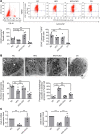
Figure 2. HFD induces a decrease in Paneth cells and antimicrobial peptide prevented by cofeeding with cholestyramine.
(A) HFD induced a reduction in lysozyme+ Paneth cells (n = 6, P = 0.055) as well as lysozyme+TGR5+ Paneth cell expression (n = 6) in the crypts. Administration of cholestyramine prevented this decrease in lysozyme+ Paneth cells (n = 6). Concurrent oral cholic acid also induced a significant decrease in both lysozyme+ Paneth cells or lysozyme+TGR5+Paneth cell (n = 5–6). (B) HFD causes a decrease in Paneth cell secretory granules. Transmission electron micrograph of small intestinal Paneth cells showed a 34% and 53% decrease in Paneth cell secretory granules (white arrowhead) from rats fed a HFD and cholic acid diet, respectively (n = 6, **P < 0.01). An increase in percentage of secretory granules with vacuoles (white arrow) was also observed in Paneth cell from rats fed a HFD, which was prevented by cofeeding with cholestyramine (n = 6). A similar increase in vacuoles was observed in rats given cholic acid (n = 6). Scale bar: 5 μm. (C) Reduction in gene expression of α-defensin 5 (Defa5) and 6 (Defa6) in rats given a HFD. A 68% and 67% decrease in gene expression of Defa5 and Defa6, respectively, in the crypts of ileum in rats fed a HFD (n = 5). These decreases in Defa5 and Defa6 gene expression were prevented by concurrent oral feeding with cholestyramine (n = 5). CHO, cholestyramine. All data are shown as mean ± SEM. P values were determined by 1-way ANOVA. **P < 0.01.
Figure 3
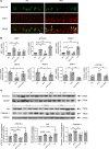
Figure 3. HFD induces protein and gene expression of ER stress, autophagy, and DNA damage.
(A) Immunocytochemical staining of ileal specimen from rats fed regular chow (RC) or HFD (lysozyme, green; XBP-1 [ER stress marker], red). HFD induces increased expression of XBP-1 in lysozyme+ Paneth cells. Scale bar: 50 μm. (B) High-fat feeding caused an increase in gene expression of XBP-1 and ATG16L1 in the crypts of ileum (n = 5). These increases in XBP-1 and ATG16L1 gene expression were prevented by concurrent oral feeding with cholestyramine (n = 5). Oral feeding with cholic acid caused a similar increase in ATG16L1 and PARP1 gene expression (n = 4–5). HFD induced alteration of α-defensins, autophagy, and ER stress were prevented by an ER stress inhibitor (4BPA) (n = 6). (C) Western blot showing the density of ATG16L1 (autophagy marker), BiP (ER stress marker), and caspase-3 (DNA damage marker) immunoreactive bands at 60 kDa, 75 kDa, and 19 kDa, which were observed in the ileal crypts from rats fed RC. HFD caused a significant increase in ATG16L1 and caspase-3–immunoreactive bands (n = 5–6). Oral feeding with cholic acid caused a similar increase in ATG16L1 and BiP expression (n = 3–6). These increases in BiP, ATG16L1, and caspase-3 expression in response to HFD were prevented by concurrent oral feeding with cholestyramine (n = 4–6). Part of the membrane used for protein expression studies of TGR5 in Figure 1E was used for Western blotting of ATG16L1. Hence, the 2 blots share the same GAPDH. C, RC; H, HFD; CHO, HFD + cholestyramine; CA, cholic acid. P values were determined by 1-way ANOVA. *P < 0.05, **P < 0.01.
Figure 4
Figure 4. DCA and CA increase ER stress and reduction of α-defensins in cultured ileum (in vitro).
(A) DCA (100 μM) and (B) CA (100 μM) caused a reduction in α-defensin and ER stress in whole-mount cultured ileum. Pretreatment with TGR5 antibody (4 μg/mL) or 4PBA (10 mM) prevented the DCA- or CA-induced alteration of α-defensins and ER stress (n = 5–7). P values were determined by 1-way ANOVA. *P < 0.05, **P < 0.01.
Figure 5
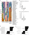
Figure 5. HFD-induced dysbiosis at the phylum level in the ileum mucosa is prevented by cholestyramine.
(A) Relative abundance of OTUs classified at the level of phylum. (B) Relative abundance of Firmicutes, Verrucomicrobia, and Bacteroidetes. (C and D) Relative abundance of (C) Gram-negative and (D) -positive communities. (E) Among Gram-negative bacteria, Verrucomicrobia (Akkermansia muciniphila) was increased by 9-fold (percentage of total bacteria). These changes were prevented by concurrent feeding with CHO. (n = 6–8 per group). CHO, cholestyramine. P values were determined by 1-way ANOVA test. *P < 0.05 versus RC; # P < 0.05 versus HFD.
Figure 6
Figure 6. HFD-induced dysbiosis in the ileum mucosa is prevented by cholestyramine.
(A) Relative abundance of OTUs classified at the level of family. (B and C) OTUs were significantly depleted or enriched after HFD (LEfSe, n = 7 or 8 per group). (D) As a group, the relative abundance of Lactobacillaceae and Verrucomicrobiaceae was altered after HFD+CHO compared with HFD (n = 6 or 8 per group). P values were determined by unpaired 2-tailed Student’s t test (2 groups) or by 1-way ANOVA (more than 2 groups). *P < 0.05 versus RC; #P < 0.05 versus HFD. CHO, cholestyramine.
Figure 7
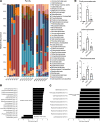
Figure 7. HFD-induced dysbiosis at phylum level in the ileum mucosa is prevented by 4PBA.
(A) Relative abundance of OTUs classified at the level of phylum. (B) Relative abundance of Firmicutes, Bacteroidetes, and Verrucomicrobia. (C and D) Relative abundance of (C) Gram-negative and (D) -positive communities. (E) Among Gram-negative bacteria, Bacteroidetes was increased by 31.62-fold, from 1.05% ± 0.50% to 33.28% ± 4.75% (percentage of total bacteria) (n = 6 per group). These changes were prevented by 4PBA (ER stress inhibitor). P values were determined by 1-way ANOVA. *P < 0.05 versus RC; #P < 0.05 versus HFD.
Figure 8
Figure 8. HFD-induced dysbiosis in the ileum mucosa is prevented by 4PBA.
(A) Relative abundance of OTUs classified at the level of family. (B and C) OTUs were significantly depleted or enriched after HFD (LEfSe, n = 6 per group). (D) As a group, the relative abundance of Porphyromonadaceae, Lachnospiraceae, and Ruminococcaceae was altered after HFD+4PBA compared with HFD. P values were determined by unpaired 2-tailed Student’s t test (2 groups) or by 1-way ANOVA (more than 2 groups). *P < 0.05 versus RC; # P < 0.05 versus HFD.
Figure 9
Figure 9. HFD altered genes from microbial community, which was prevented by 4PBA.
(A) The relative abundance of metabolic gene pathways was altered in HFD group. The abundance of metabolic gene pathways was analyzed using PICRUSt and is based on the 16S rRNA sequencing data. Only the relative gene pathway abundances that were identified as being significantly different for RC, HFD, and HFD+4PBA are shown (P < 0.01, for all pairwise comparisons, by 1-way ANOVA). Note that, genes for Gram-positive cell wall synthesis (peptidoglycan biosynthesis) were decreased in the HFD group, and this was reversed by 4PBA treatment. (B) Compared with the regular chow (RC), HFD increased many genes, mainly, for amino acid and energy metabolism. Genes responsible for synthesis of the Gram-negative cell wall (lipopolysaccharide biosynthesis, lipopolysaccharide biosynthesis proteins) were enriched in the HFD group, and this was also reversed by 4PBA.
Figure 10
Figure 10. A schematic diagram depicting the mechanism by which bile acids cause gut dysbiosis through Paneth cells.
(A) HFD lead to increased bile acid production and high concentrations of DCA. The activation of TGR5 by DCA causes ER stress, autophagy, and reduction of α-defensins in Paneth cell, which favors the growth of Gram-negative bacteria and results in mucosal microbial dysbiosis. (B) These changes are prevented by 4PBA.

"V体育安卓版" References

    1. Albenberg LG, Wu GD. Diet and the intestinal microbiome: associations, functions, and implications for health and disease. Gastroenterology. 2014;146(6):1564–1572. doi: 10.1053/j.gastro.2014.01.058. - "V体育安卓版" DOI - PMC - PubMed
    1. Graf D, et al. Contribution of diet to the composition of the human gut microbiota. Microb Ecol Health Dis. 2015;26:26164. - PMC - PubMed
    1. Llewellyn SR, et al. Interactions between diet and the intestinal microbiota alter intestinal permeability and colitis severity in mice. Gastroenterology. 2018;154(4):1037–1046.e2. doi: 10.1053/j.gastro.2017.11.030. - DOI - PMC - PubMed
    1. Martinez-Medina M, et al. Western diet induces dysbiosis with increased E coli in CEABAC10 mice, alters host barrier function favouring AIEC colonisation. Gut. 2014;63(1):116–124. doi: 10.1136/gutjnl-2012-304119. - DOI - PubMed
    1. Just S, et al. The gut microbiota drives the impact of bile acids and fat source in diet on mouse metabolism. Microbiome. 2018;6(1):134. doi: 10.1186/s40168-018-0510-8. - VSports - DOI - PMC - PubMed

Publication types

MeSH terms (VSports app下载)

Supplementary concepts