Skip to main page content
U.S. flag

An official website of the United States government

Dot gov

The . gov means it’s official. Federal government websites often end in . gov or . mil. Before sharing sensitive information, make sure you’re on a federal government site. VSports app下载.

Https

The site is secure V体育官网. The https:// ensures that you are connecting to the official website and that any information you provide is encrypted and transmitted securely. .

. 2020 Nov;69(11):2467-2480.
doi: 10.2337/db20-0585. Epub 2020 Sep 4.

A Multifunctional Role of Leucine-Rich α-2-Glycoprotein 1 in Cutaneous Wound Healing Under Normal and Diabetic Conditions

Affiliations

A Multifunctional Role of Leucine-Rich α-2-Glycoprotein 1 in Cutaneous Wound Healing Under Normal and Diabetic Conditions

Chenghao Liu et al. Diabetes. 2020 Nov.

"V体育官网" Abstract

Delayed wound healing is commonly associated with diabetes. It may lead to amputation and death if not treated in a timely fashion. Limited treatments are available partially due to the poor understanding of the complex disease pathophysiology. Here, we investigated the role of leucine-rich α-2-glycoprotein 1 (LRG1) in normal and diabetic wound healing. First, our data showed that LRG1 was significantly increased at the inflammation stage of murine wound healing, and bone marrow-derived cells served as a major source of LRG1. LRG1 deletion causes impaired immune cell infiltration, reepithelialization, and angiogenesis. As a consequence, there is a significant delay in wound closure VSports手机版. On the other hand, LRG1 was markedly induced in diabetic wounds in both humans and mice. LRG1-deficient mice were resistant to diabetes-induced delay in wound repair. We further demonstrated that this could be explained by the mitigation of increased neutrophil extracellular traps (NETs) in diabetic wounds. Mechanistically, LRG1 mediates NETosis in an Akt-dependent manner through TGFβ type I receptor kinase ALK5. Taken together, our studies demonstrated that LRG1 derived from bone marrow cells is required for normal wound healing, revealing a physiological role for this glycoprotein, but that excess LRG1 expression in diabetes is pathogenic and contributes to chronic wound formation. .


Figures

Figure 1
Figure 1
LRG1 is elevated in cutaneous wounds. A: Immunohistochemical detection of LRG1 (brown) showed low expression of LRG1 in normal mouse skin. Scale bar: 100 μm. B: Representative Western blot (left) and densitometry analysis (right) of wounds harvested at different time points. C: Immunofluorescence staining detecting LRG1 (green), CD11b (red), or DAPI (blue) in day 1 mouse wounds. Scale bars: 120 μm and 20 μm. D: qRT-PCR analysis of day 1 wounds demonstrated reduced Lrg1 expression in irradiated wild-type mice with Lrg1−/− BMC transplantation in comparison with wild-type mice receiving wild-type BMCs. All images are representative; data are represented as mean (95% CI; P) of n ≥ 5 mice per group. Significance was determined by one- or two-way ANOVA followed by Tukey multiple comparisons test. *P < 0.05, ***P < 0.001. WB, Western blot; WT, wild type.
Figure 2
Figure 2
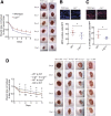
Absence of Lrg1 leads to delayed wound healing. A: Quantification (left) and representative images (right) of wound size in wild-type and Lrg1−/− mice revealed a delayed wound closure in the absence of Lrg1. *P < 0.05. Scale bar: 1 mm. B: Representative immunofluorescence staining of MPO (green) and DAPI (blue) (top) and quantification of the presentation of MOP+ cells (bottom) in day 1 wounds of wild-type and Lrg1−/− mice; 5–10 fields per wound were analyzed. *P < 0.05. Scale bar: 30 μm. C: Representative immunofluorescence staining of F4/80 (green) and DAPI (blue) (top) and quantification of F4/80+ cells (bottom) of day 5 mouse wounds of wild-type and Lrg1−/− mice; 5–10 fields per wound were analyzed. *P < 0.05. Scale bar: 30 μm. D: Quantification (left) and representative images (right) of wound size in irradiated wild-type (WT) mice receiving BMCs from wild-type mice (WT to WT), irradiated Lrg1−/− mice receiving BMCs from Lrg1−/− mice (Lrg1−/− to Lrg1−/−), irradiated Lrg1−/− mice receiving BMCs from wild-type mice (WT to Lrg1−/− ), and irradiated wild-type mice receiving BMCs from Lrg1−/− mice (Lrg1−/− to WT). *P < 0.05: Lrg1−/− to Lrg1−/− vs. WT to WT; #P < 0.05: Lrg1−/− to WT vs. WT to WT; @P < 0.05, @@P < 0.01: WT to Lrg1−/− vs. Lrg1−/− to Lrg1−/−. Scale bar: 1 mm. All images are representative; data are represented as mean (95% CI; P) of n ≥ 6 mice per group. Significance was determined by unpaired, two-tailed Student t test between wild-type and Lrg1−/− or wound size at different time points.
Figure 3
Figure 3
LRG1 mediates neutrophil adhesion. A: Representative images (left) and quantification (right) of neutrophil adhesion assay demonstrated rhLRG1 induced dH60 cell (labeled with CMFDA dye) adhesion to HDMECs. Scale bar: 200 μm. B: Representative images (left) and quantification (right) of TNFα-induced neutrophil adhesion by use of hHL-60 cells (labeled with CMFDA dye) subjected to siRNA-mediated LRG1 knockdown. Scale bar: 200 μm. C: Representative Western blot (left) and densitometry analysis (right) of L-selectin and GAPDH in rhLRG1-treated dHL-60 cells at different time points. D: Quantification of flow cytometry demonstrated an increase in L-selectinHigh population following rhLRG1treatment. All images are representative; data are presented as the mean (95% CI; P) of n ≥ 3 independent experiments per group. Significance was determined by one- or two-way ANOVA followed by Tukey multiple comparisons test or unpaired, two-tailed Student t test. *P < 0.05, **P < 0.01, ***P < 0.001. WB, Western blot.
Figure 4
Figure 4
LRG1 regulates reepithelialization during wound healing. A: Representative H-E staining (left) and quantification of reepithelialization (right) of day 4 wounds of wild-type and Lrg1−/− mice. Scale bar: 125 μm. B: Representative H-E staining (left) and quantification of epithelium thickness (right) of day 5 wounds of wild-type and Lrg1−/− mice. Scale bar: 25 μm. C: Representative images (left) and quantification of wound gap (right) in scratch wound healing assay. Scale bar: 100 μm. D: Representative Western blot (left) and densitometry analysis (right) of FN1, N-cad, and GAPDH in rhLRG1-treated HaCaT cells. E: Representative immunofluorescence staining (top) and quantification (bottom) of Ki67 (red) and DAPI (blue) in day 3 wounds. Scale bar: 30 μm. F: Quantification of viable HaCaT cells in Trypan blue exclusion assay (G). Representative Western blot (top) and densitometry analysis (bottom) of cyclin D1 and GAPDH in rhLRG1-treated HaCaT cells. All images are representative, and data are represented as mean (95% CI; P) of n ≥ 5 mice or n ≥ 3 independent experiments per treatment group. Significance was determined by unpaired, two-tailed Student t test. *P < 0.05, **P < 0.01, ***P < 0.001. WB, Western blot.
Figure 5
Figure 5
LRG1 modulates wound angiogenesis during wound healing. A: Representative immunofluorescence staining of CD31 (green) and DAPI (blue) (top) and quantification of vessel density (bottom) in day 7 wounds of wild-type and Lrg1−/− mice. Scale bar: 15 μm. B: Representative images of immunofluorescence staining detecting Ki67 (red) and DAPI (blue) (top) and quantification of percentage of Ki67+ cells (bottom) in HDMECs. Scale bar: 50 μm. C: Representative images (top) and quantification (bottom) of Matrigel tube formation. Scale bar: 125 μm. D: Representative images (top) and quantification (bottom) of Transwell migration assay. Scale bar: 100 μm. E: Representative Western blot (left) and densitometry analysis (right) of endothelial TGFβ-Smad1/5 signaling in HDMECs treated with rhLRG1 in the absence and presence of ALK1 inhibitor (LDN193189) or ALK5 inhibitor (SB431542). All images are representative, and data are represented as mean (95% CI; P) of n ≥ 6 mice or n ≥ 3 independent experiments per group. Significance was determined by unpaired, two-tailed Student t test. *P < 0.05, **P < 0.01, ***P < 0.001. WB, Western blot.
Figure 6
Figure 6
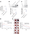
Elevated LRG1 expression is observed in diabetic humans and mice. A: ELISA analysis of LRG1 in serum from venous ulcer patients and DFU patients. B: Representative Western blot (top) and densitometry analysis (bottom) of LRG1 and GAPDH in human patients with venous ulcer and DFU. C: Representative Western blot (top) and densitometry analysis (bottom) of LRG1 and GAPDH in normal and diabetic wounds of C57BL/6 mice. D: qRT-PCR analysis of normal and diabetic wounds of C57BL/6 mice. E: Representative images (left) and quantification (right) of wound size revealed a delayed wound closure in C57BL/6 mice with STZ-induced diabetes. Scale bar: 1 mm. All images are representative, and data are represented as mean (95% CI; P) of n ≥ 6 patients or mice per group. Significance was determined by unpaired, two-tailed Student t test. *P < 0.05, **P < 0.01. WB, Western blot.
Figure 7
Figure 7
LRG1 mediates NETosis. A: Representative images (left) and quantification (right) of wound size in wild-type and Lrg1−/− mice with STZ-induced diabetes. Scale bar: 1 mm. B: Representative Western blot (top) and densitometry analysis (bottom) of H3Cit, histone H3 (H3), and GAPDH in day 3 wounds from wild-type and Lrg1−/− mice with STZ-induced diabetes. C: Representative Western blot (top) and densitometry analysis (bottom) of H3Cit, H3, and GAPDH in calcium ionophore–treated wild-type and Lrg1−/− neutrophils. D: Representative immunofluorescence staining detecting H3Cit (green) and DAPI (blue) (left) and quantification of percentage of H3Cit+ cells (right) in calcium ionophore–treated wild-type and Lrg1−/− neutrophils. Scale bar: 80 μm. E: SYTOX Green assay on calcium ionophore–treated wild-type and Lrg1−/− neutrophils. F: SYTOX Green assay on calcium ionophore–treated dHL-60 cells. G: Representative Western blot (left) and densitometry analysis (right) of H3Cit, H3, AKT, phospho-AKT (pAKT), and GAPDH in rhLRG1- and/or MK2206-treated dHL-60 cells. H: Representative Western blot (left) and densitometry analysis (right) of H3Cit, H3, AKT, phospho-AKT, and GAPDH in rhLRG1 with or without SB431542-treated dHL-60 cells. All images are representative, and data are represented as mean (95% CI; P) of n ≥ 5 mice or n ≥ 3 independent experiments per group. Significance was determined by one- or two-way ANOVA followed by Tukey multiple comparisons test or unpaired, two-tailed Student t test. *P < 0.05, **P < 0.01, ***P < 0.001. CaI, calcium ionophore; WB, Western blot.

References

    1. Gurtner GC, Werner S, Barrandon Y, Longaker MT. Wound repair and regeneration. Nature 2008;453:314–321 - PubMed
    1. Armstrong DG, Boulton AJM, Bus SA. Diabetic foot ulcers and their recurrence. N Engl J Med 2017;376:2367–2375 - VSports在线直播 - PubMed
    1. Morbach S, Furchert H, Gröblinghoff U, et al. . Long-term prognosis of diabetic foot patients and their limbs: amputation and death over the course of a decade. Diabetes Care 2012;35:2021–2027 - PMC - PubMed
    1. Gregg EW, Li Y, Wang J, et al. . Changes in diabetes-related complications in the United States, 1990-2010. N Engl J Med 2014;370:1514–1523 - PubMed
    1. Wild S, Roglic G, Green A, Sicree R, King H. Global prevalence of diabetes: estimates for the year 2000 and projections for 2030. Diabetes Care 2004;27:1047–1053 - PubMed (V体育平台登录)

Publication types

MeSH terms

Associated data (VSports在线直播)