Skip to main page content
U.S. flag

An official website of the United States government

Dot gov

The . gov means it’s official. Federal government websites often end in VSports app下载. gov or . mil. Before sharing sensitive information, make sure you’re on a federal government site. .

Https

The site is secure V体育官网. The https:// ensures that you are connecting to the official website and that any information you provide is encrypted and transmitted securely. .

. 2021 Aug 1;106(8):2131-2146.
doi: 10.3324/haematol.2019.242990.

Bile acids regulate intestinal antigen presentation and reduce graft-versus-host disease without impairing the graft-versus-leukemia effect

Affiliations

Bile acids regulate intestinal antigen presentation and reduce graft-versus-host disease without impairing the graft-versus-leukemia effect

Eileen Haring et al. Haematologica. .

Abstract

Acute graft-versus-host disease causes significant mortality in patients undergoing allogeneic hematopoietic cell transplantation. Immunosuppressive treatment for graft-versus-host disease can impair the beneficial graft-versus-leukemia effect and facilitate malignancy relapse. Therefore, novel approaches that protect and regenerate injured tissues without impeding the donor immune system are needed. Bile acids regulate multiple cellular processes and are in close contact with the intestinal epithelium, a major target of acute graft-versus-host disease. Here, we found that the bile acid pool is reduced following graft-versus-host disease induction in a preclinical model. We evaluated the efficacy of bile acids to protect the intestinal epithelium without reducing anti-tumor immunity. We observed that application of bile acids decreased cytokine-induced cell death in intestinal organoids and cell lines. Systemic prophylactic administration of tauroursodeoxycholic acid, the most potent compound in our in vitro studies, reduced graft-versus-host disease severity in three different murine transplantation models. This effect was mediated by decreased activity of the antigen presentation machinery and subsequent prevention of apoptosis of the intestinal epithelium. Moreover, bile acid administration did not alter the bacterial composition in the intestine suggesting that its effects are cell-specific and independent of the microbiome. Treatment of human and murine leukemic cell lines with tauroursodeoxycholic acid did not interfere with the expression of antigen presentation-related molecules. Systemic T cell expansion and especially their cytotoxic capacity against leukemic cells remained intact. This study establishes a role for bile acids in the prevention of acute graft-versus-host disease without impairing the graft-versus-leukemia effect. In particular, we provide a scientific rationale for the systematic use of tauroursodeoxycholic acid in patients undergoing allogeneic hematopoietic cell transplantation VSports手机版. .


"VSports注册入口" Figures

Figure 1.
Figure 1.
Allogeneic hematopoietic cell transplantation induces changes in bile acid levels and bile acid receptor expression. (A) Simplified schematic overview of bile acid metabolism and the enterohepatic circulation. (B) Experimental flow for metabolomic profiling of the bile acid pool from untreated BALB/c mice and mice developing graft-versus-host disease (GvHD). (C) Overview of the bile acids quantified by liquid chromatography - mass spectrometry. (D) Quantification of the bile acid composition in liver, ileum, ileal contents and serum of untreated mice and on day 7 after bone marrow transplantation (BMT). Data were pooled from five individual mice per group and are presented as mean ± standard error of the mean for each metabolite. P-values were calculated for the total bile acid pool using the two-tailed unpaired Student’s t-test; ns: not significant. (E) Quantitative real-time polymerase chain reaction analysis of the mRNA expression of the bile acid receptors Nr1h4 (encodes for the FXR protein) and Gpbar1 (encodes for the TGR5 protein) in the small intestine of untreated mice and mice developing GvHD. Data are pooled from n=10 mice in the untreated, GvHD d7 and GvHD d14 groups and n=5 mice in the GvHD day 4 group. The P-values were calculated using the ordinary one-way ANOVA test with correction for multiple comparisons.
Figure 2.
Figure 2.
Treatment with bile acids reduces cell death in small intestinal organoids and MODE-K cells. (A) Experimental setting for the treatment of organoids with tumor necrosis factor (TNF) (20 ng/mL), interferon (IFN) (2.5 ng/mL) and the respective bile acids. (B) Representative images of BALB/c intestinal organoids treated with TNF as described in (A). (C) Quantification of damaged organoids treated with TNF as in (A) performed by manual microscope counting. Data were normalized to the TNF plus (TNF+)vehicle group. Statistical analysis of n=4 biologically independent experiments. The P-value was calculated using an ordinary one-way ANOVA test with correction for multiple comparisons. (D and E) Intestinal organoids were cultured as described in (A). Organoids were digested and the proportion of dead cells was determined by flow cytometry. (D) Representative flow cytometry dot plots. (E) Quantification of the percentages of dead cells. Data were normalized to the TNF+vehicle group. Statistical analysis of n=3 biologically independent experiments. The P-value was calculated using the ordinary one-way ANOVA test with correction for multiple comparisons. (F) Representative images of BALB/c intestinal organoids treated with IFNγ as described in (A). (G) Quantification of damaged organoids treated with IFNas in (A) performed by manual microscope counting. Data were normalized to the IFNγ+vehicle group. Statistical analysis of n=3 biologically independent experiments. The P-value was calculated using an ordinary one-way ANOVA test with correction for multiple comparisons. (H and I) Analysis of MODE-K cell viability after treatment with TNF ± chenodeoxycholic acid (CDCA), ursodeoxycholic acid (UDCA), 6-ethylchenodeoxycholic acid (OCA) or tauroursodeoxycholic acid (TUDCA) for 48 hours performed by flow cytometry. (H) Quantification of the percentages of dead cells. Data were normalized to the TNF+vehicle group. Statistical analysis of n=3 biologically independent experiments performed in technical duplicates or triplicates. The P-values were calculated using the ordinary oneway ANOVA test with correction for multiple comparisons. (I) Representative flow cytometry dot plots.
Figure 3.
Figure 3.
Bile acid treatment improves acute graft-versus-host disease outcome in mice. (A) Transplantation model with BALB/c (H-2kd) as donor and C57BL/6 (H- 2kb) as recipient. Recipient animals were treated with 200 mg/kg body weight tauroursodeoxycholic acid (TUDCA) or an equal volume of vehicle from day 0 until day 10 after bone marrow transplantation (BMT) by a daily intraperitoneal injection. (B) Survival of C57BL/6 mice transplanted as shown in (A). Numbers (N) represent individual mice, the P-value was calculated using the two-sided Mantel-Cox test. (C) Transplantation model with C57BL/6 (H-2kb) as donor and BALB/c (H-2kd) as recipient. Recipient animals were treated with 200 mg/kg body weight TUDCA or an equal volume of vehicle from day 0 until day 10 after BMT by a daily intraperitoneal injection. (D) Survival of BALB/c mice transplanted as described in (C). Data were pooled from three independent experiments, numbers (N) represent individual mice. The P-value was calculated using the two-sided Mantel-Cox test. (E) Graft-versus-host disease (GvHD) histopathology scores of liver, small intestine and colon assessed on day 7 after BMT (C57BL/6 in BALB/c model). Data were pooled from two independent experiments, numbers (N) represent individual mice. P-values were calculated using the ordinary one-way ANOVA test with correction for multiple comparisons. (F) Serum cytokine concentrations in untreated mice and transplanted mice determined on day 14 after BMT (C57BL/6 in BALB/c model). Numbers (N) represent individual mice. P-values were calculated using the ordinary one-way ANOVA test with correction for multiple comparisons. (G and I) Fecal samples were collected for microbial analysis on day 5 after BMT. Numbers (N) represent individual mice. Data were pooled from two independent experiments. P-values were calculated using the ordinary one-way ANOVA test with correction for multiple comparisons; ns: not significant. (G) The Shannon index as a surrogate parameter for microbial diversity. (H) The reversed Simpson index as a surrogate parameter for microbial diversity. (I) Relative abundance of specified bacterial genera. (J) C57BL/6 mice were treated with an antibiotic cocktail comprising 1 mg/mL cefoxitin, metronidazole, neomycin and gentamycin for 2 weeks before they underwent BMT as described in (A). (K) Survival of C57BL/6 mice transplanted and treated as described in (J). Numbers (N) represent individual mice. Data were pooled from two independent experiments. Statistical analysis was performed using the two-sided Mantel-Cox test.
Figure 4.
Figure 4.
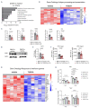
Tauroursodeoxycholic acid reduces intestinal antigen presentation. (A to G) Small intestinal samples were isolated from recipient mice treated with vehicle or tauroursodeoxycholic acid (TUDCA), on day 14 after bone marrow transplantation (BMT) (C57BL/6 to BALB/c model). (A) Identification of significantly downregulated Gene Ontology terms in animals treated with TUDCA. The dotted line corresponds to P=0.05 (log -0.05=1.29). (B) Heat map based on microarray analysis showing the differentially regulated genes (q-value<0.05) that belong to the term ‘antigen processing and presentation’ from the Gene Ontology database. Data were pooled from two independent experiments, n=6 mice per group, P=0.004. The color code represents the Z-score log2 intensity. (C) Quantitative real-time polymerase chain reaction (PCR) analysis of the mRNA expression of selected genes with Actb as a reference gene. Data were pooled from two independent experiments, numbers (N) represent individual mice. P-values were calculated using the unpaired two-tailed Student’s t-test. (D) Expression of TAP1 protein quantified by western blot. Representative western blot from n=3 mice per group. (E) Quantification of TAP1 protein expression. Data were pooled from two independent experiments, numbers (N) represent individual mice. P-values were calculated using the unpaired two-tailed Student’s t-test. (F) Flow cytometric quantification of major histocompatibility complex (MHC) class I expression on CD326+ (EpCAM+) cells (left panel) and MHC class II expression on CD45 MHC class II+ cells (right panel). Data were pooled from two independent experiments, numbers (N) represent individual mice. P-values were calculated using the unpaired two-tailed Student’s t-test. (G) Heat map based on microarray analysis showing the differentially regulated genes (q-value<0.05) that belong to the term ‘Response to interferon ’ from the Gene Ontology database. Data were pooled from two independent experiments, n=6 mice per group, P=6.49x10⁻⁹. The color code represents the Z-score log2 intensity. (H and I) Quantitative real-time PCR analysis of the mRNA expression of Tap1 (panel H) and Tap2 (panel I) with Actb as a reference gene in MODE-K cells treated with TNF ± chenodeoxycholic acid (CDCA), ursodeoxycholic acid (UDCA), 6-ethylchenodeoxycholic acid (obeticholic acid, OCA) and TUDCA for 48 hours. Representative data from one of two independent experiments with n=3 replicates/group are presented. P-values were calculated using the ordinary one-way ANOVA test with correction for multiple comparisons; ns: not significant.
Figure 5.
Figure 5.
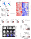
Tauroursodeoxycholic acid administration changes the transcriptional signature of T cells in the intestine but preserves their systemic expansion. (A and B) Flow cytometric analysis of T cells isolated from the small intestinal lamina propria of recipient animals on day 14 after bone marrow transplantation (BMT) (C57BL/6 in BALB/c model). (A) Representative flow cytometry dot plots. (B) Relative (left panel) and absolute (right panel) quantification of CD8+ and CD4+ T cells. Representative data from one of two biologically independent experiments, numbers (N) represent individual mice. P-values were calculated using the two-tailed unpaired Student’s t-test; ns: not significant. (C) Heat map based on microarray analysis (performed as in Figure 4) showing the differentially regulated genes (qvalue< 0.05) that belong to the term ‘T-cell activation’ from the Gene Ontology database. Data were pooled from two independent experiments, n=6 mice per group, P=4.6x10⁻¹¹. The color code represents the Z-score log2 intensity. (D) Log2 RNA intensity values for selected genes belonging to the GO term “T-cell activation”. Data were pooled from two independent experiments, numbers (N) indicate individual mice. An adjusted P-value calculated as described in the Methods is presented. (E) BALB/c mice underwent transplantation as described in Figure 3C using luciferase-transgenic T cells. Representative bioluminescence images for T-cell trafficking on different time points after transplantation. (F and G) Quantification of the bioluminescence measurement performed as described in (D). Signal was quantified either from the whole body (F) or from the gastrointestinal region only (G). One of three independent experiments with n=5 mice per group is shown. P-values were calculated using the ordinary one-way ANOVA test with correction for multiple comparisons.
Figure 6.
Figure 6.
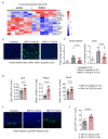
Tauroursodeoxycholic acid decreases intestinal apoptosis. (A to D) Small intestinal samples were isolated from recipient mice which were treated with vehicle or tauroursodeoxycholic acid (TUDCA), on day 14 after bone marrow transplantation (BMT) (C57BL/6 to BALB/c model). (A) Heat map based on microarray analysis showing the differentially regulated genes (q-value<0.05) from the gene set ‘apoptosis’ from the CONSENSUS database (small intestine, day 14 after BMT). Data were pooled from two independent experiments, n=6 mice per group, P=0.001. The color code represents the Z-score log2 intensity. (B) Representative immunofluorescence images of TUNEL-stained paraffin sections from the small intestine (day 14 after BMT, blue: DAPI, green: apoptotic TUNEL staining). Scale bars 200 m. (C) Quantification of apoptotic nuclei in the small intestine and the colon was obtained using the Olympus ScanR analysis software (day 14 after BMT). Data were pooled from two independent experiments, numbers (N) indicate individual mice. P-values were calculated using the ordinary one-way ANOVA test with correction for multiple comparisons. (D) Quantitative real-time polymerase chain reaction (PCR) analysis of the mRNA expression of the intestinal stem cell markers Lgr5 and Phlda1 and the goblet cell marker Muc2 with Actb as a reference gene (small intestine, day 14 after BMT). Data were pooled from two independent experiments, numbers (N) indicate individual mice. P-values were calculated using the unpaired two-tailed Student’s t-test. (E and F) B6.129P2-Lgr5tm1(cre/ERT2)Cle/J mice (H-2kb) were transplanted and treated with TUDCA as described in Figure 3A. On day 14 after BMT, the small intestine was analyzed by immunofluorescence for the number of green fluorescent protein positive (GFP+) intestinal stem cells. (E) Representative images obtained using confocal microscopy (blue: DAPI, green: GFP). Scale bars 50 m. (F) Quantification of data pooled from two independent experiments, N numbers represent individual mice. The P-value was calculated using the unpaired twotailed Student’s t-test.
Figure 7.
Figure 7.
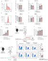
The graft-versus-leukemia/lyphoma effect is preserved despite tauroursodeoxycholic acid administration. (A to F) MV-4-11 and RMB-1 cells were cultured for 72 hours (h) with or without addition of tauroursodeoxycholic acid (TUDCA). One representative result from three independent experiments performed in technical triplicates is shown for panel (A) and (D). In panels (B), (C), (E) and (F) data were pooled from three independent experiments performed in technical duplicates or triplicates. P-values were calculated using the two-tailed unpaired Student’s t-test; ns: not significant. (A) Representative histograms from flow cytometric analysis of human leukocyte antigen (HLA) A, B, C expression on MV-4-11 cells. (B) Quantification of HLA A, B and C expression on MV-4-11 cells. (C) Quantitative polymerase chain reaction (PCR) analysis of the expression of the antigen presentation-related genes TAP1 and TAP2 in MV-4-11 cells with glyceraldehyde 3-phosphate dehydrogenase (GAPDH) as reference gene. (D) Representative histograms from a flow cytometric analysis of major histocompatibility complex (MHC) class I expression on RMB-1 cells. (E) Quantification of MHC class I expression on RMB-1 cells. (F) Quantitative PCR analysis of the expression of the antigen presentation-related genes Tap1 and Tap2 in RMB-1 cells with Actb as reference gene. (G) C57BL/6 CD8+ T cells were activated with allogeneic (BALB/c) dendritic cells for 72 h and treated with 500 M TUDCA or vehicle prior to incubation with A20 lymphoma cells. The percentage of dead A20 cells after 24 h of incubation was analyzed by flow cytometry. Representative data from one of two biologically independent experiments performed with three to four technical replicates respectively. (H) Experimental model for assessing the graft-versus-ligand (GvL) response of allogeneic T cells ex vivo. BALB/c mice underwent bone marrow transplantation (BMT) as described in Figure 3C and T cells from spleens were isolated for subsequent co-culture with A20 cells on day 14 after BMT. (I) Flow cytometric quantification of dead A20 lymphoma cells co-cultured with CD4+ and CD8+ T cells re-isolated from the spleens of recipient mice on day 14 after BMT as described in (H). Representative data from one of two biologically independent experiments performed with four to five technical replicates respectively. P-values were calculated using the ordinary one-way ANOVA test with correction for multiple comparisons, ns: not significant. (J) Experimental model for assessing the GvL response in vivo. BALB/c mice underwent BMT with additional injection of green fluorescent protein positive (GFP+) Ba/F3-ITD leukemia cells. Allogeneic T cells were transferred two days later and animals were treated with 200 mg/kg body weight TUDCA or vehicle for another 10 days. (K) Flow cytometric analysis of spleen and bone marrow for the percentage of GFP+ cells on day 12 after tumor injection. N numbers represent individual mice. Left panel: representative flow cytometry plots. Right panel: quantification, numbers (N) represent individual mice. P-values were calculated using the ordinary one-way ANOVA test with correction for multiple comparisons, ns: not significant.
Figure 8.
Figure 8.
Tauroursodeoxycholic acid leads to a reduction of acute graft-versus-host disease. A model, in which this positive effect is achieved by two distinct mechanisms: enhancing the viability during exposure to pro-inflammatory cytokines and reduction of antigen presentation in the intestine with a consequent decrease in apoptosis.

References

    1. de Lima M, Porter DL, Battiwalla M, et al. . Proceedings from the National Cancer Institute's Second International Workshop on the biology, prevention, and treatment of relapse after hematopoietic stem cell transplantation: part III. Prevention and treatment of relapse after allogeneic transplantation. Biol Blood Marrow Transplant. 2014;20(1):4-13. - PMC - PubMed
    1. Zeiser R, Blazar BR. Acute Graft-versus- Host Disease - Biologic Process, Prevention, and Therapy. N Engl J Med. 2017; 377(22):2167-2179. - VSports在线直播 - PMC - PubMed
    1. McDonald GB. How I treat acute graft-versus- host disease of the gastrointestinal tract and the liver. Blood. 2016;127(12):1544-1550. - "V体育2025版" PMC - PubMed
    1. Takashima S, Kadowaki M, Aoyama K, et al. . The Wnt agonist R-spondin1 regulates systemic graft-versus-host disease by protecting intestinal stem cells. J Exp Med. 2011;208(2):285-294. - V体育安卓版 - PMC - PubMed
    1. Eriguchi Y, Takashima S, Oka H, et al. . Graft-versus-host disease disrupts intestinal microbial ecology by inhibiting Paneth cell production of alpha-defensins. Blood. 2012;120(1):223-231. - PubMed

Publication types

Substances