<style draggable="Mv4kD"><noframes date-time="9hgIT"><code dropzone="1qfXc"></code>
Skip to main page content
U.S. flag

An official website of the United States government

Dot gov

The . gov means it’s official. Federal government websites often end in . gov or . mil. Before sharing sensitive information, make sure you’re on a federal government site VSports app下载. .

Https

The site is secure. The https:// ensures that you are connecting to the official website and that any information you provide is encrypted and transmitted securely V体育官网. .

. 2020 Jun 30;21(13):4658.
doi: 10.3390/ijms21134658.

The Effect of Lycium barbarum Polysaccharides on Pyroptosis-Associated Amyloid β1-40 Oligomers-Induced Adult Retinal Pigment Epithelium 19 Cell Damage (V体育安卓版)

Affiliations

The Effect of Lycium barbarum Polysaccharides on Pyroptosis-Associated Amyloid β1-40 Oligomers-Induced Adult Retinal Pigment Epithelium 19 Cell Damage

"VSports" Ming Yang et al. Int J Mol Sci. .

Abstract

Age-related macular degeneration (AMD) is a sight-threatening disease with limited treatment options VSports手机版. We investigated whether amyloid β1-40 (Aβ1-40) could cause pyroptosis and evaluated the effects of Lycium barbarum polysaccharides (LBP) on Aβ1-40 oligomers-induced retinal pigment epithelium 19 (ARPE-19) damage, which is an in vitro AMD model. Aβ1-40 oligomers verified by Western blot were added to ARPE-19 cells with or without 24 h LBP treatment. Aβ1-40 oligomers significantly decreased ARPE-19 cell viability with obvious morphological changes under light microscopy. SEM revealed swollen cells with a bubbling appearance and ruptured cell membrane, which are morphological characteristics of pyroptosis. ELISA results showed increased expression of IL-1β and IL-18, which are the final products of pyroptosis. LBP administration for 24 h had no toxic effects on ARPE-19 cells and improved cell viability and morphology while disrupting Aβ1-40 oligomerization in a dose-dependent manner. Furthermore, Aβ1-40 oligomers up-regulated the cellular immunoreactivity of pyroptosis markers including NOD-like receptors protein 3 (NLRP3), caspase-1, and membrane N-terminal cleavage product of GSDMD (GSDMD-N), which could be reversed by LBP treatment. Taken together, this study showed that LBP effectively protects the Aβ1-40 oligomers-induced pyroptotic ARPE-19 cell damages by its anti-Aβ1-40 oligomerization properties and its anti-pyroptotic effects. .

Keywords: cell death; drusen; eye disease; retina; traditional Chinese medicine (TCM) V体育安卓版. .


Conflict of interest statement

The authors declare no conflict of interest.

Figures
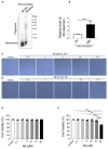
Figure 1
Figure 1
Confirmation of Aβ1-40 oligomerization and the effect of Aβ1-40 oligomers on Aβ1-40 oligomers-induced retinal pigment epithelium 19 (ARPE-19) cell viability. (A) Representative Western blot showing Aβ1-40 monomers and oligomers at 0 h and 48 h after oligomerization. (B) Semi-quantification of Aβ1-40 oligomers showing an increase in Aβ1-40 oligomers after oligomerization (n = 3, ***p < 0.001). (C) Light microscope images of ARPE-19 cells after exposure to Aβ1-40 oligomers (0.1 μM to 20 μM) for 24 h revealed no noticeable changes. (D) Light microscope images of ARPE-19 cells after exposure to Aβ1-40 oligomers (0.1 to 20 μM) for 48 h. There was a significant decrease in the cell number and change in cell shape in cells exposed to 20 mM Aβ1-40 oligomers. (E) Cell Counting Kit-8 (CCK-8) assay showed that the cell viability of ARPE-19 cells after the exposure to Aβ1-40 oligomers (0.1 μM to 20 μM) for 24 h did not change significantly (n = 4). (F) After exposure to Aβ1-40 oligomers for 48 h, ARPE-19 cell viability determined by CCK-8 assay was decreased after exposure to 20 μM Aβ1-40 oligomers for 48 h (n = 4, ***p < 0.001). Scale bar = 100 µm.
Figure 2
Figure 2
The effect of Lycium barbarum polysaccharides on ARPE-19 cell viability and morphology. (A) ARPE-19 cell viability after Lycium barbarum polysaccharides (LBP) treatment at various concentrations were determined by CCK-8 assay. No significant changes were observed for LBP treatment up to 14 mg/L. However, cell viability started to decrease when LBP was administered at 17.5 up to 35 mg/L. (n = 4, ***p < 0.001). (B) Exposure to Aβ1-40 oligomers decreased ARPE-19 cell viability (column 2). Administration of LBP at both low (3.5 mg/L) and high (14 mg/L) dosage were able to reverse the decreased cell viability imposed by Aβ1-40 oligomers (columns 3 and 4) (n = 4, ***p < 0.001). (C) Protective effects of LBP at low and high dosages were confirmed by trypan blue assay, which revealed that the ARPE-19 cell death rate was decreased by LBP treatment (n = 4, ***p < 0.001). (D) The morphological changes of ARPE-19 cells upon Aβ1-40 oligomers exposure with/without LBP treatment were examined by light microscopy. There was a decrease in cell number together with rounded cells and debris after Aβ1-40 oligomers exposure. The cells retained their normal morphology with LBP treatment (scale bar = 100 µm).
Figure 3
Figure 3
The effect of Lycium barbarum polysaccharides on Aβ1-40 oligomerization. (A) Representative Western blot illustrating the presence of Aβ1-40 monomers and oligomers with or without LBP incubation. The amount of the higher molecular weight Aβ1-40 oligomers generated after 48 h incubation decreased with increasing LBP concentration. (B) Semi-quantification of the relative level of Aβ1-40 oligomers based on the Western blot results (n = 3, ***p < 0.001).
Figure 4
Figure 4
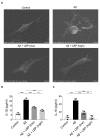
1-40 oligomers-induced cell death and release of pyroptotic products (IL-1β and IL-18) were alleviated by LBP treatment. (A). Scanning electron microscopy images revealed the morphology of ARPE-19 cells upon Aβ1-40 oligomers exposure with/without LBP treatment. Aβ1-40 oligomers exposure resulted in cell swelling and bubbling with a ruptured membrane while surrounded by membrane fragments and debris. LBP treatment recovered their normal morphology. (B). The concentration of IL-1β secreted by ARPE-19 cells was determined by ELISA. A significant increase of IL-1β in the cell culture supernatant was observed after Aβ1-40 oligomers exposure (column 2). Nevertheless, the level of IL-1β began to decrease when LBP was added (columns 3 and 4). LBP (low) represents a low concentration of LBP (3.5 mg/L) and LBP (high) stands for a high concentration of LBP (14 mg/L). (n = 4, ***p < 0.001). (C) IL-18 ELISA results showed that Aβ1-40 oligomers’ exposure resulted in a rise of IL-18 (column 2). However, LBP significantly reduced the level of IL-18 at both low (3.5 mg/L) and high (14 mg/L) concentration (columns 3 and 4). (n = 4, ***p < 0.001).
Figure 5
Figure 5
Lycium barbarum polysaccharides lessened the changes in expression of pyroptosis markers (NOD-like receptors protein 3 (NLRP3), caspase-1, and membrane N-terminal cleavage product of GSDMD (GSDMD-N)) in Aβ1-40 oligomers-exposed ARPE-19 cells. (A) Representative immunofluorescence images of NLRP3 (green fluorescence) and DAPI (blue fluorescence) in ARPE-19 cells under different treatments. Aβ1-40 oligomers exposure increased the cellular expressions of NLRP3 (second row). However, the increased expression was subsequently decreased by LBP treatment with both low (3.5 mg/L) and high (14 mg/L) concentration (third row and the fourth row). (B) The histogram indicated the average fluorescence intensity of NLRP3 based on the immunofluorescence results (n = 3, ***p < 0.001). (C) Representative immunofluorescence images of caspase-1 (green fluorescence) and DAPI (blue fluorescence) of ARPE-19 cells in different treatment groups. The expression of caspase-1 in ARPE-19 increased after Aβ1-40 oligomers exposure (second row). Nevertheless, the elevated expression was reduced by LBP treatment with both low (3.5 mg/L) and high (14 mg/L) concentration (third and fourth row). (D) The histogram for the average fluorescence intensity of caspase-1 based on the immunofluorescence data (n = 3, ***p < 0.001). (E) Representative immunofluorescence images showing the expression membrane GSDMD-N (green fluorescence) and DAPI (blue fluorescence) in ARPE-19 cells. There was a remarkable increase in GSDMD-N expression after Aβ1-40 oligomers exposure (second row). Nonetheless, LBP reduced the increased expression at both low (3.5 mg/L) and high (14 mg/L) concentration (third and fourth row). (F) The histogram for the average fluorescence intensity of GSDMD-N based on the immunofluorescence images (n = 3, ***p < 0.001). Scale bar = 100 µm.
Figure 6
Figure 6
Schematic diagram of the proposed mechanism of amyloid β1-40 oligomer-induced ARPE-19 cell damage and LBP’s effects by anti-Aβ1-40 oligomerization and anti-pyroptosis. (A) In ARPE-19 cells, Aβ1-40 oligomers activate the NLRP3 inflammasome (NLRP3, the adaptor ASC, and pro-caspase-1), which subsequently leads to the generation of caspase-1. Caspase-1 cleaves Pro-IL-1β and Pro-IL-18, yielding IL-1β and IL-18, respectively. At the same time, C-GSDMD-N is also cleaved by caspase-1 thereby generating GSDMD-N and GSDMD-C. GSDMD-N moves to the cell membrane and forms ‘‘pores’’, allowing the newly generated IL-1β and IL-18 to be released from the cell. (B) Lycium barbarum polysaccharide (LBP) inhibits Aβ1-40 oligomerization and reverses the activation of pyroptosis, thereby protecting ARPE-19 cells from Aβ1-40 oligomers-induced cell death.

References

    1. Wong W.L., Su X., Li X., Cheung C.M., Klein R., Cheng C.Y., Wong T.Y. Global prevalence of age-related macular degeneration and disease burden projection for 2020 and 2040: A systematic review and meta-analysis. Lancet Glob. Health. 2014;2:e106–e116. doi: 10.1016/S2214-109X(13)70145-1. - DOI - PubMed
    1. Lim L.S., Mitchell P., Seddon J.M., Holz F.G., Wong T.Y. Age-related macular degeneration. Lancet. 2012;379:1728–1738. doi: 10.1016/S0140-6736(12)60282-7. - DOI (VSports最新版本) - PubMed
    1. Mitchell P., Liew G., Gopinath B., Wong T.Y. Age-related macular degeneration. Lancet. 2018;392:1147–1159. doi: 10.1016/S0140-6736(18)31550-2. - DOI - PubMed
    1. Pedrosa A.C., Reis-Silva A., Pinheiro-Costa J., Beato J., Freitas-da-Costa P., Falcao M.S., Falcao-Reis F., Carneiro A. Treatment of neovascular age-related macular degeneration with anti-VEGF agents: Retrospective analysis of 5-year outcomes. Clin. Ophthalmol. 2016;10:541–546. doi: 10.2147/OPTH.S90913. - DOI - PMC - PubMed
    1. Evans J.R., Lawrenson J.G. Antioxidant vitamin and mineral supplements for preventing age-related macular degeneration. Cochrane Database Syst. Rev. 2017;7:CD000253. doi: 10.1002/14651858.CD000253.pub4. - DOI - PMC - PubMed

MeSH terms

V体育2025版 - LinkOut - more resources