Skip to main page content
U.S. flag

An official website of the United States government

Dot gov

The . gov means it’s official. Federal government websites often end in . gov or VSports app下载. mil. Before sharing sensitive information, make sure you’re on a federal government site. .

Https

The site is secure. The https:// ensures that you are connecting to the official website and that any information you provide is encrypted and transmitted securely V体育官网. .

. 2020 Aug 20;48(14):7801-7817.
doi: 10.1093/nar/gkaa536.

The chromatin landscape at the HIV-1 provirus integration site determines viral expression (VSports在线直播)

Affiliations

The chromatin landscape at the HIV-1 provirus integration site determines viral expression

Gerlinde Vansant et al. Nucleic Acids Res. .

Abstract (V体育ios版)

HIV-1 persists lifelong in memory cells of the immune system as latent provirus that rebounds upon treatment interruption. Therefore, the latent reservoir is the main target for an HIV cure. Here, we studied the direct link between integration site and transcription using LEDGINs and Barcoded HIV-ensembles (B-HIVE). LEDGINs are antivirals that inhibit the interaction between HIV-1 integrase and the chromatin-tethering factor LEDGF/p75. They were used as a tool to retarget integration, while the effect on HIV expression was measured with B-HIVE. B-HIVE tracks insert-specific HIV expression by tagging a unique barcode in the HIV genome. We confirmed that LEDGINs retarget integration out of gene-dense and actively transcribed regions. The distance to H3K36me3, the marker recognized by LEDGF/p75, clearly increased. LEDGIN treatment reduced viral RNA expression and increased the proportion of silent provirus. Finally, silent proviruses obtained after LEDGIN treatment were located further away from epigenetic marks associated with active transcription VSports手机版. Interestingly, proximity to enhancers stimulated transcription irrespective of LEDGIN treatment, while the distance to H3K36me3 only changed after treatment with LEDGINs. The fact that proximity to these markers are associated with RNA expression support the direct link between provirus integration site and viral expression. .


Figures

Figure 1.
Figure 1.
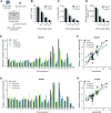
LEDGINs inhibit integration and reduce chromosomal preference. (A) 40 000 Jurkat or SupT1 cells were transduced in a 96-well plate with barcoded HIV in the presence of varying concentrations of LEDGIN CX014442. Cells were cultured for two weeks before DNA and RNA were extracted. (B) The percentage of GFP positive cells two weeks post transduction of SupT1 (black) and Jurkat (gray) cells in the presence of LEDGIN CX014442. Bars represent the average of flow cytometry measurement in duplicate with standard deviation. (C) The number of integrated copies two weeks post transduction, as determined by Alu-LTR nested qPCR on gDNA that was normalized to CCR5. Bars represent the average of qPCR in duplicate with standard deviation. (D) The number of retrieved integration sites in SupT1 and Jurkat cells. (E, G) Relative number of mapped insertions/Mb plotted for each chromosome in SupT1 (E) and Jukat (G) cells whereby the sum of all chromosomes per condition is 100. Different colors represent different concentrations of LEDGINs added during transduction. (F, H) XY-plot showing the relative number of mapped insertions/Mb (y-axis, log2 scale) over the gene density of each chromosome (x-axis, log2 scale) in SupT1 (F) and Jurkat (H) cells. Thus, each value on the x-axis corresponds to a certain chromosome (see also Supplementary Table S1 for gene densities) and to four y-values, one for each condition. The lines are the result of regression analysis (see also Supplementary Table S2). Two experiments were performed in Jurkat cells and two in SupT1 cells. Results are shown for one representative experiment, referred to as experiment A, in which SupT1 and Jurkat cells were transduced in parallel. GFP; Green Fluorescent Protein, Mb; megabase.
Figure 2.
Figure 2.
LEDGIN treatment retargets integration away from active genes. Integration sites retrieved in each condition in SupT1 (A) and Jurkat (B) cells were divided in four genomic categories and plotted as relative proportions: silent genes (SG), intergenic regions (IR), regulatory elements containing enhancers and promoters (RE) and active genes (AG).
Figure 3.
Figure 3.
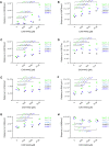
LEDGIN treatment retargets integration away from H3K36me3 in SupT1 and Jurkat cells. The median distance in base pairs (bp) between the integration site and certain features is plotted for two experiments in SupT1 cells and three in Jurkat cells. Panels A-H plot the distance to: (A) H3K36me3, (B) H3K79me3, (C) H3K79me2, (D) H3K27ac, (E) H3K4me1, (F) RNAPII, (G) H3K4me3 and (H) H3K9me3 (See also Supplementary Table S3 for explanation of different markers). Statistical significance was calculated by the Kruskal–Wallis test, * P< 0.05, ** P< 0.01, *** P< 0.001, **** P< 0.0001. RNAPII; RNA polymerase II.
Figure 4.
Figure 4.
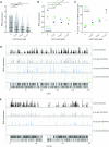
LEDGIN treatment reduces RNA expression. Two weeks post transduction in the presence of varying concentrations of LEDGIN CX014442, mRNA was extracted and reverse transcribed to cDNA to determine RNA expression. (A) Expression in Jurkat cells (experiment B) was determined using the B-HIVE method. The expression score is calculated for each unique barcode (dot) by normalizing the barcode counts in the RNA to the corresponding DNA counts. Error bars represent median and interquartile range. (B) Median RNA expression scores for two independent experiments in Jurkat cells (experiment A and B) and one in SupT cells (experiment A). (C) The percentage of DNA barcodes that did not show RNA expression was calculated relative to the total number of barcodes found in each condition in SupT1 and Jurkat cells. (D) Expression landscape of barcodes integrated in chromosome 2. Each bar represents an integration site and the height of the bar corresponds to the expression level. (E) Expression landscape of barcodes integrated in chromosome 19. Statistical significance was calculated by the Kruskal–Wallis test, **** P< 0.0001, *P< 0.05.
Figure 5.
Figure 5.
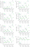
Silent barcoded provirus is targeted away from epigenetic features associated with active transcription. The distance in base pairs (bp) of integration sites to certain features was determined for either ‘all’ retrieved insertion sites or the ‘no RNA’ sites in each condition (0, 6.25, 15.62 or 31.25 μM of CX014442). The median distance (bp) is plotted on the y-axis for two independent experiments in Jurkat cells (experiment A blue, experiment B black) and one experiment in SupT1 cells (experiment A green). Panels A-H plot the distance to: (A) H3K36me3, (B) H3K79me3, (C) H3K79me2, (D) H3K27ac, (E) H3K4me1, (F) RNAPII, (G) H3K4me3 and (H) H3K9me3. Statistical significance was calculated by the Kruskal–Wallis test, * P< 0.05, ** P< 0.01, *** P< 0.001, **** P< 0.0001. bp; base pairs, RNAPII; RNA polymerase II.
Figure 6.
Figure 6.
Effect of LEDGINs on super-enhancer markers Med1 and CBP. (A, B) Integration site analysis in SupT1 and Jurkat cells determined the distance in base pairs of HIV provirus to super-enhancer markers Med1 (A) and CBP (B) in each condition (0, 6.25, 15.62 or 31.25 μM of CX014442). The median distance (bp) is plotted on the y-axis for two independent experiments in Jurkat cells (experiment A blue, experiment B black) and two in SupT1 cells (experiment A green, experiment B gray). (C, D) The distance in base pairs (bp) of integration sites to Med1 (C) and CBP (D) was determined for ‘no RNA’ sites in each condition (0, 6.25, 15.62 and 31.25 μM of CX014442) and plotted next to ‘all sites’. The median distance (bp) is plotted on the y-axis for two independent experiments in Jurkat cells and one experiment in SupT1 cells. Statistical significance was calculated by the Kruskal–Wallis test, *P< 0.05, **P< 0.01, ***P< 0.001, ****P< 0.0001. bp; base pairs, Med1; Mediator 1, CBP; CREB-binding protein.
Figure 7.
Figure 7.
Model for HIV-1 integration and transcription. (A) HIV-1 integration is mainly determined by LEDGF/p75 binding to H3K36me3 in active genes. HIV-1 also integrates near enhancer regions characterized by H3K27ac and H3K4me1. Integration in these areas is associated with high RNA expression. (B) LEDGIN treatment retargets integration away from actively transcribed regions to silent genes and intergenic regions, resulting in provirus with lower RNA expression. However, some LEDGF/p75-independent integrations might still occur near regulatory elements, explaining few high residual expressors even in the presence of LEDGINs.

References (V体育2025版)

    1. Finzi D., Hermankova M., Pierson T., Carruth L.M., Buck C., Chaisson R.E., Quinn T.C., Chadwick K., Margolick J., Brookmeyer R. et al. .. Identification of a reservoir for HIV-1 in patients on highly active antiretroviral therapy. Science. 1997; 278:1295–1300. - PubMed
    1. Finzi D., Blankson J., Siliciano J.D., Margolick J.B., Chadwick K., Pierson T., Smith K., Lisziewicz J., Lori F., Flexner C. et al. .. Latent infection of CD4+ T cells provides a mechanism for lifelong persistence of HIV-1, even in patients on effective combination therapy. Nat. Med. 1999; 5:512. - PubMed
    1. Chomont N., El-Far M., Ancuta P., Trautmann L., Procopio F.A., Yassine-Diab B., Boucher G., Boulassel M.-R., Ghattas G., Brenchley J.M. et al. .. HIV reservoir size and persistence are driven by T cell survival and homeostatic proliferation. Nat. Med. 2009; 15:893. - PMC (VSports app下载) - PubMed
    1. Chun T.-W., Stuyver L., Mizell S.B., Ehler L.A., Mican J.A.M., Baseler M., Lloyd A.L., Nowak M.A., Fauci A.S.. Presence of an inducible HIV-1 latent reservoir during highly active antiretroviral therapy. Proc. Natl. Acad. Sci. U.S.A. 1997; 94:13193–13197. - PMC - PubMed
    1. Chun T.-W., Carruth L., Finzi D., Shen X., DiGiuseppe J.A., Taylor H., Hermankova M., Chadwick K., Margolick J., Quinn T.C. et al. .. Quantification of latent tissue reservoirs and total body viral load in HIV-1 infection. Nature. 1997; 387:183–188. - PubMed

V体育官网入口 - Publication types

MeSH terms