Skip to main page content
U.S. flag

An official website of the United States government

Dot gov

The . gov means it’s official. Federal government websites often end in . gov or . mil VSports app下载. Before sharing sensitive information, make sure you’re on a federal government site. .

Https

The site is secure. The https:// ensures that you are connecting to the official website and that any information you provide is encrypted and transmitted securely V体育官网. .

. 2021 Apr;37(2):151-175.
doi: 10.1007/s10565-020-09537-1. Epub 2020 Jun 14.

Drug-induced hepatic steatosis in absence of severe mitochondrial dysfunction in HepaRG cells: proof of multiple mechanism-based toxicity

Affiliations

Drug-induced hepatic steatosis in absence of severe mitochondrial dysfunction in HepaRG cells: proof of multiple mechanism-based toxicity

Julien Allard et al. Cell Biol Toxicol. 2021 Apr.

VSports最新版本 - Abstract

Steatosis is a liver lesion reported with numerous pharmaceuticals. Prior studies showed that severe impairment of mitochondrial fatty acid oxidation (mtFAO) constantly leads to lipid accretion in liver. However, much less is known about the mechanism(s) of drug-induced steatosis in the absence of severe mitochondrial dysfunction, although previous studies suggested the involvement of mild-to-moderate inhibition of mtFAO, increased de novo lipogenesis (DNL), and impairment of very low-density lipoprotein (VLDL) secretion. The objective of our study, mainly carried out in human hepatoma HepaRG cells, was to investigate these 3 mechanisms with 12 drugs able to induce steatosis in human: amiodarone (AMIO, used as positive control), allopurinol (ALLO), D-penicillamine (DPEN), 5-fluorouracil (5FU), indinavir (INDI), indomethacin (INDO), methimazole (METHI), methotrexate (METHO), nifedipine (NIF), rifampicin (RIF), sulindac (SUL), and troglitazone (TRO). Hepatic cells were exposed to drugs for 4 days with concentrations decreasing ATP level by less than 30% as compared to control and not exceeding 100 × Cmax. Among the 12 drugs, AMIO, ALLO, 5FU, INDI, INDO, METHO, RIF, SUL, and TRO induced steatosis in HepaRG cells. AMIO, INDO, and RIF decreased mtFAO. AMIO, INDO, and SUL enhanced DNL VSports手机版. ALLO, 5FU, INDI, INDO, SUL, RIF, and TRO impaired VLDL secretion. These seven drugs reduced the mRNA level of genes playing a major role in VLDL assembly and also induced endoplasmic reticulum (ER) stress. Thus, in the absence of severe mitochondrial dysfunction, drug-induced steatosis can be triggered by different mechanisms, although impairment of VLDL secretion seems more frequently involved, possibly as a consequence of ER stress. .

Keywords: Endoplasmic reticulum stress; Fatty acid oxidation; Lipogenesis; Mitochondria; Steatosis; Very low-density lipoprotein V体育安卓版. .


Conflict of interest statement

J. A. , S. B. , J. M. , P. -J. F V体育ios版. , D. L. G. , Y. D. , Y. L. , K. B. , and B. F. declare that they have no conflict of interest in relation to this work. N. B. and A. B. -S. are co-founders of MITOLOGICS S. A. S. R. L. is an employee of MITOLOGICS S. A. S.

Figures

Fig. 1
Fig. 1
Effects of drugs on neutral lipids. HepaRG cells were treated for 4 consecutive days with different concentrations (μM) of amiodarone, allopurinol, 5-fluoruracil, indinavir, indomethacin, methotrexate, rifampicin, sulindac, and troglitazone. Results are means ± SD for 5 to 9 independent cultures. The horizontal dotted line represents 100% of the control values. Statistical significance of treated vs. control cells, determined by one-way ANOVA, is indicated by an asterisk (P < 0.05)
Fig. 2
Fig. 2
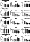
Effects of drugs on mtFAO, DNL, and apoB secretion. HepaRG cells were treated for 4 consecutive days with different concentrations (μM) of amiodarone (a), allopurinol (b), 5-fluoruracil (c), indinavir (d), or indomethacin (e) in order to determine their respective effects on mitochondrial fatty acid oxidation (mtFAO), de novo lipogenesis (DNL), and apoB secretion in the culture medium. Results are means ± SD for 5 to 9 independent cultures for mtFAO and DNL and 5 independent cultures for apoB secretion. The horizontal dotted line represents 100% of the control values. Statistical significance of treated vs. control cells, determined by one-way ANOVA, is indicated by an asterisk (P < 0.05)
Fig. 3
Fig. 3
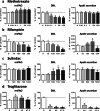
Effects of drugs on mtFAO, DNL, and apoB secretion. HepaRG cells were treated for 4 consecutive days with different concentrations (μM) of methotrexate (a), rifampicin (b), sulindac (c), or troglitazone (d) in order to determine their respective effects on mitochondrial fatty acid oxidation (mtFAO), de novo lipogenesis (DNL), and apoB secretion in the culture medium. Results are means ± SD for 5 to 9 independent cultures for mtFAO and DNL and 5 independent cultures for apoB secretion. The horizontal dotted line represents 100% of the control values. Statistical significance of treated vs. control cells, determined by one-way ANOVA, is indicated by an asterisk (P < 0.05)
Fig. 4
Fig. 4
Effects of drugs on mRNA level of DNL enzymes. HepaRG cells were treated for 4 consecutive days with two concentrations (μM) of amiodarone (a), indomethacin (b), or sulindac (c) in order to determine their respective effects on the mRNA level of four enzymes playing a key role in DNL, namely ATP citrate lyase (ACLY), acetyl-CoA carboxylase alpha (ACACA), fatty acid synthase (FASN), and stearoyl-CoA desaturase 1 (hSCD1). Results are means ± SD for 5 independent cultures and are shown as fold change of control cells. The horizontal dotted line represents control values set at 1. Statistical significance of treated vs. control cells, determined by one-way ANOVA, is indicated by an asterisk (P < 0.05)
Fig. 5
Fig. 5
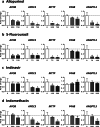
Effects of drugs on mRNA level of proteins and enzymes involved in VLDL assembly. HepaRG cells were treated for 4 consecutive days with two concentrations (μM) of allopurinol (a), 5-fluorouracil (b), indinavir (c), or indomethacin (d) in order to determine their respective effects on the mRNA level of five structural proteins and enzymes playing a significant role in VLDL assembly, namely, apolipoprotein B (APOB), apolipoprotein C3 (APOC3), microsomal triglyceride transfer protein (MTTP), prolyl 4-hydroxylase subunit beta (P4HB, also known as PDI), and angiopoietin-like 3 (ANGPTL3). Results are means ± SD for 5 independent cultures and are shown as fold change of control cells. The horizontal dotted line represents control values set at 1. Statistical significance of treated vs. control cells, determined by one-way ANOVA, is indicated by an asterisk (P < 0.05)
Fig. 6
Fig. 6
Effects of drugs on mRNA level of proteins and enzymes involved in VLDL assembly. HepaRG cells were treated for 4 consecutive days with two concentrations (μM) of rifampicin (a), sulindac (b), or troglitazone (c) in order to determine their respective effects on the mRNA level of five structural proteins and enzymes playing a significant role in VLDL assembly, namely, apolipoprotein B (APOB), apolipoprotein C3 (APOC3), microsomal triglyceride transfer protein (MTTP), prolyl 4-hydroxylase subunit beta (P4HB, also known as PDI), and angiopoietin-like 3 (ANGPTL3). Results are means ± SD for 5 independent cultures and are shown as fold change of control cells. The horizontal dotted line represents control values set at 1. Statistical significance of treated vs. control cells, determined by one-way ANOVA, is indicated by an asterisk (P < 0.05)
Fig. 7
Fig. 7
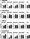
Effects of drugs on mRNA level of ER stress markers. HepaRG cells were treated for 4 consecutive days with two concentrations (μM) of allopurinol (a), 5-fluorouracil (b), indinavir (c), or indomethacin (d) in order to determine their respective effects on the mRNA level of five different proteins and transcription factors classically induced upon ER stress, namely heat shock protein family A member 5 (HSPA5, also known as BIP), DNA damage inducible transcript 3 (DDIT3, also known as CHOP), endoplasmic reticulum to nucleus signaling 1 (ERN1, also known as IRE1α), eukaryotic translation initiation factor 2 alpha kinase 3 (EIF2AK3, also known as PERK), and activating transcription factor 6 (ATF6). Results are means ± SD for 5 independent cultures and are shown as fold change of control cells. The horizontal dotted line represents control values set at 1. Statistical significance of treated vs. control cells, determined by one-way ANOVA, is indicated by an asterisk (P < 0.05)
Fig. 8
Fig. 8
Effects of drugs on mRNA level of ER stress markers. HepaRG cells were treated for 4 consecutive days with two concentrations (μM) of rifampicin (a), sulindac (b), or troglitazone (c) in order to determine their respective effects on the mRNA level of five different proteins and transcription factors classically induced upon ER stress, namely heat shock protein family A member 5 (HSPA5, also known as BIP), DNA damage inducible transcript 3 (DDIT3, also known as CHOP), endoplasmic reticulum to nucleus signaling 1 (ERN1, also known as IRE1α), eukaryotic translation initiation factor 2 alpha kinase 3 (EIF2AK3, also known as PERK), and activating transcription factor 6 (ATF6). Results are means ± SD for 5 independent cultures and are shown as fold change of control cells. The horizontal dotted line represents control values set at 1. Statistical significance of treated vs. control cells, determined by one-way ANOVA, is indicated by an asterisk (P < 0.05)
Fig. 9
Fig. 9
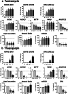
Effects of tunicamycin and thapsigargin on gene expression, apoB secretion, neutral lipids, mtFAO, and DNL. HepaRG cells were treated for 4 consecutive days with different concentrations (μM) of tunicamycin (a) or thapsigargin (b), two prototypical ER stress inducers, in order to determine their respective effects on the mRNA level of three proteins classically induced upon ER stress, namely heat shock protein family A member 5 (HSPA5, also known as BIP), DNA damage inducible transcript 3 (DDIT3, also known as CHOP), and endoplasmic reticulum to nucleus signaling 1 (ERN1, also known as IRE1α), mRNA level of five structural proteins and enzymes playing a significant role in VLDL assembly, namely, apolipoprotein B (APOB), apolipoprotein C3 (APOC3), microsomal triglyceride transfer protein (MTTP), prolyl 4-hydroxylase subunit beta (P4HB, also known as PDI), and angiopoietin-like 3 (ANGPTL3), apoB secretion in the culture medium, accumulation of neutral lipids, mitochondrial fatty acid oxidation (mtFAO), and de novo lipogenesis (DNL). Results are means ± SD for 3 independent cultures for mRNA abundance, 5 independent cultures for apoB secretion and neutral lipids and 5 to 6 independent cultures mtFAO and DNL. Results for mRNA level are shown as fold change of control cells. The horizontal dotted line represents control values set at 1 for gene expression, or 100% of the control values for other data. Statistical significance of treated vs. control cells, determined by one-way ANOVA, is indicated by an asterisk (P < 0.05)
Fig. 10
Fig. 10
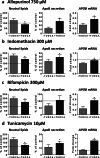
Effects of TUDCA on neutral lipids, apoB secretion, and APOB mRNA level in cells cotreated with allopurinol, indomethacin, rifampicin, and tunicamycin. HepaRG cells were treated for 4 consecutive days without (−) or with (+) TUDCA and with 750 μM allopurinol (a), 300 μM indomethacin (b), 300 μM rifampicin (c), or 10 μM tunicamycin (d). TUDCA effects were then determined for neutral lipids, apoB secretion, and APOB mRNA level. Results are means ± SD for 5 independent cultures for neutral lipids and 3 independent cultures for apoB secretion and APOB mRNA level. Results for mRNA level are shown as fold change of untreated control cells. The horizontal dotted line represents untreated control values set at 1 for gene expression, or 100% of the untreated control values for other data. Statistical significance of treated vs. control cells, determined by Student’s t test, is indicated by an asterisk (P < 0.05)

References

    1. Allard J, Le Guillou D, Begriche K, Fromenty B. Drug-induced liver injury in obesity and nonalcoholic fatty liver disease. Adv Pharmacol. 2019;85:75–107. - PubMed
    1. Amacher DE, Chalasani N. Drug-induced hepatic steatosis. Semin Liver Dis. 2014;34(2):205–214. - V体育2025版 - PubMed
    1. Anthérieu S, Rogue A, Fromenty B, Guillouzo A, Robin MA. Induction of vesicular steatosis by amiodarone and tetracycline is associated with up-regulation of lipogenic genes in HepaRG cells. Hepatology. 2011;53(6):1895–1905. - PubMed
    1. Baiceanu A, Mesdom P, Lagouge M, Foufelle F. Endoplasmic reticulum proteostasis in hepatic steatosis. Nat Rev Endocrinol. 2016;12(12):710–722. - PubMed
    1. Basseri S, Austin RC. Endoplasmic reticulum stress and lipid metabolism: mechanisms and therapeutic potential. Biochem Res Int. 2012;2012:841362. - PMC - PubMed

Publication types

MeSH terms

LinkOut - more resources