- to an NK+ maturational stage to give rise to NKT1 cells. Maturation takes place in the thymus or after emigration of NK- NKT cells to the periphery. In this study, w …">- to an NK+ maturational stage to give rise to NKT1 cells. Maturation takes place in the thymus or after emigration of NK- NKT cells to the periphery. In this study, w …">
"V体育2025版" Skip to main page content
U.S. flag

An official website of the United States government

Dot gov

The . gov means it’s official. Federal government websites often end in . gov or . mil. Before sharing sensitive information, make sure you’re on a federal government site. VSports app下载.

Https

The site is secure V体育官网. The https:// ensures that you are connecting to the official website and that any information you provide is encrypted and transmitted securely. .

. 2020 May 7:11:815.
doi: 10.3389/fimmu.2020.00815. eCollection 2020.

Cortical Thymocytes Along With Their Selecting Ligands Are Required for the Further Thymic Maturation of NKT Cells in Mice (V体育平台登录)

Affiliations

Cortical Thymocytes Along With Their Selecting Ligands Are Required for the Further Thymic Maturation of NKT Cells in Mice (V体育安卓版)

Jihene Klibi et al. Front Immunol. .

Abstract

Following positive selection, NKT cell precursors enter an "NK-like" program and progress from an NK- to an NK+ maturational stage to give rise to NKT1 cells. Maturation takes place in the thymus or after emigration of NK- NKT cells to the periphery. In this study, we followed the fate of injected NKT cells at the NK- stage of their development in the thymus of a series of mice with differential CD1d expression VSports手机版. Our results indicate that CD1d-expressing cortical thymocytes, and not epithelial cells, macrophages, or dendritic cells, are necessary and sufficient to promote the maturation of thymic NKT1 cells. Migration out of the thymus of NK- NKT cells occurred in the absence of CD1d expression, however, CD1d expression is required for maturation in peripheral organs. We also found that the natural ligand Isoglobotriosylceramide (iGb3), and the cysteine protease Cathepsin L, both localizing with CD1d in the endosomal compartment and crucial for NKT cell positive selection, are also required for NK- to NK+ NKT cell transition. Overall, our study indicates that the maturational transition of NKT cells require continuous TCR/CD1d interactions and suggest that these interactions occur in the thymic cortex where DP cortical thymocytes are located. We thus concluded that key components necessary for positive selection of NKT cells are also required for subsequent maturation. .

Keywords: CD1d; NKT1; cathepsin L; development; iGb3; plck; thymus V体育安卓版. .


V体育官网入口 - Figures

FIGURE 1
FIGURE 1
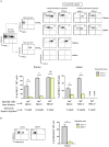
CD1d expression is required for maturation of post-selected NKT cells. (A) CD1d-tetramer-positive (tet+) and NK1.1 negative (NK1.1), or tet+NK1.1+ subsets (as gated) were FACS-sorted from CD8-negative thymocyte preparations obtained from 2 to 4 week-old congenic CD45.1 C57BL/6 mice. Cells were injected into the thymus of NKT-deficient CD45.2 Jα18–/– or CD1d–/– recipient mice, both on C57BL/6 background. One or four weeks after injection, expression of the NK1.1 marker was assessed on CD45.1+ tet+ NKT cells in the thymus and spleen. Histograms show the frequency of tet+ NKT cells expressing NK1.1 in various recipient mice, and times. (B) Cells were FACS-sorted like in (A) and then injected intrathymically into CD45.2 NKT-deficient Jα18–/– or CD1d–/– recipient mice. Three weeks after injection, the expression of NKG2A/C/E was assessed on CD45.1+ tet+ NKT cells in the thymus of injected mice. Histograms show the frequency of tet+ NKT cells positive for NKG2A/C/E. Data are from 3 to 6 experiments, using 4 to 5 recipient mice per experiment. Statistical significance is represented by an asterisk and was evaluated by applying a non-parametric Mann-Whitney U test. A p-value < 0.05 was considered significant. NS, non-significant. Numbers in dot plots represent percentages of cells in the associated quadrant.
FIGURE 2
FIGURE 2
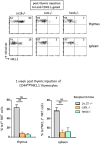
CD1d-expression on cortical thymocytes, but not antigen presenting cells, promotes maturation of NKT cells. CD8-depleted thymocyte preparations from CD45.1 C57BL/6 mice were stained with HSA, CD44, and NK1.1. HSAlowCD44highNK1.1 cells were FACS-sorted for intrathymic injection into CD45.2 NKT-deficient Jα18–/–, CD1d–/–, pEαCD1d or pLck-CD1d/Cα–/– recipients. One week after injection, the expression of the NK1.1 marker was assessed on CD45.1+ tet+ NKT cells in the thymus and spleen of injected mice. Histograms show the frequency of tet+ NKT cells expressing NK1.1 in the different recipient mice. Data are from 4 to 5 experiments, using 3 to 5 recipient mice per experiment. Statistical significance is represented by an asterisk and was assessed by a non-parametric Mann-Whitney U test. A p-value < 0.05 was considered significant. NS, non-significant. Numbers in dot plots represent percentages of cells in the associated quadrant.
FIGURE 3
FIGURE 3
Ligands required for NKT cell selection are also required for subsequent maturation. CD45.1+HSAlowCD44highNK1.1 cells were FACS-sorted like in Figure 2, and then injected intrathymically into CD45.2 NKT-deficient Jα18–/–, catL–/– or hexb–/– recipient mice. One week after injection, expression of the NK1.1 marker was assessed on CD45.1+ tet+ NKT cells in the thymus and in the spleen of recipient mice. Histograms show the frequency of tet+ NKT cells expressing NK1.1 in the different recipient mice. Data are from 4 to 5 experiments, using 3 to 5 recipient mice per experiment. Statistical significance was assessed by a non-parametric Mann-Whitney U test. A p-value < 0.05 was considered significant. NS, non-significant. Numbers in dot plots represent percentages of cells in the associated quadrant.
FIGURE 4
FIGURE 4
Model of NKT cell development involving CD1d/TCR interaction in post-selection NKT cell maturation. Cortical DP thymocytes expressing the canonical Vα14–Jα18 TCR are selected by CD1d-expressing neighboring DP thymocytes. These HSAhigh NKT0 NKT cells lose HSA expression and acquire CCR7 expression, giving rise to NKT cell precursors, NKTp. Some NKTp will commit to the NKT1 lineage by expressing T-bet (NKT1c) but no other markers of the NK lineage. During their progression to the cortex, NKT1c cells continuously sense ligands exposed on cortical thymocytes. This sensing is required for their progression to the NK1.1 + NKT1 stage in the medulla, and long-term residency in the thymus. These CD1d/TCR interactions appear not to be essential for NKT1c migration to the periphery. CMJ, cortico-medullary junction.

References

    1. Bendelac A, Savage PB, Teyton L. The biology of NKT cells. Annu Rev Immunol. (2007) 25:297–336. - PubMed
    1. Bendelac A, Rivera MN, Park SH, Roark JH. Mouse CD1-specific NK1 T cells: development, specificity, and function. Annu Rev Immunol. (1997) 15:535–62. - PubMed
    1. Benlagha K, Weiss A, Beavis A, Teyton L, Bendelac A. In vivo identification of glycolipid antigen-specific T cells using fluorescent CD1d tetramers. J Exp Med. (2000) 191:1895–903. - PMC (VSports在线直播) - PubMed
    1. Benlagha K, Kyin T, Beavis A, Teyton L, Bendelac A. A thymic precursor to the NK T cell lineage. Science. (2002) 296:553–5. - PubMed
    1. Townsend MJ, Weinmann AS, Matsuda JL, Salomon R, Farnham PJ, Biron CA, et al. T-bet regulates the terminal maturation and homeostasis of NK and Valpha14i NKT cells. Immunity. (2004) 20:477–94. - PubMed

Publication types

MeSH terms (VSports注册入口)

"V体育安卓版" LinkOut - more resources