V体育官网 - Skip to main page content
U.S. flag

An official website of the United States government

Dot gov

The VSports app下载. gov means it’s official. Federal government websites often end in . gov or . mil. Before sharing sensitive information, make sure you’re on a federal government site. .

Https

The site is secure. The https:// ensures that you are connecting to the official website and that any information you provide is encrypted and transmitted securely. V体育官网.

. 2020 May 12;10(1):7856.
doi: 10.1038/s41598-020-64521-3.

Metallothioneins regulate ATP7A trafficking and control cell viability during copper deficiency and excess

Affiliations

Metallothioneins regulate ATP7A trafficking and control cell viability during copper deficiency and excess

Nikita Gudekar et al. Sci Rep. .

Abstract (VSports手机版)

Copper (Cu) is an essential, yet potentially toxic nutrient, as illustrated by inherited diseases of copper deficiency and excess VSports手机版. Elevated expression of the ATP7A Cu exporter is known to confer copper tolerance, however, the contribution of metal-binding metallothioneins is less clear. In this study, we investigated the relative contributions of ATP7A and the metallothioneins MT-I and MT-II to cell viability under conditions of Cu excess or deficiency. Although the loss of ATP7A increased sensitivity to low Cu concentrations, the absence of MTs did not significantly affect Cu tolerance. However, the absence of all three proteins caused a synthetic lethal phenotype due to extreme Cu sensitivity, indicating that MTs are critical for Cu tolerance only in the absence of ATP7A. A lack of MTs resulted in the trafficking of ATP7A from the trans-Golgi complex in a Cu-dependent manner, suggesting that MTs regulate the delivery of Cu to ATP7A. Under Cu deficiency conditions, the absence of MTs and / or ATP7A enhanced cell proliferation compared to wild type cells, suggesting that these proteins compete with essential Cu-dependent pathways when Cu is scarce. These studies reveal new roles for ATP7A and metallothioneins under both Cu deficiency and excess. .


Conflict of interest statement

The authors declare no competing interests.

Figures
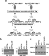
Figure 1
Figure 1
Derivation and characterization of cell lines lacking Atp7a, MtI and MtII genes. (a) Primary fibroblasts were isolated from the lungs of Atp7afl/Y;MtI+/+/MtII+/+ and Atp7afl/Y;MtI−/−/MtII−/− mice and then immortalized by transfection with a plasmid expressing the SV40 large T antigen (SV40 Tag) resulting in WT and MT- cells, respectively. An adenoviral vector encoding CRE recombinase was used to delete Atp7a in WT and MT- cells to obtain ATP7A- and ATP7A-/MT- cells, respectively. (b) PCR analysis of genomic DNA was used to confirm deletion of MtI and MtII genes in both the MT- and ATP7A-/MT- cell lines. Expected PCR product sizes: MtI gene (WT = 161 bp; knockout = 176 bp); MtII gene (WT = 282 bp; knockout = 299 bp). (c) Immunoblot analysis was used to confirm the loss of ATP7A protein in both ATP7A- and ATP7A-/MT- cell lines. Tubulin was detected as a loading control. Images of full-length gels and immunoblots are provided in the supplementary data.
Figure 2
Figure 2
Disruption of Atp7a and MtI/II results in a loss of cell viability that is suppressed by Cu chelation. (a) The rescue of ATP7A-/MT- cells by the Cu chelator BCS is suppressed by copper, but not zinc or iron. ATP7A-/MT- cells were grown for 5 days in basal media containing 1 µM BCS with or without equimolar concentrations of CuCl2, ZnCl2 or FeCl2. Cell survival was then determined using the Crystal Violet assay and imaged. (b,c) Cu concentrations in each cell line were determined by ICP-MS. Cells were grown for 2 days in medium containing 50 µM of the copper chelator BCS and then exposed for 24 h to either basal media or media supplemented with 50 µM BCS (mean ± SEM; *p < 0.05; **p < 0.01; ***p < 0.001; ns = not significant).
Figure 3
Figure 3
Relative contributions of ATP7A and metallothioneins to Cu tolerance. (a) Cu sensitivity of WT, ATP7A-, MT- and ATP7A-/MT- cells. For each cell line, 103 cells/well were seeded in 6-well plates containing basal medium, or basal medium containing 1 µM BCS with or without the indicated concentrations of CuCl2. After 6 days, cell survival was determined using the Crystal Violet assay and imaged. (b) Quantification of Crystal Violet staining. Data are expressed as percent cell survival for each cell line normalized against its growth in BCS (mean ± SEM). (c) Complementation of ATP7A-/MT- cells with plasmids encoding human cDNAs for ATP7A ( + ATP7A), MTI ( + MTI) or MTII ( + MTII). Cells were transfected with each plasmid and then selected in basal medium supplemented with 1 µM CuCl2. Equal numbers of surviving cells (103 cells/well) were then seeded into 6-well plates containing basal medium, 1 µM BCS or 1 µM BCS plus the indicated concentrations of Cu. After 6 days, cell survival was determined using the Crystal Violet assay and imaged. (d) Quantification of Crystal Violet staining. Data are expressed as percent cell survival for each cell line normalized against its growth in BCS (mean ± SEM).
Figure 4
Figure 4
Selection for Cu tolerance in the absence of ATP7A increases ATP7B expression. (a) Immunoblot detection of ATP7B in ATP7A-/MT- cells that were selected for survival in either basal media (Basal) or media containing 20 µM Cu (CuR20). The control sample is ATP7A-/MT- cells propagated in medium containing 50 µM BCS. Each cell line was grown for 16 h in basal media prior to immunoblot analysis. Tubulin was detected as a loading control. (b) Densitometry analysis of ATP7B protein levels normalized against control samples (mean ± SEM; *p < 0.05; ****p < 0.0001). Data were calculated from at least 3 independent experiments. (c) Immunoblot detection of ATP7A and ATP7B proteins in parental ATP7A + /MT- cells (Basal) or ATP7A + /MT- cells that were selected in media containing 900 µM Cu (CuR900). Both cell lines were grown for 16 h in basal media prior to immunoblot analysis. Tubulin was detected as a loading control. (d,e) Densitometry analysis of ATP7A and ATP7B protein levels normalized against Basal samples (mean ± SEM; *p < 0.05; ***p < 0.001). Data were calculated from at least 3 independent experiments. Images of full-length immunoblots are provided in the supplementary data.
Figure 5
Figure 5
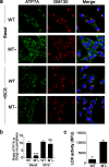
Metallothionein regulates Cu-stimulated ATP7A trafficking and function. (a) Immunofluorescence microscopy was performed to detect endogenous ATP7A (green) in WT and MT- cells cultured for 24 h in basal medium alone or basal medium supplemented with 50 µM BCS. Antibodies against GM130 were used to label the Golgi complex (red) and DAPI was used to stain the nuclei (blue). (b) The ATP7A staining intensity in the Golgi region was expressed relative to the ATP7A staining intensity within the non-Golgi region for each cell line. Values were calculated using ImageJ software (mean ± SEM; ***p < 0.001; ns = not significant; n = 24 cells for WT and MT- cells for each condition). (c) Lysyl oxidase (LOX) activity was measured in the media collected from WT and MT- cells cultured for 48 h (mean ± SEM; *p < 0.05).
Figure 6
Figure 6
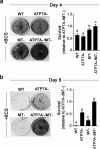
ATP7A and MTs decrease cell viability during Cu starvation. (a) WT, ATP7A-, MT- and ATP7A-/MT- cells were seeded into 6-well plates (2 ×10⁴ cells/well) and cultured in medium supplemented with 50 µM BCS. After 4 days, cells were stained with Crystal Violet and imaged. (b) A second batch of identically treated cells was passaged for an additional 4 days in BCS-containing medium and then stained with Crystal Violet (i.e., after 8 days of Cu chelation). Crystal Violet staining in both (a) and (b) was quantified as a measure of cell survival. Values were normalized against the survival of the ATP7A-/MT- cell line in BCS at Day 4 and Day 8 (mean ± SEM). Different letters indicate values that are significantly different from each other. Note that the loss of ATP7A or MTI/II enhances the ability of cells to propagate in the presence of the BCS copper chelator.

"V体育安卓版" References

    1. Nevitt T, Ohrvik H, Thiele DJ. Charting the travels of copper in eukaryotes from yeast to mammals. Biochim Biophys Acta. 2012;1823:1580–1593. doi: 10.1016/j.bbamcr.2012.02.011. - DOI - PMC - PubMed
    1. Burkhead JL, Reynolds KA, Abdel-Ghany SE, Cohu CM, Pilon M. Copper homeostasis. New Phytol. 2009;182:799–816. doi: 10.1111/j.1469-8137.2009.02846.x. - DOI (VSports) - PubMed
    1. Arioz C, Wittung-Stafshede P. Folding of copper proteins: role of the metal? Q Rev Biophys. 2018;51:e4. doi: 10.1017/S0033583518000021. - DOI - PubMed
    1. Smith AD, Logeman BL, Thiele DJ. Copper Acquisition and Utilization in Fungi. Annu Rev Microbiol. 2017;71:597–623. doi: 10.1146/annurev-micro-030117-020444. - DOI - PMC - PubMed
    1. Maryon EB, Molloy SA, Kaplan JH. Cellular glutathione plays a key role in copper uptake mediated by human copper transporter 1. Am J Physiol Cell Physiol. 2013;304:C768–779. doi: 10.1152/ajpcell.00417.2012. - DOI - PMC - PubMed

Publication types

MeSH terms