Skip to main page content
U.S. flag

An official website of the United States government

Dot gov

The VSports app下载. gov means it’s official. Federal government websites often end in . gov or . mil. Before sharing sensitive information, make sure you’re on a federal government site. .

Https

The site is secure. The https:// ensures that you are connecting to the official website and that any information you provide is encrypted and transmitted securely V体育官网. .

. 2020 Apr;26(4):618-629.
doi: 10.1038/s41591-020-0769-8. Epub 2020 Feb 24.

Broad immune activation underlies shared set point signatures for vaccine responsiveness in healthy individuals and disease activity in patients with lupus

Affiliations

Broad immune activation underlies shared set point signatures for vaccine responsiveness in healthy individuals and disease activity in patients with lupus

Yuri Kotliarov et al. Nat Med. 2020 Apr.

Abstract

Responses to vaccination and to diseases vary widely across individuals, which may be partly due to baseline immune variations. Identifying such baseline predictors of immune responses and their biological basis is of broad interest, given their potential importance for cancer immunotherapy, disease outcomes, vaccination and infection responses. Here we uncover baseline blood transcriptional signatures predictive of antibody responses to both influenza and yellow fever vaccinations in healthy subjects VSports手机版. These same signatures evaluated at clinical quiescence are correlated with disease activity in patients with systemic lupus erythematosus with plasmablast-associated flares. CITE-seq profiling of 82 surface proteins and transcriptomes of 53,201 single cells from healthy high and low influenza vaccination responders revealed that our signatures reflect the extent of activation in a plasmacytoid dendritic cell-type I IFN-T/B lymphocyte network. Our findings raise the prospect that modulating such immune baseline states may improve vaccine responsiveness and mitigate undesirable autoimmune disease activity. .


"VSports最新版本" Conflict of interest statement

Competing Interests: all authors declare that there are no competing interests.

Figures

Extended Data Fig. 1
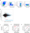
Extended Data Fig. 1. Identification and characterization of the CD19+CD20+CD38++ B cell population, a baseline, pre-vaccination cell frequency-based signature (CBSig) of antibody responses to influenza vaccination.
a, Flow cytometric gating strategy for the CD19+CD20+CD38++ B cell population. Populations 1–4 are further described in (Fig. 1b). b, Box plots (top) showing the frequency of CD19+CD20+CD38++ cells (CBSig; y-axis) at the three baseline time points from ref. (days −7 and 0 are prior to vaccination and day 70 is after vaccination) in low and high responders (x-axis) to the seasonal and pandemic H1N1 influenza vaccines as defined by the Adjusted Maximum Fold Change (adjMFC) metric (see ref.). There are 11 low and 12 high responders for day −7 and 0, and 10 low and 11 high responders for day 70. P values from the Wilcoxon one-tailed test results are shown on the boxplots (based on results from ref. our hypothesis was that the high responders have higher frequencies of these cells than low responders). Boxplots’ center line corresponds to the median value, lower and upper hinges correspond to the first and third quartiles (the 25th and 75th percentiles); lower and upper whiskers extend from the box to the smallest or largest value correspondingly, but no further than 1.5x inter-quantile range. (Bottom) Corresponding receiver operator curves (ROC) for vaccine response at each of the above baseline time points and their AUC (area under the curve) and corresponding permutation based one-tailed p value are shown. c, Dot plots (CD38 vs. CD10 of CD19+CD20+ B cells) for example high and low responders.
Extended Data Fig. 2
Extended Data Fig. 2. Derivation of TGSig, a transcriptional surrogate signature for CBSig.
a, Step 1: Identification of genes with high temporal stability across the three baseline time points (days −7 and 0 are prior to vaccination and day 70 is after vaccination) in the NIH influenza study. The middle box shows the distribution of the temporal stability metric (TSM) across all the genes. The boxes on the right and left show examples of genes with high and low temporal stability, respectively; each line corresponds to an individual. Genes with high temporal stability (≥0.75) across the three baseline time points (depicted to the right of the red dashed line) were subsequently evaluated for correlation with CD19+CD20+CD38++ B cell frequency. b, Step 2: 726 temporally stable genes were ranked by their “robust” correlation with CD19+CD20+CD38++ B cell frequency. Robustness is evaluated using all 231 random samplings of 20 subjects out of the cohort of 22 subjects (i.e., two random subjects were dropped out from each sampling); the mean Pearson correlation coefficient divided by the standard deviation across the samplings (x-axis left panel) was used to rank the genes. Top genes are shown together with the predictive performance of each gene evaluated at day 0 (AUC; right panel). The red dashed line in the right panel corresponds to AUC = 0.50 (prediction performance as expected by chance); the top 10 genes were selected in TGSig (the black dashed line; see Supplementary Table 1 for full list of ranked genes) based on (c). c, Performance (AUC; y-axis) of the gene signature by baseline time point (different lines) and number of top genes included in computing the signature score (x-axis). The vertical dashed line corresponds to a gene signature (TGSig) containing the top 10 genes achieving the best AUC across all three time-points. d, Schema for gene signature score calculation. Gene expression data is standardized through calculation of Z-scores for each gene (i.e., each gene would have mean 0 and standard deviation 1). The Z-scores for each gene in the signature are then averaged to generate the gene signature score. e, Distribution of predictive performance (AUC; x-axis) of 500 random top-10 gene signatures generated from subject-label shuffled gene expression data using the robust correlation metric approach described above. The dashed red line indicates the observed AUC of TGSig at each of the baseline time points. One-tailed empirical p-values are shown. f, Relative rank (rank position divided by the total number of genes) of the top 20 genes from (b) and Supplementary Table 1 at different TSM thresholds. The black line shows TSM cutoff = 0.75, the value used in selecting the top 10 genes for inclusion in TGSig (boxed). g, Change in AUC (x-axis) of TGSig score following removal of the indicated gene in the signature (y-axis) at each of the three baseline time points in the NIH influenza study.
Extended Data Fig. 3
Extended Data Fig. 3. Comparison among response classes by including middle responders and evaluating TGSig in the influenza datasets from Emory.
a, similar to Figs. 1e and 1f but including middle responders (for day 0 n=11/19/13 for low/middle/high responders, respectively; for day −7 n=10/22/14 and for day 70 n=11/21/12); p values from the Jonckheere trend test (with an a priori alternative hypothesis that the high responders >= middle responders >= low responders.) For all boxplots the center line corresponds to the median value, lower and upper hinges correspond to the first and third quartiles (the 25th and 75th percentiles); lower and upper whiskers extend from the box to the smallest or largest value correspondingly, but no further than 1.5x inter-quantile range. b, Similar to (a) but related to Fig. 2a; note that Yale 2012 does not have middle responders based on data retrieved from ImmuneSpace (n=9/10/10 (Stanford 2008), 7/3/6 (Yale 2010), 7/0/8 (Yale 2012)). c, Similar to Fig. 2a but testing TGSig using influenza datasets from Emory University over four years (n=14/8 low/high responders for year 2008, 8/8 for 2009, 11/11 for 2010, 12/10 for 2011). Box plots (top) showing the TGSig score (y-axis) in low and high responders (x-axis) as defined by adjMFC in the indicated season. P values shown on the boxplots were obtained from the Wilcoxon one-tailed test. (bottom) Corresponding receiver operator curves (ROC) for vaccine response and the AUC (area under the curve) and corresponding permutation-based one-tailed p values are shown. d, Similar to (a) but for yellow fever and related to Fig. 2b.
Extended Data Fig. 4
Extended Data Fig. 4. Further evaluation of TGSig in yellow fever and influenza datasets.
a, (top) Box plots (top) showing the TGSig score applied to pre-vaccination PBMC expression data (y-axis) between low and high responders (x-axis) to yellow fever vaccine in trial #2 (x-axis; 4 high vs. 3 low responders). (bottom) Corresponding receiver operator curves (ROC) for vaccine response and the AUC (area under the curve) and corresponding one-tailed permutation based p value are shown. This vaccination cohort included 10 subjects (see Fig. 2b for results on a first, larger trial with 15 subjects). Boxplots’ center line corresponds to the median value, lower and upper hinges correspond to the first and third quartiles (the 25th and 75th percentiles); lower and upper whiskers extend from the box to the smallest or largest value correspondingly, but no further than 1.5x inter-quantile range. P-value is from Wilcoxon one-tailed test. b, Forest plot showing the meta effect sizes (Hedge’s g reflecting correlation strength with adjMFC) of the TGSig genes from a meta-analysis of four influenza datasets (Stanford 2008, NIH 2009, Yale 2011, Yale 2012; genes in gray were not present in all datasets); the bars represent the 95% CI. c, Similar to (b) but for yellow fever vaccination (computed from trial #1).
Extended Data Fig. 5
Extended Data Fig. 5. SLE patient phenotypes based on DA-associated transcriptional signatures and assessing the association between TGSig (at low DA) and DaCP after the removal of patients with lower DA-associated plasmablast signature scores.
a, Recreation of Figure 6a from ref. but as a robustness check here we used a different method: for each patient we ranked genes based on the difference between their average expression at high/middle and low DA time points and then performed GSEA analysis using the blood transcriptomic modules (columns) to fingerprint the patient. The colors on the heatmap denote the statistical significance (−log10 of BH-adjusted p-values from CERNO) of the gene set enrichment test. Here we kept the order of patients (rows) and modules (columns) the same as in the original heatmap, although some patients from the original heatmap were removed due to the absence of low DA time points. The original patient groups were used to annotate the patients (rows). The overall fingerprinting pattern is visually highly consistent with the original heatmap. This heatmap/matrix was then used to construct Fig. 2c by averaging individual patient values within each patient and phenotype group combination. b, DaCP versus average plasmablast score difference between high (or middle if no high was available) and low DA time points in SLE patients from patient groups 2, 3 (in blue, n=19) or groups 2, 3, and 4 (group 4 in grey, n=12) from ref. (see patient groups in Fig. 2c). Spearman rho for groups 2 and 3 is 0.87 (two-tailed p=2.2×10⁻¹⁶); and for groups 2, 3 and 4 is 0.85 (two-tailed p=4.9×10⁻⁷). c, Similar to Fig. 2e, but here patients are from patient groups 2 and 3 only (n=22) and are shaded based on their DaCP. Note that there are more patients here for analysis from groups 2 and 3 than those shown in (a) and (b) because there we required that every patient has at least one low DA and one high/mid DA sample, while here (and in Fig. 2) the DaCP was estimated using all patients with at least one sample including those without high/mid DA time-points (see Methods). Pearson correlation coefficient and two-tailed p values are shown. Spearman correlation coefficient is 0.47 (p=0.029 two-tailed). d, Evaluating the correlation between DaCP and mean TGSig score at low DA time-points (as in (c)) by removing patients with DaCP below the indicated cutoff (y-axis). The first panel shows the number of patients in the evaluation given the threshold; the second and third panels show the corresponding Pearson r and two-tailed p value (shown as −log10(p)).
Extended Data Fig. 6
Extended Data Fig. 6. Evaluating the predictive capacity and information overlap among the TGSig, SLE-Sig, and IFN-I-DCact signatures.
a-d, The predictive profile of SLE-Sig (Fig. 3e) (a), TGSig (b), IFN-I-DCact (Fig. 3f) (c), and the non-leading edge genes from the brown module (Fig. 3e) (d) used as the sole predictor in logistic regression models of high versus low influenza vaccination responder status. Influenza vaccination data pooled from four datasets (Stanford 2008, NIH 2009, Yale 2011, Yale 2012) were used (n = 71 high and low responders). Note that for the brown module (Fig. 3a–d), most of the predictive information come from the leading-edge genes since the signature score of genes outside of the leading edge is not predictive (shown in (d)). e, f when both TGSig and SLE-Sig were used as predictors in the logistic regression (e) or when both TGSig and IFN-I-DCact score were used as predictors (f). In these graphs, the predictor scores are shown on the x axis and the probability that a high responder falls within the predictor score bin is shown on the y axis. The error bars correspond to 95% CIs. The two-tailed p value indicates the probability that the coefficient (“effect”) of the term (e.g., TGSig score) in the logistic regression is 0.
Extended Data Fig. 7
Extended Data Fig. 7. RNA-seq analysis of CD19+CD20+CD38++ B cells sorted from healthy individuals.
a, Sorting strategy and approach: CD19+CD20+ and CD20+CD38++ B cell populations were isolated by FACS from peripheral blood samples of six healthy donors. RNA-seq libraries were prepared for each isolated sample using the Nugen Ovation SoLo low-input RNAseq library preparation kit. b, Differential gene expression between the CD20+CD38++ B cells and parental CD19+CD20+ B cells from six paired samples was compared using DESeq2. Plot shows the log2 fold change versus the log2 of the mean normalized counts across all samples for each gene. TGSig genes are shown in red; genes from the differentially expressed gene set (BH-adjusted two-tailed p-value < 1%, log of mean normalized counts > 1; total genes in set: 105) that fall into the top enriched Gene Ontology Biological Processes category “Cell Activation” (enrichment analysis done using ToppGene) are shown in cyan (21 genes). c, Enrichment of the 87 SLE-Sig genes in genes ranked by differential expression between CD19+CD20+CD38++ versus CD19+CD20+ cells. The p value shown was computed from the GSEA test. d, Similar to (c) but instead of the SLE-Sig genes here the top k (k=10 (TGSig), 30, 50) genes correlated with the frequency of CD20+CD38++ B cells is assessed (only 713 temporally stable genes with TSM≥ 0.75 were included in the analysis); also see Extended Data Fig. 2. e, Similar to (d) but using 7731 genes with TSM ≥ 0.5. A lower/more relaxed TSM cutoff was used to evaluate whether by starting with more genes (therefore potentially more statistical power for enrichment analysis) an enrichment signal can be detected.
Extended Data Fig. 8
Extended Data Fig. 8. Supporting data for CITE-seq single cell analysis to dissect the cellular origin of baseline signatures.
a, Single cell scatterplots of key markers in the CD4+ T cell clusters. C0.0.0 (Naïve), C1.0.0 (Central/Transitional Memory), and C1.1.0 (TEMRA/Effector memory) clusters show different distributions of CD62L, CD45RA, CD27, and CD28. b, Distributions of key markers in the CD8+ Memory T cell clusters. CD45RA vs. CD62L expression are shown in the top panels, ridge plots of CD45RO and CD28 are shown in lower panels. Similar to CD4+ cells, these CD8+ clusters show differences in CD62L and CD45RA distributions; C4.0.1 (TEMRA/Effector Memory) are mainly CD62L negative and CD28 negative, with CD45RA+ (TEMRA) and CD45RO+ (Effector Memory) subsets within this cluster. C4.0.0 and C4.0.3 show highly similar protein expression, with C4.0.3 being defined by high CD103 expression (upper right panel). c, Unconventional T cells (C7 clusters) show variable CD161, CD8, and CD56 expression. C7.0.0 and C7.0.1 are both CD3+/CD161+, with C7.0.1 being CD8 positive while C7.0.0 is CD8 negative. C4.0.2 (NKT-like) are also CD3 positive, but express CD57/56 and are CD8/CD161 negative or low. The C4.0.2 cluster also showed distinctly low CD27 and high in CD45RA compared to the C7 clusters, making it more similar to the C4.0.1 TEMRA/Effector memory CD8+ subset, except expressing CD56/CD57; this could also be consistent with a Terminal Effector phenotype. d, Hand gating strategy for CD20+CD38++ B cells using CITE-seq data (number of cells in the gate shown in red). e, Boxplot comparing the hand gated frequency of CD20+CD38++ B cells between 10 high and 10 low responders; p value from Wilcoxon one-tailed test. Boxplots’ center line corresponds to the median value, lower and upper hinges correspond to the first and third quartiles (the 25th and 75th percentiles); lower and upper whiskers extend from the box to the smallest or largest value correspondingly, but no further than 1.5x inter-quantile range. f, Similar to Fig. 5b (10 high versus 10 low responders) but for an independently obtained Type I IFN signature gene set (Supplementary Table 6: IFN gene set; see Methods). One or two asterisks denote significance with p<0.05 or p<0.01, respectively (Wilcoxon one-tailed test because we are interested in assessing whether the high responders are higher than the low responders; see also Supplementary Table 7). g, Results of the “drop out” analysis using CITE-seq data. The goal was to assess which cell clusters (or combination of clusters) that were individually significant delineators of high versus low responders were essential for prediction using the baseline signatures in bulk (i.e., simulating transcriptional data from PBMCs). The “pseudo bulk” results (average across all single cells for every subject in 10 low and 10 high responders) of the three signatures tested are shown on the first row. For subsequent rows cells from the indicated cell cluster(s) were dropped before repeating the pseudo-bulk analysis as in row 1. P values obtained from one-tailed Wilcoxon test: *: p<0.05; **: p<0.01.
Extended Data Fig. 9
Extended Data Fig. 9. Evaluating CMV correlates and pDC surface expression phenotypes strongly influenced by genetics from ref. in high versus low responders.
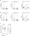
Since CMV status is not available for the cohorts we evaluated, we evaluated whether CMV correlates are significantly different between high and low responders in the NIH influenza vaccination cohort. a, Boxplots comparing the frequency of CD4+ TEMRA cells between 10 low and 10 high responders using CITE-seq (left panel) and between 9 low and 8 high responders using flow cytometry (center panel) data. Wilcoxon two-tailed p values are shown. The third panel is a scatter plot of CITE-seq versus the flow cytometry cell frequencies (n=17). Pearson correlation and two-tailed p values are shown. b, Same as (a) but for CD8+ TEMRA cells. c, Boxplots comparing the relative surface protein expression of CD86 and HLA-DR in pDCs (cluster C9) between 10 low and 10 high responders using CITE-seq data. Wilcoxon two-tailed p values are shown. For all boxplots the center line corresponds to the median value, lower and upper hinges correspond to the first and third quartiles (the 25th and 75th percentiles); lower and upper whiskers extend from the box to the smallest or largest value correspondingly, but no further than 1.5x inter-quantile range.
Extended Data Fig. 10
Extended Data Fig. 10. Relationship between sex and baseline signatures.
a, Box plots comparing the TGSig and SLE-Sig scores in females versus males; here only subjects with CITE-seq data are included to indicate that sex was not a driver of the differences between 10 high and 10 low responders emerged from CITE-seq data analysis (see Fig. 4). Wilcoxon two-tailed p values comparing 11 females and 9 males are shown. For all boxplots the center line corresponds to the median value, lower and upper hinges correspond to the first and third quartiles (the 25th and 75th percentiles); lower and upper whiskers extend from the box to the smallest or largest value correspondingly, but no further than 1.5x inter-quantile range. b, Same as (a) but including all high and low responders from original NIH study. (day 0: 12 females and 12 males; day −7: 13 females and 11 males; day 70: 11 females and 12 males) c, same as (b) but including middle responders (i.e., all subjects in the study: day 0: 27 females and 16 males; day −7: 30 females and 16 males; day 70: 27 females and 17 males). d, Box plots comparing TGSig scores among low, middle, and high responders in males only (all subjects in the original NIH study used: day 0: 6/4/6 for low/middle/high responders, respectively; day −7: 5/5/6; day 70: 6/5/6). e, same as (d) but in females only (day 0: 5/15/17 for low/middle/high responders, respectively; day −7: 5/17/8; day 70: 5/16/6). All p values shown for two-group comparison were from the Wilcoxon two-tailed test; one-tailed p values shown for three-group comparison were from the Jonckheere trend test (with an a priori alternative hypothesis that the high responders >= middle responders >= low responders).
Figure 1:
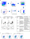
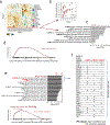
Figure 1:. Study questions and the derivation of a baseline, pre-vaccination signature predictive of response using an influenza fever vaccination cohort.
a, Overview of the study and research questions. b, Prediction performance for antibody response in the NIH influenza study using the frequency of several B cell subsets (y-axis) (see Methods and gating strategy outlined in Extended Data Fig. 1a). The left panel shows the AUC (area under receiver operator curve; x-axis) for predicting high and low responders (n = 23 with flow cytometry data) to the seasonal and pandemic H1N1 influenza vaccines in ref.. The right panel shows the temporal stability metric (TSM) (x-axis); higher TSM indicates greater temporal stability over the three baseline time points (days −7 and 0 prior to vaccination and day 70 after vaccination) using 136 samples from 51 subjects. Population 2 (red box) is the CD19+CD20+CD38++ B cell population. c, Flow chart showing the steps to derive the gene expression-based surrogate signature (TGSig). d, Top temporally stable genes correlated with the frequency of CD19+CD20+CD38++ B cell and the selected genes in TGSig (red box). 22 high and low responders (those with both gene expression and flow cytometry data) are used to assess correlations and rank genes. Genes are ranked based on the average Spearman correlation divided by the standard deviation obtained from 231 iterations (as a safeguard against noise we iterated over all sub-cohorts containing 20 subjects by taking out 2 random subjects at a time [i.e., excluding 2 out of 22 subjects] to assess the correlation). See Extended Data Fig. 2a–d and Methods for further details about temporal stability, gene selection, and signature score calculation. e, Top: box plots comparing the TGSig score (y-axis) at day 0 (pre-vaccination) between low (n=11) and high (n=13) responders (x-axis) (Wilcoxon one-tailed p value shown); bottom: receiver operating curve (ROC) for assessing predictive capacity (area under the curve (AUC) and one-tailed permutation test p value shown). Boxplots’ center line corresponds to the median value, lower and upper hinges correspond to the first and third quartiles (the 25th and 75th percentiles); lower and upper whiskers extend from the box to the smallest or largest value correspondingly, but no further than 1.5x inter-quantile range. f, Similar to (e) but for the other two baseline time points: days −7 (10 low and 14 high responders) and day −70 (11 low and 12 high responders).
Figure 2:
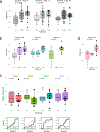
Figure 2:. Assessing TGSig in independent influenza vaccination, yellow fever vaccination, and SLE datasets.
a, Same as Fig. 1e in terms of statistical tests and plot types but here showing the predictive performance of TGSig (evaluated at baseline/pre-vaccination) in the indicated independent datasets: Stanford 2008 (purple boxes; 10 high vs. 8 low responders), Yale 2011 (turquoise boxes; 6 high vs. 7 low responders) and Yale 2012 (green boxes; 8 high vs. 7 low responders) with the corresponding ROC shown at the bottom. b, Similar to (a) but for yellow fever vaccination. This cohort (trial #1) included 6 high and 5 low responders; see Extended Data Fig. 4a for results on a second, smaller cohort (trial #2) with 10 subjects (4 high and 3 low responders) from the same publication). See also Extended Data Fig. 3 for boxplots that include middle responders. c, SLE patient groups determined based on the blood gene expression signatures associated with disease activity/flares (see also Extended Data Fig. 5a and Methods.) The color and size of the circle denote the average statistical significance (−log10 of BH-adjusted p-values from the CERNO test) of the association across patients in the group. The patient group IDs (columns) are listed with the number of patients in each group in parentheses. The same phenotypic annotations from ref are used: ER = erthryopoiesis; IFN = IFN response/neutrophils; ML = myeloid linage/neutrophils; PB = plasmablasts; LL = Lymphoid lineage. The three groups with prominent PB signatures are boxed in red. d, Overview of the analysis approach. The dynamics of disease activity as measured by the SLEDAI score of an actual patient is shown. Individual visits are shown as dots (low DA (blue dots) = SLEDAI < 3, medium DA (grey dots) = SLEDAI: 3–7, high DA (red dots) = SLEDAI ≥ 8)). The TGSig score was computed from the low DA time points and the DaCP was computed from all time-points using a mixed-effect model accounting for treatment effects (see Methods); the correlation between TGSig (averaged across low DA time-points) and DaCP was then evaluated. e, Scatterplot showing the relationship between the DaCP (y-axis) and the mean TGSig score at low DA time points (x-axis) in (left panel) patient groups 2, 3, and 4 (34 subjects) and (right panel) subjects whose DA tended not to be associated with a plasmablast signature (patient groups 1, 5, 6, and 7; 27 subjects). Pearson correlation coefficient and two-tailed p values are shown, and for the left panel separately for patient groups 2 and 3 only (blue dots, n=22) or groups 2, 3, 4 (blue and grey dots; n=34). As a robustness check we also computed the Spearman correlation: rho=0.47 (p=0.029 two-tailed test) for groups 2 and 3; rho=0.36 (p=0.038 two-tailed test) for groups 2, 3 and 4; rho=−0.12 (p=0.56 two-tailed test) for the rest of the patient groups. f, Same as in (e) but the plasmablast (PB) signature score was computed from the low DA time-points instead of TGSig. Groups 2,3: Spearman rho=0.014 (p=0.95 two-tailed test); groups 2,3,4: rho=−0.006 (p=0.97 two-tailed test).
Figure 3:
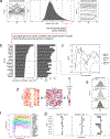
Figure 3:. A transcriptional correlate of plasmablast-associated disease activity in SLE is also associated with influenza vaccination responses and functionally related to TGSig.
a, Identification of 18 transcriptomic modules from genes whose expression was temporally stable across low DA time-points in SLE patients from patient groups 2, 3, and 4. The heatmap shows the eigengene of each module averaged across low-DA time-points of each patient. Rows and columns correspond to modules and SLE patients, respectively. The number of genes in each module is shown in Supplementary Fig. 1a. b, Scatterplot showing the relationship between the brown module score and the DaCP for patients in groups 2, 3, and 4 as above (n=34; Pearson correlation=0.31, p=0.04 based on a one-tailed permutation test (Supplementary Fig. 1b)). Spearman rho=0.29 (p=0.05 one-tailed permutation test) c, Top enriched blood transcriptome modules (BTMs) of the brown module (370 genes) based on the hypergeometric test with BH-adjusted p values (FDR) shown; the red line corresponds to 1% FDR (no additional BTMs were identified at 5% FDR cutoff). Complete results of the enrichment analysis can be found in Supplementary Fig. 1c. d, Gene set enrichment analysis (GSEA) of the brown module genes (370 genes). 6563 genes were ranked by their magnitude of association with antibody responses based on a meta-analysis of four influenza vaccination datasets (the enrichment P value shown was computed from the GSEA test). The tick marks denote the location of the genes in the brown module. e, (top) Enrichment analysis of blood transcriptome modules and the brown module in the temporally stable genes from the NIH influenza study as ranked by their correlation with the frequency of CD20+CD38++ B cells. A temporal stability score (see Methods) cutoff of 0.5 is used here to define 7889 stable genes; the enrichment results are robust to the threshold used. 5% and 1% FDR are indicated by the red dashed lines (BH-adjusted p values shown were computed from the GSEA test). (bottom) GSEA enrichment plot for the brown module. The top 87 genes in the brown module (based on the gene rank on the x axis) were identified by GSEA as the “leading edge genes” (i.e., the main driver of the enrichment signal) (we called this gene set SLE-Sig). The P value shown was computed from the GSEA test. The full list of leading-edge genes can be found in Supplementary Table 5. f, Genes in the brown module that are also in at least one of the three top Type I IFN /antiviral/dendritic cell activation BTMs from (e); black dots indicate that the gene is present in the indicated gene set.
Figure 4:
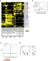
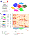
Figure 4:. CITE-seq (simultaneous protein and transcriptome expression profiling in single cells) analysis of high and low influenza vaccine responders.
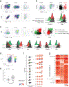
a, Experiment and analysis overview: single PBMCs from 10 high (red) and 10 low (blue) responders (as defined by adjMFC from ref.) were profiled by CITE-seq (measuring 82 cell surface proteins and transcriptome). Cells from all subjects were clustered together using only surface protein expression profile at three increasingly detailed clustering resolutions (referred herein as levels 1–3, denoting the lowest to the highest resolutions; see Methods). 10 cell clusters (C0-C9) were identified at level 1 and shown in different colors in the tSNE plot. b, Cell clusters from levels 1–3 are shown in three columns and depicted as circles (size is proportional to the number of cells in the cluster). The edges denote containment relationship between the clusters at neighboring resolutions: an edge connecting one cluster to another cluster indicates that some fraction (or all) of the cells in the former are found in the latter. Annotations are provided for levels 1 (1st column) and 3 (3rd column) clusters. The clusters/circles are colored, matching those in the tSNE visualization. c, A heatmap showing the average expression of selected protein markers (columns) in each of the cell clusters (rows) derived from the three different clustering resolutions. The cell cluster names are color matched with those in (b). See Extended Data Fig. 8a–c and Supplementary Fig. 2 for additional details. d, Boxplot comparing the TGSig score between high (solid dot; n=10) and low (empty dot; n=10) responders using “pseudo bulk” data (average across all single cells within each subject; see Methods); p value from Wilcoxon one-tailed test. Boxplots’ center line corresponds to the median value, lower and upper hinges correspond to the first and third quartiles (the 25th and 75th percentiles); lower and upper whiskers extend from the box to the smallest or largest value correspondingly, but no further than 1.5x inter-quantile range. e, Same as (d) but for SLE-Sig.
Figure 5:
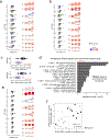
Figure 5:. Dissecting the cellular origin of baseline signatures.
a, Evaluating the difference in TGSig score between high (n=10) and low (n=10) responders in each cell cluster from Fig. 4b (see Methods). Left panel: boxplot comparing high (solid dot) and low (empty dot) responders in each of the level 1 (1st column) clusters; each dot corresponds to the signature score of a subject. Red asterisks denote significance with p<0.05 (Wilcoxon one-tailed test; see also Supplementary Table 7). Right panel uses the same visualization as in Fig. 4b but here the color reflects the average normalized difference in TGSig signature score between the high and low responders (shown here as a t statistic). One or two asterisks denote significance with p<0.05 or p<0.01, respectively (Wilcoxon one-tailed test because we are interested in assessing whether the high responders are higher than the low responders; see also Supplementary Table 7). For all boxplots the center line corresponds to the median value, lower and upper hinges correspond to the first and third quartiles (the 25th and 75th percentiles); lower and upper whiskers extend from the box to the smallest or largest value correspondingly, but no further than 1.5x inter-quantile range. b, Same as (a) but for SLE-Sig. c, Similar to the boxplots in the left panel of (a) (10 high versus 10 low responders) but for the signature score of the LI.M165 BTM (top panel: enriched for dendritic cell activation) and the IFN-I-DCact (bottom panel; see Fig. 3f) gene sets evaluated for cells in the pDC cluster only (cluster C9). d, Enrichment analysis result of the CD40act gene set (Supplementary Table 6; 49 genes) using the hyper-geometric test against the BTMs from ref. All 32,738 detected genes were used as a background. BTMs with an adjusted one-tailed p-value (FDR computed using the BH method) of 0.05 (red line) or lower are shown. e, Same as (a) but for CD40act. f, Scatterplot (based on ranks since Spearman correlation is being evaluated) assessing the correlation between the frequency of CD20+CD38++ B cells (see Fig. 1b) and the CD40 activation signature score in the switched B cell cluster (C3.1.0 – see Fig. 4b,c). Spearman correlation and two-tailed p value are shown (based on 9 high and 9 low responders because not all 20 subjects assessed by CITE-seq have corresponding flow cytometry data). Detailed test statistics for data shown in (a), (b), and (e) can be found in Supplementary Table 7.
Figure 6:
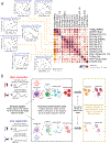
Figure 6:
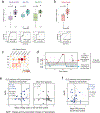
a, Matrix heatmap showing the pairwise Spearman correlation among the select signature scores across subjects. Example scatterplots similar to the one in Fig. 5f are shown for assessing the correlation between the original TGSig (computed using microarray data generated from PBMCs – see Figs. 1c–e) and the cell cluster based signature scores found to be significantly different between high (n=10) and low (n=10) responders. Spearman correlation and two-tailed p value are shown. The name of the cell cluster (see Fig. 4a,b) for which the indicated signature score was computed is in parentheses. Each example scatterplot corresponds to a highlighted (yellow) entry in the matrix on the right. The matrix is symmetrical: row and column profiles are identical. The size and shade of the circle indicate the correlation strength (Spearman rho) and asterisks denote significance level (two-tailed test) as shown in legend below. Note that SLE-Sig (PBMC) was computed using the original bulk microarray data (the same as TGSig (PBMC)). b, Model describing the molecular/cellular underpinnings and differences between high versus low responders. Activation of this entire circuit (including the components in the plasmablast/plasma cell box on the right) typically follows infection, vaccination, or during autoimmune disease flares. Here we propose that the high responders tend to have more activated pDCs and thus more Type I IFNs and activated B and T cells at baseline, but only upon additional antigenic and/or inflammatory co-stimulation (and flare trigger in the case of SLE patients) does the system mount a full-blown plasmablast/plasma cell response cumulating in the generation of antibodies. Open questions include: 1) What sets the system into such temporally stable “activated” states in pDCs, lymphocytes, and other myeloid cells?; 2) What constrains the activated immune baselines from mounting full-blown plasmablast/plasma cell responses?; 3) What is the antigen specificity repertoire of the activated lymphocytes at baseline?

Comment in

References (VSports app下载)

    1. Kotas ME & Medzhitov R Homeostasis, inflammation, and disease susceptibility. Cell 160, 816–827 (2015). - PMC - PubMed
    1. Chen DS & Mellman I Elements of cancer immunity and the cancer-immune set point. Nature 541, 321–330 (2017). - PubMed
    1. Tsang JS Utilizing population variation, vaccination, and systems biology to study human immunology. Trends Immunol. 36, 479–493 (2015). - PMC - PubMed
    1. Willis JCD & Lord GM Immune biomarkers: the promises and pitfalls of personalized medicine. Nat. Rev. Immunol 15, 323 (2015). - PubMed
    1. Brodin P & Davis MM Human immune system variation. Nat. Rev. Immunol 17, 21–29 (2017). - PMC - PubMed

Publication types

MeSH terms

Substances