Skip to main page content (VSports app下载)
U.S. flag

An official website of the United States government

Dot gov

The . gov means it’s official. Federal government websites often end in . gov or . mil. Before sharing sensitive information, make sure you’re on a federal government site VSports app下载. .

Https

The site is secure. The https:// ensures that you are connecting to the official website and that any information you provide is encrypted and transmitted securely. V体育官网.

. 2020 Apr 3;295(14):4428-4437.
doi: 10.1074/jbc.AC119.012178. Epub 2020 Feb 21.

The ubiquitin ligase FBXW7 targets the centriolar assembly protein HsSAS-6 for degradation and thereby regulates centriole duplication

Affiliations

The ubiquitin ligase FBXW7 targets the centriolar assembly protein HsSAS-6 for degradation and thereby regulates centriole duplication

Binshad Badarudeen (VSports手机版) et al. J Biol Chem. .

Abstract (VSports最新版本)

Formation of a single new centriole from a pre-existing centriole is strictly controlled to maintain correct centrosome number and spindle polarity in cells VSports手机版. However, the mechanisms that govern this process are incompletely understood. Here, using several human cell lines, immunofluorescence and structured illumination microscopy methods, and ubiquitination assays, we show that the E3 ubiquitin ligase F-box and WD repeat domain-containing 7 (FBXW7), a subunit of the SCF ubiquitin ligase, down-regulates spindle assembly 6 homolog (HsSAS-6), a key protein required for procentriole cartwheel assembly, and thereby regulates centriole duplication. We found that FBXW7 abrogation stabilizes HsSAS-6 and increases its recruitment to the mother centriole at multiple sites, leading to supernumerary centrioles. Ultrastructural analyses revealed that FBXW7 is broadly localized on the mother centriole and that its presence is reduced at the site where the HsSAS-6-containing procentriole is formed. This observation suggested that FBXW7 restricts procentriole assembly to a specific site to generate a single new centriole. In contrast, during HsSAS-6 overexpression, FBXW7 strongly associated with HsSAS-6 at the centriole. We also found that SCFFBXW7 interacts with HsSAS-6 and targets it for ubiquitin-mediated degradation. Further, we identified putative phosphodegron sites in HsSAS-6, whose substitutions rendered it insensitive to FBXW7-mediated degradation and control of centriole number. In summary, SCFFBXW7 targets HsSAS-6 for degradation and thereby controls centriole biogenesis by restraining HsSAS-6 recruitment to the mother centriole, a molecular mechanism that controls supernumerary centrioles/centrosomes and the maintenance of bipolar spindles. .

Keywords: E3 ubiquitin ligase; F-Box and WD repeat domain-containing 7 (FBXW7); cell division; centriole; centrosome; human SAS-6 centriolar assembly protein (HsSAS6); mitosis; mitotic spindle; procentriole; ubiquitin. V体育安卓版.


Conflict of interest statement

The authors declare that they have no conflicts of interest with the contents of this article

Figures (V体育ios版)

Figure 1.
Figure 1.
FBXW7 controls centriole duplication by regulating HsSAS-6 at the centrosome. a, representative confocal images showing supernumerary centrioles (more than four) in DLD-1 FBXW7 KO cells. b, plot of the percentage of cells versus centriole number in DLD-1 FBXW7 WT versus FBXW7 KO; n ≈ 80–100 cells. c, G1/S synchronized FBXW7 WT versus FBXW7 KO DLD-1 cells stained for HsSAS-6 and CP110. d, plot of centrosomal HsSAS-6 intensity in FBXW7 KO versus FBXW7 WT DLD-1 cells. Data are mean ± S.D. (error bars). n ≈ 20 cells. e, FBXW7 KO versus FBXW7 WT DLD-1 cells at G1/S were stained for Odf2 and HsSAS-6. f, regions of individual centrosomes are indicated by boxes with numbers (1,2,3) and are displayed in enlarged forms on the right. The plot in g shows percentage bipolar versus multipolar cells. n ≈ 50 mitotic cells.
Figure 2.
Figure 2.
FBXW7 overexpression abrogates new centriole formation by diminishing HsSAS-6 at the centrosome. a, U2OS cells transfected with mCherry-FBXW7-WT stained for HsSAS-6 and γ-tubulin. b, plot of HsSAS-6 intensity at centrosome with two centrioles in FBXW7-WT versus mCherry-FBXW7-FboxΔ condition. n ≈ 30. c, mitotic synchronized U2OS cells transfected with mCherry-tagged FBXW7-WT or FBXW7-FboxΔ were stained for CP110 and α-tubulin. Loss of centrioles at the spindle pole is indicated by an arrow. d, plot for percentage mitotic cells with bipolar (four centrioles), defective bipolar (less than four centrioles), and multipolar (more than four centrioles) spindles. n ≈ 50 mitotic cells in each. Data are mean ± S.D. (error bars) (three experiments) in all of the plots.
Figure 3.
Figure 3.
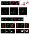
Organization of FBXW7 at the centrosome. a, reconstructed SIM images of centrioles of a G1/S synchronized centrin-1-eGFP RPE1 cell stained for HsSAS-6 and FBXW7. HsSAS-6–stained procentrioles are shown by an arrow. b, a cartoon representing FBXW7 (red) and HsSAS-6 (light blue) on mother centrioles. The centriole lumen is shown by light violet, and centrin-1 localization in the distal lumen is shown by green. HsSAS-6 staining at one centriole appeared dimmer than the other, presumably due to different orientations of the centrioles. c, reconstructed three-dimensional view SIM images of centrioles of RPE-1 centrin-1-eGFP cell showing FBXW7, HsSAS-6, and centrin-1 localization. Images of the same cell (as shown in a) in three different orientations are shown. Scale bar, 1 μm. d, reconstructed SIM images of centrin-1-eGFP stably expressed RPE1 cell stained for centrobin and FBXW7. e, reconstructed SIM image in two different orientations (i and ii) of eGFP-HsSAS-6–overexpressed HeLa Kyoto cell showing co-localization of eGFP-HsSAS-6 with endogenous FBXW7. f, SIM images of HsSAS-6 localization on mother centrioles in FBXW7 WT versus KO DLD-1 cells. Cells were stained with Odf2 and HsSAS-6 antibodies. Unless otherwise mentioned, scale bars of all SIM images are 500 nm.
Figure 4.
Figure 4.
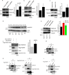
HsSAS-6 is a novel FBXW7 substrate. HeLa (a) or U2OS cells (b) after transfection with FBXW7 esiRNA, followed by synchronization at G1/S, were analyzed for HsSAS-6. c, lysates of DLD-1 FBXW7 WT versus KO cells were immunoblotted for HsSAS-6. Plots of relative HsSAS-6 levels with respect to control in a–c are shown. d, representative (n = 3) HsSAS-6 levels in FBXW7 KO versus FBXW7 WT DLD-1 cells at different cell cycle stages. e, HsSAS-6 levels in the lysates of G1/S synchronized FBXW7 WT, FBXW7 KO, and FLAG-FBXW7–expressed FBXW7 KO DLD-1 cells. f, U2OS cells were co-transfected with Myc-HsSAS-6 and FLAG-FBXW7 or FLAG-FBXW7-WDΔ for 24 h followed by G1/S synchronization were immunoblotted for Myc-HsSAS-6. g, co-IP of FBXW7 in HeLa cells shows the presence of HsSAS-6. h, lysates of HEK293T cells expressed with FLAG-FBXW7 (left) or FLAG-FBXW7-WDΔ (right) were subjected to FLAG-pulldown and probed for endogenous HsSAS-6 with FLAG-FBXW7 versus FLAG-FBXW7-WDΔ. i, co-IP of HsSAS-6 in HEK293T cells transfected with FLAG-FBXW7 (24 h) followed by G1/S synchronization and immunoblotted for FLAG-FBXW7. In all of the plots, data are mean ± S.D. (error bars) (n = 3). Data were compared considering the control value as 1.
Figure 5.
Figure 5.
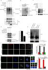
SCFFBXW7 ubiquitylates HsSAS-6. a, HEK293T cells were transfected with HA-Ub and FLAG-FBXW7 or FLAG-FBXW7-WDΔ (24 h) followed by G1/S synchronization by thymidine (18 h) and then MG-132 (6 h). Co-IPs of HsSAS-6 were probed for ubiquitylated proteins. b, recombinant MBP-tagged HsSAS-6 (1×, 2×, and 3×) bound with amylose resin was incubated separately with a mixture of SCF-FBXW7 components, Myc-Skp-1, Myc-Cul-1, HA-Rbx-1, and FLAG-FBXW7, followed by E1, E2, Ub, and ATP. Samples were immunoblotted for HsSAS-6 and ubiquitin. c, MBP-tagged HsSAS-6 mixed with either Ub WT or UB K63R or Ub K48R mutants probed for ubiquitination as described in b. d, lysates of HEK293T cells transfected with FLAG-FBXW7 together with either Myc-HsSAS-6 WT or Myc-HsSAS-6 2A (S111A/T495A) were subjected to FLAG pulldown. e, Myc-HsSAS-6 protein (WT versus mutant) exogenously expressed and pulled down was mixed with SCF-FBXW7 and probed for ubiquitination (top). The same samples were probed for Myc-HsSAS-6 (bottom). f, confocal images of HeLa Kyoto cells transfected with HsSAS-6 3′-UTR siRNA followed by transfection with mCherry-HsSAS-6 WT or mCherry-HsSAS6 2A and then G1/S synchronized. Scale bar, 5 μm. The percentage of cells with centriole numbers in these conditions is plotted. g, HeLa Kyoto cells were transfected with HsSAS-6 3′-UTR siRNA followed by mCherry-HsSAS-6 WT or mCherry-HsSAS6 2A, and the mitotic cells were imaged for CP110 and γ-tubulin. The plot shows percentage bipolar versus multipolar cells. n ≈ 70 cells in f and ∼50 mitotic cells in g. In all of the plots, data are mean ± S.D. (error bars) (n = 3).

"VSports手机版" References

    1. Levine M. S., Bakker B., Boeckx B., Moyett J., Lu J., Vitre B., Spierings D. C., Lansdorp P. M., Cleveland D. W., Lambrechts D., Foijer F., and Holland A. J. (2017) Centrosome amplification is sufficient to promote spontaneous tumorigenesis in mammals. Dev. Cell 40, 313–322.e5 10.1016/j.devcel.2016.12.022 - VSports在线直播 - DOI - PMC - PubMed
    1. Nigg E. A., and Holland A. J. (2018) Once and only once: mechanisms of centriole duplication and their deregulation in disease. Nat. Rev. Mol. Cell Biol. 19, 297–312 10.1038/nrm.2017.127 - DOI - PMC - PubMed
    1. Nigg E. A., and Raff J. W. (2009) Centrioles, centrosomes, and cilia in health and disease. Cell 139, 663–678 10.1016/j.cell.2009.10.036 - DOI - PubMed
    1. Doxsey S., Zimmerman W., and Mikule K. (2005) Centrosome control of the cell cycle. Trends Cell Biol. 15, 303–311 10.1016/j.tcb.2005.04.008 - DOI - PubMed
    1. Fırat-Karalar E. N., and Stearns T. (2014) The centriole duplication cycle. Philos. Trans. R. Soc. Lond. B Biol. Sci. 369, 20130460 10.1098/rstb.2013.0460 - DOI - PMC - PubMed

"VSports在线直播" Publication types

MeSH terms (V体育2025版)

"V体育官网" LinkOut - more resources