Skip to main page content
U.S. flag

An official website of the United States government

Dot gov

The . gov means it’s official. Federal government websites often end in . gov or VSports app下载. mil. Before sharing sensitive information, make sure you’re on a federal government site. .

Https

The site is secure. The https:// ensures that you are connecting to the official website and that any information you provide is encrypted and transmitted securely. V体育官网.

. 2020 Feb 16;12(4):3156-3174.
doi: 10.18632/aging.102790. Epub 2020 Feb 16.

V体育平台登录 - LncRNA MEG3 targeting miR-424-5p via MAPK signaling pathway mediates neuronal apoptosis in ischemic stroke

Affiliations

LncRNA MEG3 targeting miR-424-5p via MAPK signaling pathway mediates neuronal apoptosis in ischemic stroke

Yanxiao Xiang et al. Aging (Albany NY). .

Abstract

Emerging evidence suggests that long non-coding RNAs (lncRNAs) are significant regulators in the pathological process of ischemic stroke (IS). However, little is known about lncRNAs and their roles in IS. In this study, we aimed to screen out differentially expressed lncRNAs and revealed the underlying mechanisms in IS. The results of bioinformatic analysis showed that lncRNA MEG3 and Sema3A were over-expressed in IS samples, while miR-424-5p was lower-expressed. Correlation between MEG3/miR-424-5p, and miR-424-5p/Sema3A were predicted with miRanda and TargetScan, and verified by dual luciferase assay. Inhibition of MEG3 remarkably increased the expression of miR-424-5p and decreased the expression of Sema3A, which also led to in an increased cell viability and decreased cellular apoptosis in oxygen-glucose deprivation and reoxygenation (OGD/R) model, as well as an activated MAPK signaling pathways. Consistently, MEG3 was upregulated in MCAO mice, knockdown of MEG3 reduced the infarct volume and improved neurobehavioral outcomes in rats following MCAO. In conclusion, it was demonstrated that MEG3 accelerated the process of IS by suppressing miR-424-5p, which targeted Sema3A and the activated MAPK pathway. These results might provide useful information for exploring the potential therapeutic targets in IS. VSports手机版.

Keywords: MEG3; Sema3A; ischemic stroke; miR-424-5p. V体育安卓版.


Conflict of interest statement

CONFLICTS OF INTEREST: The authors declare that there is no conflicts of interest.

Figures

Figure 1
Figure 1
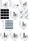
MRNA Sema3A was highly expressed in IS and correlated with cell viability and apoptosis. (A) The mRNA level of Sema3A in N2a cells climbed up with time after 6h, 12h, 24h of OGD/R treatment. *P<0.05 vs control. (B) Successful downregulation of Sema3A by si-Sema3A. *P<0.05 vs control. (C) Sema3A downregulation led to higher viability of OGD/R cells. *P<0.05 vs control, #P<0.05 vs siRNA-NC. (D, E) OGD/R cells have higher apoptosis rate compared with control groups while Sema3A downregulation is correlated with lower apoptosis level among OGD/R cells. *P<0.05 vs control, #P<0.05 vs siRNA-NC. (F) OGD/R cells tend to have higher level of c-caspase-3 as well as p-JNK and p-p38 in comparison with control group. Downregulation of Sema3A could counteract with this trend in OGD/R cells partly. *P<0.05 vs control, #P<0.05 vs siRNA-NC.
Figure 2
Figure 2
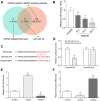
MiR-424-5p targeted with Sema3A and was lowly expressed in IS. (A) MiR-424-5p was among 11 miRNAs which both targeted Sema3A and related to MAPK signaling pathway. (B) The relative miR-424-5p expression declined with time of 6h, 12h, 24h of OGD/R treatment. *P<0.05 vs control. (C, D) The target relationship between Sema3A and miR-424-5p was predicted by Targetscan and verified by dual luciferase assay. *P<0.05 vs control. (E, F) Upregulation of miR-424-5p by miR-424-5p mimics led to lower Sema3A expression, while downregulation of miR-424-5p led to higher Sema3A expression. *P<0.05 vs control.
Figure 3
Figure 3
MEG3 was a highly expressed lncRNA in IS that bonded with miR-424-5p. (A) The heatmap from the microarray analysis of GSE22255 showed the significantly differentially expressed lncRNAs including MEG3 in the IS group. (B) MEG3 bound to miR-424-5p and was related to IS. (C) MEG3 expression was positively correlated with OGD/R intervention time. *P<0.05 vs control. (D) The target relationship between MEG3 and miR-424-5p was predicted and proved. *P<0.05 vs control (E) The effect of downregulation MEG3 by si-MEG3 was proved significant. *P<0.05 vs control. (F) MEG3 knockdown correlated with higher miR-424-5p expression. *P<0.05 vs control. (G) MEG3 knockdown correlated with lower Sema3A expression. *P<0.05 vs control.
Figure 4
Figure 4
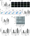
The MEG3/miR-424-5p/Sema3A axis affected cell viability and apoptosis in OGD cells via the MAPK pathway. (A) OGD/R cells have lower cell viability compared with control group. Among OGD/R cells, MEG3 downregulation led to higher cell viability, which could be offset by a simultaneous overexpression of Sema3A; miR-424-5p downregulation by its inhibitor led to a lower cell viability compared among OGD/R cells, which could be neutralized by a simultaneous downregulation of Sema3A. *P<0.05 vs control, #P<0.05 vs OGD/R NC, $P<0.05 vs si-MEG3, &P<0.05 vs inhibitor (B, C) OGD/R cells tend to have fairly high apoptosis rate compared to control group. Among OGD/R cells, MEG3 downregulation led to lower cell apoptosis level, which could be neutralized by a simultaneous overexpression of Sema3A; miR-424-5p downregulation by its inhibitor mediated more cell apoptosis, which could be neutralized by a simultaneous downregulation of Sema3A. *P<0.05 vs control, #P<0.05 vs OGD/R NC, $P<0.05 vs si-MEG3, &P<0.05 vs inhibitor. (D) OGD/R cells tend to have higher cleaved caspase-3, p-JNK and p-p38 expression in comparison with control. Among OGD/R cells, the low expression of MEG3 led to a rise in expression of cleaved caspase-3, p-JNK and p-p38, miR-424-5p inhibitor led to drop of expression of cleaved caspase-3, p-JNK and p-p38. Sema3A overexpression could reverse the rise of si-MEG3, while si-Sema3A could reverse the drop of miR-424-5p inhibitor. *P<0.05 vs control, #P<0.05 vs OGD/R NC, $P<0.05 vs si-MEG3, &P<0.05 vs inhibitor.
Figure 5
Figure 5
MEG3 expression accelerated the process of IS by promoting Sema3A expression in vivo. (A) Expression level of MEG3 increased gradually at 4h, 8h, 24h point in the process of ischemia reperfusion (I/R stands for ischemic reperfusion). *P<0.05 vs sham. (B) Downregulation of MEG3 expression by si-MEG3 was proved significant. Simultaneous upregulation of Sema3A had little influence on the MEG3 expression. *P<0.05 vs control. (C) Sema3A expression was suppressed by si-MEG3, and the decrease was neutralized by manual Sema3A overexpression. *P<0.05 vs control, #P<0.05 vs si-MEG3. (D) Representative image of brain slices of three groups of rats. Normal tissues were a pink or red color, whereas the ischemic tissues were white. MCAO rats tend to have a bigger ischemic area in contrast with Sham group. Among MCAO rats, si-MEG3 group with MEG3 down-regulated have smaller ischemic area while it could be reversed by overexpression of Sema3A. (E) Quantified average percentage of the infarction area in the whole brain, the percentage was higher in MCAO rats compared with sham group. Among MCAO rats, the infarct volume decreased in si-MEG3 group, while simultaneous overexpression of Sema3A weakened the decrease. *P<0.05 vs sham, #P<0.05 vs MCAO, $P<0.05 vs MCAO+si-MEG3. (F) Neurological score rose in MCAO rats compared with sham rats. Among MCAO rats, si-MEG3 group had lower neurological score than MCAO control, while simultaneous overexpression of Sema3A neutralized the decrease of score. *P<0.05 vs sham, #P<0.05 vs MCAO, $P<0.05 vs MCAO+si-MEG3. (G) Expression of cleaved caspase-3, p-JNK and p-p38 increased significantly in MCAO group compared with control group. In si-MEG3 group, expression of cell apoptosis markers cleaved caspase-3 and phosphorylated levels of JNK and p38 decreased. Overexpression of Sema3A reversed the change. *P<0.05 vs sham, #P<0.05 vs MCAO, $P<0.05 vs MCAO+si-MEG3.

V体育官网入口 - References

    1. Bao MH, Szeto V, Yang BB, Zhu SZ, Sun HS, Feng ZP. Long non-coding RNAs in ischemic stroke. Cell Death Dis. 2018; 9:281. 10.1038/s41419-018-0282-x - V体育官网 - DOI - PMC - PubMed
    1. Randolph SA. Ischemic Stroke. Workplace Health Saf. 2016; 64:444. 10.1177/2165079916665400 - V体育2025版 - DOI - PubMed
    1. Hao L, Zou Z, Tian H, Zhang Y, Zhou H, Liu L. Stem cell-based therapies for ischemic stroke. Biomed Res Int. 2014; 2014:468748. 10.1155/2014/468748 - DOI - PMC - PubMed
    1. Young RS, Ponting CP. Identification and function of long non-coding RNAs. Essays Biochem. 2013; 54:113–26. 10.1042/bse0540113 - DOI (V体育官网) - PubMed
    1. Dykstra-Aiello C, Jickling GC, Ander BP, Shroff N, Zhan X, Liu D, Hull H, Orantia M, Stamova BS, Sharp FR. Altered Expression of Long Noncoding RNAs in Blood After Ischemic Stroke and Proximity to Putative Stroke Risk Loci. Stroke. 2016; 47:2896–903. 10.1161/STROKEAHA.116.013869 - DOI - PMC - PubMed

"VSports手机版" Publication types

MeSH terms