Skip to main page content
U.S. flag

An official website of the United States government

Dot gov

The VSports app下载. gov means it’s official. Federal government websites often end in . gov or . mil. Before sharing sensitive information, make sure you’re on a federal government site. .

Https

The site is secure. The https:// ensures that you are connecting to the official website and that any information you provide is encrypted and transmitted securely V体育官网. .

. 2020 Jan 13;12(1):193.
doi: 10.3390/cancers12010193.

Polyphyllin VI Induces Caspase-1-Mediated Pyroptosis via the Induction of ROS/NF-κB/NLRP3/GSDMD Signal Axis in Non-Small Cell Lung Cancer

Affiliations

Polyphyllin VI Induces Caspase-1-Mediated Pyroptosis via the Induction of ROS/NF-κB/NLRP3/GSDMD Signal Axis in Non-Small Cell Lung Cancer

Jin-Feng Teng et al. Cancers (Basel). .

Abstract

Trillium tschonoskii Maxim (TTM), a traditional Chinese medicine, has been demonstrated to have a potent anti-tumor effect. Recently, polyphyllin VI (PPVI), a main saponin isolated from TTM, was reported by us to significantly suppress the proliferation of non-small cell lung cancer (NSCLC) via the induction of apoptosis and autophagy in vitro and in vivo. In this study, we further found that the NLRP3 inflammasome was activated in PPVI administrated A549-bearing athymic nude mice. As is known to us, pyroptosis is an inflammatory form of caspase-1-dependent programmed cell death that plays an important role in cancer. By using A549 and H1299 cells, the in vitro effect and action mechanism by which PPVI induces activation of the NLRP3 inflammasome in NSCLC were investigated. The anti-proliferative effect of PPVI in A549 and H1299 cells was firstly measured and validated by MTT assay. The activation of the NLRP3 inflammasome was detected by using Hoechst33324/PI staining, flow cytometry analysis and real-time live cell imaging methods. We found that PPVI significantly increased the percentage of cells with PI signal in A549 and H1299, and the dynamic change in cell morphology and the process of cell death of A549 cells indicated that PPVI induced an apoptosis-to-pyroptosis switch, and, ultimately, lytic cell death. In addition, belnacasan (VX-765), an inhibitor of caspase-1, could remarkably decrease the pyroptotic cell death of PPVI-treated A549 and H1299 cells. Moreover, by detecting the expression of NLRP3, ASC, caspase-1, IL-1β, IL-18 and GSDMD in A549 and h1299 cells using Western blotting, immunofluorescence imaging and flow cytometric analysis, measuring the caspase-1 activity using colorimetric assay, and quantifying the cytokines level of IL-1β and IL-18 using ELISA, the NLRP3 inflammasome was found to be activated in a dose manner, while VX-765 and necrosulfonamide (NSA), an inhibitor of GSDMD, could inhibit PPVI-induced activation of the NLRP3 inflammasome VSports手机版. Furthermore, the mechanism study found that PPVI could activate the NF-κB signaling pathway via increasing reactive oxygen species (ROS) levels in A549 and H1299 cells, and N-acetyl-L-cysteine (NAC), a scavenger of ROS, remarkably inhibited the cell death, and the activation of NF-κB and the NLRP3 inflammasome in PPVI-treated A549 and H1299 cells. Taken together, these data suggested that PPVI-induced, caspase-1-mediated pyroptosis via the induction of the ROS/NF-κB/NLRP3/GSDMD signal axis in NSCLC, which further clarified the mechanism of PPVI in the inhibition of NSCLC, and thereby provided a possibility for PPVI to serve as a novel therapeutic agent for NSCLC in the future. .

Keywords: NSCLC; ROS/NF-κB/NLRP3/GSDMD; polyphyllin VI; pyroptosis V体育安卓版. .


Conflict of interest statement

The authors declare no conflict of interest.

Figures (V体育ios版)

Figure 1
Figure 1
Polyphyllin VI (PPVI) activates the NLRP3 inflammasome in A549 bearing athymic nude mice. (A) Chemical structure of PPVI. (B) Tumor tissue lysates were analyzed by Western blot for NLRP3, caspase-1, IL-1β, GSDMD and β-actin. Bar chart indicates the relative density of the protein to β-actin; bars, S.D. ** p ≤ 0.01; *** p ≤ 0.001. The full-length Western blotting images are shown in Figure S4. (C) The expression of NLRP3, caspase-1, IL-1β and GSDMD in the tumor tissue of A549-bearing athymic nude mice were analyzed by the immunohistochemistry method. Magnification: 40×, Scale bar: 40 µm.
Figure 2
Figure 2
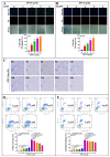
PPVI induces lytic cell death in A549 and H1299 cells. A549 cells (A) and H1299 cells (B) were treated with PPVI for 24 h. The cells were then stained with 2 μg/mL PI (red, staining dying cells) plus 2 μg/mL Hoechst 33342 (blue, staining all cells) for 10 min, and observed by fluorescent microscopy. Bar chart indicates the percentage of cells with PI signal to blue signal; bars, S.D. *** p ≤ 0.001. Magnification: 10×, Scale bar: 25 µm. (C) The time-lapse phase-contrast and fluorescent images of A549 cells stained with PI and Hoechst 33342 solution were taken at the indicated timepoints after the stimulation of 6 μM PPVI. The images in the red, blue and white channels were merged at the same field. Red arrow indicated the cells at 6–9 h, beginning with membrane blebbing and producing apoptotic body-like cell protrusions (termed pyroptotic bodies) prior to plasma membrane rupture. The real-time video was included in Video S1. Data shown are representative of at least three independent experiments. Magnification: 10×, Scale bar: 100 µm. Pyroptotic cells or living cells of A549 cells (D) and H1299 cells (E) treated as indicated graded concentrations were measured by flow cytometry using an annexin V-FITC/PI apoptotic detecting kit. Annexin V+/PI+ indicated the pyroptotic cells and annexin-V/PI represented living cells. Bar chart indicates the percentage of pyroptotic cells and living cells; bars, S.D. ** p ≤ 0.01, *** p ≤ 0.001.
Figure 3
Figure 3
PPVI induces cell death via the activation of caspase-1 in A549 and H1299 cells. A549 cells (A) and H1299 cells (B) were treated with 4 μM PPVI for 24 h; the activated caspase-1 were then measured by using a Caspase-1 Activity Assay Kit (Colorimetric) according to the manufacturer’s instructions. Bar chart indicated the amount of pNA in A549 and H1299 cells. A549 cells (C) and H1299 cells (D) were treated with PPVI or co-treated with PPVI and VX-765 at the indicated concentration for 24 h. After treatment, the cytotoxicity was measured by using MTT assay. Bar chart indicates the cell viability of A549 and H1299 cells; bars, S.D. * p ≤ 0.05; ** p ≤ 0.01. A549 cells (E) and H1299 cells (F) were treated with PPVI or co-treated with PPVI and VX-765 at the indicated concentration for 24 h. The cells were then stained with 2 μg/mL PI (red, staining dying cells) plus 2 μg/mL Hoechst 33342 (blue, staining all cells) for 10 min, and observed by fluorescent microscopy. Bar chart indicates the percentage of cells with PI signal compared to blue signal. Magnification: 10×, Scale bar: 100 µm. Necrotic cells, pyroptotic cells, apoptotic cells or living cells of A549 cells (G) and H1299 cells (H), treated as indicated graded concentrations, were measured by flow cytometry using an annexin V-FITC/PI apoptotic detecting kit. Annexin V-/PI+ indicated the necrotic cells, annexin V+/PI+ indicated the pyroptotic cells, annexin V+/PI represented apoptotic cells and annexin-V/PI represented living cells. Bar chart indicates the percentage of necrotic cells, pyroptotic cells, apoptotic cells and living cells; bars, S.D. ** p ≤ 0.01, *** p ≤ 0.001.
Figure 4
Figure 4
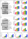
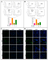
PPVI increased the expression of NLRP3, ASC and caspase-1 in A549 and H1299 cells. A549 cells (A) and H1299 cells (B) transiently transfected with EGFP-N1-NLRP3, mCherry-C1-ASC and EGFP-N1-caspase-1 plasmids for 24 h were treated with PPVI as indicated by the graded concentrations for another 24 h. After treatment, the cells were fixed with 4% PFA, and the representative images were captured using the fluorescence microscope. Magnification: 40×. Scale bar: 25 μm. A549 cells (CE) and H1299 cells (FH) transiently transfected with EGFP-N1-NLRP3, mCherry-C1-ASC and EGFP-N1-caspase-1 plasmids for 24 h were then treated with PPVI as indicated by the graded concentrations for another 24 h. After treatment, the cells were trypsinzed and collected for the analysis of fluorescence intensity by flow cytometry. Bar chart indicates the percentage of cells with GFP or mCherry signal in A549 and H1299 cells; bars, S.D. * p ≤ 0.05; ** p ≤ 0.01; *** p ≤ 0.001.
Figure 5
Figure 5
PPVI activated the NLRP3 inflammasome and promoted the release of IL-1β and IL-18. A549 cells (A) and H1299 cells (B) were treated with PPVI as indicated by the graded concentrations for 24 h. Cell protein was then harvested for detecting NLRP3, ASC, caspase-1, IL-1β, IL-18, GSDMD and β-actin by Western blot. Bar chart indicates the relative density of protein to β-actin; bars, S.D. * p ≤ 0.05; ** p ≤ 0.01. The full-length Western blotting images are shown in Figure S5. A549 cells and H1299 cells were treated with PPVI as indicated by gradient concentrations for 24 h. Culture medium was collected for centrifugation and the supernatant was used for the detection of the level of IL-1β and IL-18 by using a Human IL-1β ELISA Kit (CHE0001) and a Human IL-18 ELISA Kit (CHE0007). Bar chart indicates the level of IL-1β and IL-18 in the cell supernatant of A549 cells (C,D) and H1299 cells (E,F), respectively. bars, S.D. * p ≤ 0.05; ** p ≤ 0.01. *** p ≤ 0.001.
Figure 6
Figure 6
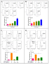
PPVI VX-765 and NSA reversed the activation of the NLRP3 inflammasome of PPVI-treated A549 and H1299 cells. A549 cells (A) and H1299 cells (B), transiently transfected with EGFP-N1-NLRP3, mCherry-C1-ASC and EGFP-N1-caspase-1 plasmids for 24 h, were treated with PPVI or co-treated with VX-765 or NSA and PPVI, as per the indicated concentration, for another 24 h. After treatment, the cells were fixed with 4% PFA, and the representative images were captured using the fluorescence microscope. Magnification: 20×. Scale bar: 50 μm. Pyroptotic cells of A549 cells (C) and H1299 cells (D) treated with PPVI or co-treated with NSA and PPVI, as indicated. Treatment concentrations were measured by flow cytometry using an annexin V-FITC/PI apoptotic detecting kit. Annexin V+/PI+ indicated the pyroptotic cells. Bar chart indicates the percentage of pyroptotic cells and living cells; bars, S.D. ** p ≤ 0.01, *** p ≤ 0.001.
Figure 7
Figure 7
PPVI increased the intracellular reactive oxygen species (ROS) levels in A549 and H1299 cells. A549 cells (A) and H1299 cells (B) were treated with PPVI as indicated graded by the concentrations for 24 h. The cells were then incubated with 5 µM H2DCFDA reagent for 30 min, and the ROS generation was determined by flow cytometry. Bar chart indicates the ROS levels in A549 and H1299 cells; bars, S.D. * p ≤ 0.05; *** p ≤ 0.001. A549 cells (C) and H1299 cells (D) were treated with PPVI or co-treated with PPVI and NAC for 24 h. The cells were then incubated with 5 µM H2DCFDA reagent for 30 min, and the ROS generation was determined by flow cytometry. Bar chart indicates the ROS levels in A549 and H1299 cells; bars, S.D. * p ≤ 0.05; ** p ≤ 0.01; *** p ≤ 0.001.
Figure 8
Figure 8
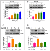
PPVI activated the ROS/NF-кB pathway in A549 and H1299 cells. A549 cells (A) and H1299 cells (B) were treated with PPVI as indicated by the graded concentrations for 24 h. A549 cells (C) and H1299 cells (D) were treated with PPVI or co-treated with NAC and PPVI, as indicated by the concentration shown, for 24 h. After treatment, cell protein was then harvested to detect p65 and β-actin by Western blot. Bar chart indicates the relative density of p65 to β-actin; bars, S.D. p ≤ 0.05; * p ≤ 0.01. The full-length Western blotting images are shown in Figure S6.
Figure 9
Figure 9
NAC inhibits the expression of NLRP3 and caspase-1 in PPVI treated A549 cells and H1299 cells. A549 cells (A) and H1299 cells (B) transiently transfected with EGFP-N1-NLRP3 plasmid for 24 h, treated with PPVI or co-treated with NAC and PPVI, at indicated concentrations, for another 24 h. After treatment, the cells were trypsinzed and collected for the analysis of fluorescence intensity by flow cytometry. Bar chart indicates the percentage of cells with GFP signal in A549 and H1299 cells; bars, S.D. ** p ≤ 0.01; *** p ≤ 0.001. A549 cells (C) and H1299 cells (D) transiently transfected with EGFP-N1-caspase-1 for 24 h were treated with PPVI or co-treated with NAC and PPVI at indicated concentrations for another 24 h. After treatment, the cells were fixed with 4% PFA, and the representative images were captured using the fluorescence microscope. Magnification: 40×. Scale bar: 25 μm.
Figure 10
Figure 10
BAY-inhibited, PPVI-induced pyroptotic cell death in A549 cells and H1299 cells. A549 cells (A) and H1299 cells (B) treated with PPVI or co-treated with PPVI and BAY at the indicated concentration for 24 h. After treatment, the cytotoxicity was measured by MTT method, and the bar chart indicates the cell viability of A549 and H1299 cells; bars, S.D. ** p ≤ 0.01; *** p ≤ 0.001. A549 cells (C) and H1299 cells (D) treated with PPVI or co-treated with PPVI and BAY at the indicated concentration for 24 h. The cells were then stained with 2 μg/mL PI (red, staining dying cells) plus 2 μg/mL Hoechst 33342 (blue, staining all cells) for 10 min, and then observed by fluorescent microscopy. Bar chart indicates the percentage of cells with PI signal compared to blue signal; bars, S.D. *** p ≤ 0.001. Magnification: 10×, Scale bar: 100 µm.
Figure 11
Figure 11
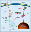
A schematic diagram of the molecular mechanism by which PPVI induced pyroptotic cell death via the activation of the ROS/NF-κB/NLRP3/GSDMD signal axis in A549 and H1299 cells.

"V体育安卓版" References

    1. Magnuson W.J., Yeung J.T., Guillod P.D., Gettinger S.N., Yu J.B., Chiang V.L. Impact of Deferring Radiation Therapy in Patients With Epidermal Growth Factor Receptor-Mutant Non-Small Cell Lung Cancer Who Develop Brain Metastases. Int. J. Radiat. Oncol. Biol. Phys. 2016;95:673–679. doi: 10.1016/j.ijrobp.2016.01.037. - VSports app下载 - DOI - PubMed
    1. Suwinski R., Giglok M., Galwas-Kliber K., Idasiak A., Jochymek B., Deja R., Maslyk B., Mrochem-Kwarciak J., Butkiewicz D. Blood serum proteins as biomarkers for prediction of survival, locoregional control and distant metastasis rate in radiotherapy and radio-chemotherapy for non-small cell lung cancer. BMC Cancer. 2019;19:427. doi: 10.1186/s12885-019-5617-1. - V体育官网 - DOI - PMC - PubMed
    1. Fan T.W.M., Zhang X., Wang C., Yang Y., Kang W.Y., Arnold S., Higashi R.M., Liu J., Lane A.N. Exosomal lipids for classifying early and late stage non-small cell lung cancer. Anal. Chim. Acta. 2018;1037:256–264. doi: 10.1016/j.aca.2018.02.051. - DOI (VSports最新版本) - PMC - PubMed
    1. Zhang P.T., Yu M.W., Yang Z.Y. Comparative study on the methods of Chinese medicine and Western medicine therapeutic evaluation for advanced non-small cell lung cancer. Zhongguo Zhong Xi Yi Jie He Za Zhi. 2010;30:702–705. - PubMed
    1. Raben D., Helfrich B.A., Chan D., Johnson G., Bunn P.A., Jr. ZD1839, a selective epidermal growth factor receptor tyrosine kinase inhibitor, alone and in combination with radiation and chemotherapy as a new therapeutic strategy in non-small cell lung cancer. Semin. Oncol. 2002;29:37–46. doi: 10.1053/sonc.2002.31521. - DOI - PubMed

"V体育安卓版" LinkOut - more resources