Skip to main page content
U.S. flag

An official website of the United States government

Dot gov

The . gov means it’s official. Federal government websites often end in . gov or VSports app下载. mil. Before sharing sensitive information, make sure you’re on a federal government site. .

Https

The site is secure. The https:// ensures that you are connecting to the official website and that any information you provide is encrypted and transmitted securely. V体育官网.

. 2019 Dec 17;4(6):e00463-19.
doi: 10.1128/mSystems.00463-19.

Gut Microbiota Dysbiosis Is Associated with Altered Bile Acid Metabolism in Infantile Cholestasis

Affiliations

Gut Microbiota Dysbiosis Is Associated with Altered Bile Acid Metabolism in Infantile Cholestasis (VSports)

VSports app下载 - Yizhong Wang et al. mSystems. .

Abstract

The co-occurrence of gut microbiota dysbiosis and bile acid (BA) metabolism alteration has been reported in several human liver diseases. However, the gut microbiota dysbiosis in infantile cholestatic jaundice (CJ) and the linkage between gut bacterial changes and alterations of BA metabolism have not been determined. To address this question, we performed 16S rRNA gene sequencing to determine the alterations in the gut microbiota of infants with CJ, and assessed their association with the fecal levels of primary and secondary BAs. Our data reveal that CJ infants show marked declines in the fecal levels of primary BAs and most secondary BAs. A decreased ratio of cholic acid (CA)/chenodeoxycholic acid (CDCA) in infants with CJ indicated a shift in BA synthesis from the primary pathway to the alternative BA synthesis pathway VSports手机版. The bacterial taxa enriched in infants with CJ corresponded to the genera Clostridium, Gemella, Streptococcus, and Veillonella and the family Enterobacteriaceae and were negatively correlated with the fecal BA level and the CDCA/CA ratio but positively correlated with the serological indexes of impaired liver function. An increased ratio of deoxycholic acid (DCA)/CA was observed in a proportion of infants with CJ. The bacteria depleted in infants with CJ, including Bifidobacterium and Faecalibacterium prausnitzii, were positively and negatively correlated with the fecal levels of BAs and the serological markers of impaired liver function, respectively. In conclusion, the reduced concentration of BAs in the gut of infants with CJ is correlated with gut microbiota dysbiosis. The altered gut microbiota of infants with CJ likely upregulates the conversion from primary to secondary BAs. IMPORTANCE Liver health, fecal bile acid (BA) concentrations, and gut microbiota composition are closely connected. BAs and the microbiome influence each other in the gut, where bacteria modify the BA profile, while intestinal BAs regulate the growth of commensal bacteria, maintain the barrier integrity, and modulate the immune system. Previous studies have found that the co-occurrence of gut microbiota dysbiosis and BA metabolism alteration is present in many human liver diseases. Our study is the first to assess the gut microbiota composition in infantile cholestatic jaundice (CJ) and elucidate the linkage between gut bacterial changes and alterations of BA metabolism. We observed reduced levels of primary BAs and most secondary BAs in infants with CJ. The reduced concentration of fecal BAs in infantile CJ was associated with the overgrowth of gut bacteria with a pathogenic potential and the depletion of those with a potential benefit. The altered gut microbiota of infants with CJ likely upregulates the conversion from primary to secondary BAs. Our study provides a new perspective on potential targets for gut microbiota intervention directed at the management of infantile CJ. .

Keywords: bile acids; cholestatic jaundice; dysbiosis; gut microbiota; infants. V体育安卓版.


Figures (V体育安卓版)

FIG 1
FIG 1
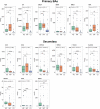
Alteration of fecal bile acid metabolism in infantile cholestasis. The levels of primary and secondary bile acids in the cohort of infants with cholestatic jaundice (CJ) or impaired hepatic function (IHF) and the health controls (HC) were measured by tandem mass spectrometry. Significant differences were determined by one-way analysis of variance (ANOVA) followed by Tukey’s post hoc test. ns, not significant; *, P < 0.05; **, P < 0.01; ***, P < 0.001. HCA, hyocholic acid; 3_DHCA, 3-dehydrocholic acid; 7_DHCA, 7-dehydrocholic acid; KLCA, ketolithocholic acid; UCA, ursocholic acid; bUCA, β-ursocholic acid; muroCA, murocholic acid; NorCA, norcholic acid.
FIG 2
FIG 2
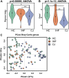
Comparison of the taxonomic diversity of the gut microbiomes among the cholestatic jaundice (CJ), impaired hepatic function (IHF), and healthy control (HC) groups. The gut microbiota alpha diversity was measured from the Shannon (A) and Chao1 (B) indexes. The Wilcoxon test was performed for pairwise comparisons. *, P < 0.05; **, P < 0.01; ***, P < 0.001. (C) PCoA of Bray-Curtis distances generated from taxa summarized at the genus level. Each point corresponds to a sample shaped and colored by diagnosis.
FIG 3
FIG 3
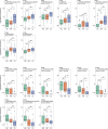
Significant differences between the gut bacterial taxa of the cholestatic jaundice (CJ), impaired hepatic function (IHF), and healthy control (HC) groups. Comparisons among the groups were performed using one-way analysis of variance (ANOVA; P < 0.05). The Wilcoxon test was performed for pairwise comparisons, *, P < 0.05; **, P < 0.01; ***, P < 0.001. g, genus; s, species; f, family.
FIG 4
FIG 4
Discrepancy in microbial community phenotypes between the cholestatic jaundice (CJ) group and the other two groups. BugBase identified phenotypes associated with aerobic bacteria (A), anaerobic bacteria (B), facultatively anaerobic bacteria (C), oxidative stress tolerance (D), Gram-negative bacteria (E), Gram-positive bacteria (F), mobile element content (G), biofilm formation (H), and pathogenesis (I). Statistical significance was identified by the Wilcoxon test with false discovery rate (FDR)-corrected pairwise P values. *, P < 0.05; **, P < 0.01; ***, P < 0.001.
FIG 5
FIG 5
Functional alterations of the gut microbiome in infants with cholestatic jaundice (CJ) and impaired hepatic function (IHF). Statistical significance was determined by using LEfSe, with a P value of <0.05 (Wilcoxon test) and a linear discriminant analysis (LDA) score (log10) of >3 being considered significant.
FIG 6
FIG 6
Correlation matrices constructed from gut bacterial taxa, fecal BAs, and serum indicators of hepatic function. Spearman correlation analysis between fecal BA serum indicators of hepatic function and the top 17 OTUs with significantly different abundances among the groups (A) and the gut microbiota functional categories (B) was employed. Four distinct OTU clusters (clusters O1 to O4) and two distinct functional clusters (clusters F1 and F2) were observed. Red and blue represent the positive and negative correlations, respectively. GHDCA, glycohyodeoxycholic acid; TCA, taurocholic acid; GCA, glycocholic acid; LCA, lithocholic acid; GLCA, glycolithocholic acid; THDCA, taurohyodeoxycholic acid; TLCA-3S, taurolithocholic acid-3-sulfate acid; GCDCA, glycochenodeoxycholic acid; GHCA, glycohyocholic acid; TUDCA, tauroursodeoxycholic acid; GDCA, glycodeoxycholic acid; THCA, taurohyocholic acid; TDCA, taurodeoxycholic acid; GUDCA, glycoursodeoxycholic acid; ALB, albumin; ALT, alanine aminotransferase; AST, aspartate aminotransferase; BA, bile acid; DB, direct bilirubin; HB, hemoglobin; GGT, gamma-glutamyltransferase; TB, total bilirubin.

References

    1. Moyer V, Freese DK, Whitington PF, Olson AD, Brewer F, Colletti RB, Heyman MB. 2004. Guideline for the evaluation of cholestatic jaundice in infants: recommendations of the North American Society for Pediatric Gastroenterology, Hepatology and Nutrition. J Pediatr Gastroenterol Nutr 39:115–128. doi:10.1097/00005176-200408000-00001. - DOI - PubMed
    1. Dick MC, Mowat AP. 1985. Hepatitis syndrome in infancy—an epidemiological survey with 10 year follow up. Arch Dis Child 60:512–516. doi:10.1136/adc.60.6.512. - DOI - PMC - PubMed
    1. Fawaz R, Baumann U, Ekong U, Fischler B, Hadzic N, Mack CL, McLin VA, Molleston JP, Neimark E, Ng VL, Karpen SJ. 2017. Guideline for the evaluation of cholestatic jaundice in infants: joint recommendations of the North American Society for Pediatric Gastroenterology, Hepatology, and Nutrition and the European Society for Pediatric Gastroenterology, Hepatology, and Nutrition. J Pediatr Gastroenterol Nutr 64:154–168. doi:10.1097/MPG.0000000000001334. - "VSports注册入口" DOI - PubMed
    1. Karpen SJ. 2002. Update on the etiologies and management of neonatal cholestasis. Clin Perinatol 29:159–180. doi:10.1016/S0095-5108(03)00069-1. - DOI - PubMed
    1. Ananth R. 2018. Neonatal cholestasis: a primer of selected etiologies. Pediatr Ann 47:e433–e439. doi:10.3928/19382359-20181018-01. - DOI - PubMed

LinkOut - more resources (V体育2025版)