Skip to main page content
U.S. flag

An official website of the United States government

Dot gov

The . gov means it’s official. Federal government websites often end in . gov or . mil. Before sharing sensitive information, make sure you’re on a federal government site. VSports app下载.

Https

The site is secure V体育官网. The https:// ensures that you are connecting to the official website and that any information you provide is encrypted and transmitted securely. .

. 2019 Nov;1(11):1074-1088.
doi: 10.1038/s42255-019-0122-z. Epub 2019 Oct 21.

Cardiac glycosides are broad-spectrum senolytics

Affiliations

Cardiac glycosides are broad-spectrum senolytics

Ana Guerrero et al. Nat Metab. 2019 Nov.

Abstract

Senescence is a cellular stress response that results in the stable arrest of old, damaged or preneoplastic cells. Oncogene-induced senescence is tumor suppressive but can also exacerbate tumorigenesis through the secretion of pro-inflammatory factors from senescent cells. Drugs that selectively kill senescent cells, termed senolytics, have proved beneficial in animal models of many age-associated diseases. Here, we show that the cardiac glycoside, ouabain, is a senolytic agent with broad activity. Senescent cells are sensitized to ouabain-induced apoptosis, a process mediated in part by induction of the pro-apoptotic Bcl2-family protein NOXA. We show that cardiac glycosides synergize with anti-cancer drugs to kill tumor cells and eliminate senescent cells that accumulate after irradiation or in old mice VSports手机版. Ouabain also eliminates senescent preneoplastic cells. Our findings suggest that cardiac glycosides may be effective anti-cancer drugs by acting through multiple mechanism. Given the broad range of senescent cells targeted by cardiac glycosides their use against age-related diseases warrants further exploration. .


Conflict of interest statement

Competing Interests J. G. owns equity and has acted as a consultant for Unity Biotechnology and Geras Bio. Unity Biotechnology funds research on senolytics in J. G. ’s laboratory. J. G. , A. G. and N. H V体育安卓版. are named inventors in a MRC patent related to senolytic therapies (PCT/GB2018/051437).

Figures
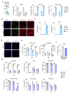
Extended Figure 1
Extended Figure 1. IMR90 ER:RAS cells as a model of OIS
a, Quantification of immunofluorescence staining for BrdU, p16INK4a, and SA-β-Galactosidase of IMR90 ER:RAS cells 6 or 8 days after treatment with 4-OHT or vehicle (DMSO) (n = 3). b, Representative immunofluorescence images. BrdU incorporation, which indicates proliferation, is stained green; p16INK4a is stained red. Scale bar, 50 μm. SA-b-Galactosidase is stained red. Scale bar, 100 μm. c, Expression levels for IL8 and IL1A of senescent and control IMR90 ER:RAS cells 6 days after 4-OHT or vehicle (DMSO) (n = 4). d, DAPI staining of senescent and control IMR90 ER:RAS cells after 1 μM ABT-263 treatment for 3 days showing reduced numbers of senescent cells after ABT-263 treatment. Scale bar, 100 μM. e, Senolytic activity of the indicated drugs in the context of oncogene-induced senescence in IMR90 ER:RAS cells (n = 4). f, Quantification of immunofluorescence staining for BrdU in IMR90 ER:RAS cells expressing E6 and E7 proteins of HPV16 (n = 3). g, Senolytic activity of the indicated drugs in IMR90 ER:RAS cells expressing E6 and E7 proteins of HPV16. All error bars represent mean ± s.d; n represents independent experiments. All statistical significances were calculated using unpaired two-tailed Student’s t-tests.
Extended Figure 2
Extended Figure 2. Senolytic drug screen in therapy-induced senescence
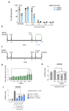
a, Quantification of immunofluorescence staining for BrdU, SA-b-Galactosidase activity, p21CIP1 and 53BP1 in IMR90 cells treated with 50 μM etoposide (n = 3). b-c, Senolytic activity of the indicated drugs in the context of therapy-induced senescence in IMR90 (n = 4, b) and oncogene-induced senescence in IMR90 ER:RAS cells (n = 4, c). All error bars represent mean ± s.d; n represents independent experiments. All statistical significances were calculated using unpaired two-tailed Student’s t-tests.
Extended Figure 3
Extended Figure 3. The glycoside chain in CGs is dispensable for their senolytic activity
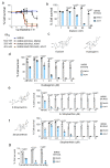
a, Dose response analysis of senolytic activity of ouabain in IMR90, control IMR90 ER:RAS cells (DMSO), senescent IMR90 ER:RAS cells (4-OHT) and IMR90 ER:RAS cells expressing E6 and E7 proteins of HPV16 (n = 3). b, Dose response analysis of senolytic activity of digitoxin in the context of oncogene-induced senescence in IMR90 ER:RAS cells (n = 3). c, Chemical structure of ouabain and its aglycone version, ouabagenin. d, Quantification of cell survival in senescent and control IMR90 ER:RAS cells after treatment with ouabagenin, the aglycone version of ouabain (n = 6). e-f, Quantification of cell survival of IMR90 ER:RAS cells undergoing OIS and the corresponding controls after treatment with the CG KStrophanthin (e) (n = 4) or its aglycone version Strophantidin (f) (n = 5). g, Quantification of cell survival in senescent and control IMR90 ER:RAS 3 days after 1 μM ABT-263 or CG treatment (50 nM ouabain, 100 nM digoxin). Senolytic drugs were added 8 days after 4-OHT or vehicle (DMSO) (n = 4). All error bars represent mean ± s.d; n represents independent experiments. All statistical significances were calculated using unpaired two-tailed Student’s t-tests.
Extended Figure 4
Extended Figure 4. Senescent cells are more sensitive to CGs due to their altered osmotic balance
a, Quantification of cell survival of senescent and control IMR90 ER:RAS cells after treatment with curcumin (n = 6). Statistical significance was calculated using unpaired two-tailed, Student’s t-test. b, Experimental design for the transcriptional profiling of senescent and control IMR90 ER:RAS cells after treatment with cardiac glycosides (CG). QVD indicates treatment with a general caspase inhibitor (Q-VD-OPh). c, IMR90 ER:RAS cells were transfected with 2 independent siRNAs targeting BCL-2 family genes at day 6 after senescence induction as indicated in the scheme. d, IMR90 ER:RAS were transfected with at least two independent siRNAs targeting BCL-2 family genes at day 6 after senescence induction (n = 3; scrambled siRNA versus three different siRNAs against NOXA, ***P < 0.001). The timeline of the experiment is shown in (c). Statistical significance was calculated using one-way ANOVA (Dunnett’s test). e, Expression levels of NOXA after knock down with three independent siRNAs (n = 3). Statistical significance was calculated using one-way ANOVA (Dunnett’s test). f, Expression levels of NOXA after knock down with four independent shRNAs (n = 3; vector versus different shRNAs against NOXA, ****P < 0.0001). Statistical significance was calculated using one-way ANOVA (Dunnett’s test). All error bars represent mean ± s.d; n represents independent experiments.
Extended Figure 5
Extended Figure 5. Ouabain eliminates liver preneoplastic senescent cells
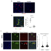
a, Representative images of immunofluorescence staining of Nras. Mice were treated with vehicle (n = 9) or ouabain (n = 12) as explained in Fig 4a. Nras is stained in red. Scale bar, 70 μm. b, Immunofluorescence staining and quantification of p21Cip1 in Nras-positive senescent hepatocytes vs Nras-negative normal hepatocytes. Nras is stained in green, p21Cip1 is stained in red. White arrows indicate Nras-positive, p21Cip1-positive cells; green arrow indicates a Nras-positive, p21Cip1-negative cells (n = 10 per group). c, SCID/beige mice were treated with saline or Digoxin (1mg/kg) on two consecutive days, 5 days after hydrodynamic transduction of Nras-GFP. Mice were culled 6 hours after the second treatment. Representative images of immunofluorescence staining of GFP and cleaved caspase-3 and quantification of intensity levels in 1/2 independent experiments (n = 200 cells). Scale bar, 50 μm. Statistical significance was calculated using unpaired two-tailed Student's t-test. Data represent mean ± s.d; n represents number of mice.
Extended Figure 6
Extended Figure 6. Ouabain eliminates preneoplastic senescent cells
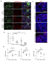
a, Representative images of immunofluorescence staining of β-catenin (green) and synaptophysin (red) in tumoral pituitaries from 18.5dpc Hesx1Cre/+;Ctnnb1lox(ex3)/+ mice that were cultured in the presence of either ABT-737 (2.5 μM), ouabain (250 nM and 500 nM) or vehicle (DMSO) (n = 10 per group). Scale bar, 50 μm. b, Quantitative analysis of the immunofluorescence in (a) demonstrates that ABT-737 and ouabain significantly reduce the number of β-catenin-positive cells. Statistical significance was calculated using Kruskal-Wallis and Dunn’s multiple comparisons test. c, Representative images of immunofluorescence staining of ACTH (adrenocorticotropic hormone; magenta). Scale bar: 50μm. d, qRT-PCR analysis revealing that the senescent marker Cdkn1a (encoding for p21Cip1) and the SASP components Il1b and Il6 are reduced in neoplastic pituitaries treated with 100 nM ouabain and 100 nM digoxin relative to vehicle controls (n = 3 per group). Statistical significance was calculated using unpaired two-tailed Student's t-test; data represent mean ± s.d; n represents number of mice.
Extended Figure 7
Extended Figure 7. Anti-cancer effect of cardiac glycosides across different human cancer cell lines
a-b, Quantification of cell survival by trypan blue staining of Huh7 cells (a) and HLF cells (b) after treatment with the indicated drug combinations (n = 3). Timeline of the experiment is shown in Supplementary Fig 4a. Statistical significance was calculated using unpaired two-tailed Student's t-test. c, Quantification of cell survival of senescent (alisertib, palbociclib) and control (DMSO) SK-Mel-5 melanoma cells (n = 4). Statistical significance was calculated using two-way ANOVA (Dunnett’s test). d, Quantification of cell survival of senescent (doxorubicin, palbociclib) and control (DMSO) MCF-7 or MCF-7 breast cancer cells infected with a shRNA against TP53 (n = 4). Statistical significance was calculated using twoway ANOVA (Dunnett’s test). e-f, mRNA expression levels of TP53 in MCF-7 cells (e) and HCT-116 cells (n = 3). Statistical significance was calculated using unpaired two-tailed, Student’s t-test. Data represent mean ± s.d; n represents independent experiments; ns, not significant.
Extended Figure 8
Extended Figure 8. Ouabain treatment reverses age-associated changes in old mice
a, Ouabain levels in plasma were assessed by ELISA 24 hours after finishing a 4-day course of daily 1mg/kg ouabain i.p. injections. (n = 6). b, Phosphate and amylase levels of young (n = 7) and old mice, either treated with vehicle (n = 6) or ouabain (n = 9), were determined in whole-blood samples at the endpoint of the experiment. Statistical significance was calculated using unpaired two-tailed Student's t-test. c, Grip strength assessment in old mice treated with vehicle (n = 7) or ouabain (n = 9) 10 weeks after the start of the experiment, referred to the basal test. Statistical significance was calculated using unpaired two-tailed Student's t-test. d, Expression levels of p16Ink4a in heart and kidney were determined by qRT-PCR following treatment with vehicle (n = 6) or ouabain (n = 7). mRNA expression levels in young mice (n = 7) were used as reference. Statistical significance was calculated using unpaired two-tailed Student's t-test. e-f, GSEA signature for chemokines, oncogene-induced senescence (e) and ageing (f). g, Quantitative analysis (left) and representative IHC pictures (right) of p21Cip1 positive hepatocytes in the liver of young (n = 6) and old mice treated with ouabain (n = 8) or vehicle (saline) (n = 6). Scale bar, 100 μm. Data represent mean ± s.e.m. Statistical significance was calculated using one-way ANOVA with Tukey’s post hoc comparison. h, Quantitative analysis (left) and representative IHC pictures (right) of LINE-1 ORF in the liver of young (n = 6) and old mice treated with ouabain (n = 8) or vehicle (saline) (n = 7). Statistical significance was calculated using one-way ANOVA with Tukey’s post hoc comparison. Scale bar, 50 μm. Data represent mean ± s.d; n represents number of mice.
Extended Figure 9
Extended Figure 9. Ouabain treatment resets immune infiltration in old mice
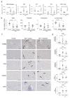
a, xCell analysis of the transcriptome data predicts changes in immune infiltration in the liver of old mice that could be reverted with ouabain. RNA-Seq data from the livers of young (n = 6) and old mice, either treated with vehicle (n = 6) or ouabain (n = 6) was used. Statistical significance was calculated using unpaired two-tailed Student's t-test. b, Blood analysis at the end of the experiment show that ouabain treatment does not change immune composition. Blood from young (n = 8) and old mice, either treated with vehicle (n = 6) or ouabain (n = 8) was used. Statistical significance was calculated using unpaired two-tailed Student's t-test. c-d, Representative IHC images (c) and quantification (d) of the indicated immune cell markers in the liver of young (n = 6) and old mice, either treated with vehicle (n = 6) or ouabain (n = 8). Scale bar: 100μm. Statistical significance was calculated using one-way ANOVA with Tukey’s post hoc comparison. Data represent mean ± s.e.m.; n represents number of mice; ns, not significant.
Figure 1
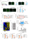
Figure 1. Drug screens identify ouabain as a broad-spectrum senolytic.
a, Experimental design for the senolytic screen on oncogene-induced senescence. b, Quantification of cell survival of senescent and control IMR90 ER:RAS cells after treatment with 1 μM ABT-263 for 3 days (n = 4). c, Screen results. LOPAC 1,280 library compounds were assessed at 10 μM for 3 days. Hits were selected based on their ability to specifically kill senescent cells. Blue dots represent library drugs, grey dots represent DMSO controls. Each dot is the mean of three replicates. d, Senolytic activity of the indicated drugs in the context of oncogene-induced senescence in IMR90 ER:RAS cells (n = 4; n = 6 for ouabain; DMSO versus 4OHT, ****P < 0.0001). e, Experimental design for the senolytic screen on therapy-induced senescence. f, Quantification of cell survival of senescent and control IMR90 cells after treatment with 1 μM ABT-263 for 3 days (n = 5). g, Screen results. LOPAC 1,280 library compounds were assessed at 10 μM for 3 days. Hits were selected based on their ability to specifically kill senescent cells. Red dots represent library drugs, grey dots represent DMSO controls. Each dot is the mean of three replicates. h, Comparison of senolytic activity for the LOPAC 1,280 library compounds in the context of OIS vs therapy-induced senescence. i, Senolytic activity of the indicated drugs in the context of therapy-induced senescence in IMR90 (n = 4; DMSO versus etoposide, ****P < 0.0001). All error bars represent mean ± s.d; n represents independent experiments. All statistical significances were calculated using unpaired two-tailed Student’s t-tests and Holm-Sidak method for multiple comparisons test.
Figure 2
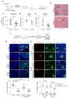
Figure 2. Cardiac glycosides induce apoptosis of senescent cells.
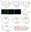
a-c, Senolytic activity of Ouabain in the context of therapy-induced senescence (doxorubicin, n = 4; palbociclib, n =3) and replicative senescence (n = 4). Statistical significance was calculated using unpaired two-tailed Student’s t-tests. d, Representative pictures (left) of immunofluorescence (IF) staining for p16INK4a in PBECs after treatment with ABT-263, Ouabain or vehicle (DMSO). p16INK4a is stained green. Scale bar, 50 μm. Quantification of total and p16INK4a-positive PBECs (right, n =6). Statistical significance was calculated using two-way ANOVA (Dunnett’s multiple comparisons test). Red stars refer to comparison of p16INK4a-positive PBECs, black stars refer to comparison of total PBEC numbers. e-g, Dose response analysis of senolytic activity of digoxin (e), digitoxin (f) and bufalin (g) in IMR90 ER:RAS cells (n = 4). h, Ouabain treatment of senescent cells induce caspase-3/7 activity. IMR90 ER:RAS were treated with 4-OHT or vehicle (DMSO) for 6 days to induce senescence. 100 nM ouabain was then added together with NucLight Rapid Red reagent for cell labelling and Caspase-3/7 reagent for apoptosis (IncuCyte). Caspase 3/7 activity was measured at 4h intervals (n = 3). i, Pan-caspase inhibition (20 μM Q-VD-OPh) rescues senolytic activity of cardiac glycosides (50 nM ouabain, 50 nM bufalin, 100 nM digoxin) and 1 μM ABT-263 on IMR90 ER:RAS cells (n = 5). Statistical significance was calculated using two-way ANOVA (Tukey’s test). All error bars represent mean ± s.d; n represents independent experiments.
Figure 3
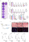
Figure 3. Mechanism explaining the senolytic properties of cardiac glycosides.
a, Chemical homeostasis GSEA signature in cells undergoing OIS compared to growing cells. b-c, Intracellular levels of Na+ (n = 5) b) and Ca2+ (n = 3) c) in IMR90 ER:RAS senescent cells compared to the corresponding controls. Representative pictures (left) and quantification (right) are shown. d, Intracellular levels of K+ in IMR90 ER:RAS senescent cells compared to the corresponding controls treated with 1 μM ouabain for 18 hours or with vehicle (DMSO) (n = 3). Representative pictures (left) and quantification (right) are shown. e, Senolytic activity of 1 μM ABT-263 and 50 nM ouabain assessed after supplementing the media with 10 mM KCl (n = 4). f, Senolytic activity of ABT-263 and ouabain assessed in IMR90 ER:RAS overexpressing the ouabain-insensitive rat ATP1A1 or an empty vector (n = 4; 4OHT + vector versus 4OHT + rAtp1a1, ****P < 0.0001). Statistical significance was calculated using unpaired two-tailed Student’s t-test (bf). g, Heatmap showing the differential expression of BCL-2 family genes in senescent compared to non-senescent cells after treatment with ouabain, digoxin or vehicle (DMSO). The timeline of the experiment is shown in Supplementary Figure 5b. 36C: cells analyzed 36h post-CG treatment on the presence of caspase inhibitor (Q-VD-OPh). h, IMR90 ER:RAS were treated with 4OHT or vehicle (DMSO) for 6 days to induce senescence. Cells were then treated with 1 μM ouabain for 6 hours, in the presence of 50 mM KCl or vehicle. Protein extracts were prepared. Immunoblots are a representative experiment out of 3. i, IMR90 ER:RAS were infected with four different doxycycline-inducible shRNAs targeting NOXA (n = 4; vector versus each different shRNA, ****P < 0.0001). Cells were subsequently treated with 4OHT or vehicle (DMSO) for 6 days to induce senescence, and with 1 μg/μl doxycycline or vehicle (DMSO) to induce NOXA knockdown. Cells were then treated with 50 nM ouabain for 48 hours. j, IMR90 ER:RAS were treated with 4OHT or vehicle (DMSO) for 6 days to induce senescence. Cells were then treated with 50 nM Ouabain for 20 hours, in the presence of 50 mM KCl, GSK3-β inhibitor, JNK inhibitor, p38MAPK inhibitor, or vehicle (DMSO). The effect of ouabain and all the different inhibitors on NOXA expression was determined by qRT-PCR (n = 5; ouabain versus ouabain + inhibitors, ****P < 0.0001). Statistical significance was calculated using one-way ANOVA (Dunnett’s test) (ij). k, IMR90 ER:RAS were treated with 4OHT or vehicle (DMSO) for 6 days to induce senescence. Cells were then treated with 50 nM ouabain for 48 hours, in the presence of GSK3-β inhibitor, JNK inhibitor, p38MAPK inhibitor or vehicle (DMSO) (n = 3). Quantification of cell survival was performed as described in methods. Statistical significance was calculated using two-way ANOVA (Tukey’s test). All error bars represent mean ± s.d; n represents independent experiments.
Figure 4
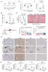
Figure 4. Ouabain selectively eliminates cells undergoing oncogene-induced senescence.
a, Experimental design for the oncogene-induced senescence experiment in the liver. b, Representative images of SA-β-Galactosidase staining in the liver. Arrows indicate examples of SA-β-Galactosidase-positive cells. Scale bar, 50 μm. c-e, Quantification of SA-β-galactosidase activity (c), Nras-positive cells (d), and p21Cip1-positive cells (e) in mice treated with vehicle (n = 9) or Ouabain (n = 12). Statistical significance was calculated using unpaired two-tailed Student's t-test. f, Experimental design for the senolytic experiment in an ACP mouse model. g, Tumoral pituitaries from 18.5dpc Hesx1Cre/+;Ctnnb1lox(ex3)/+ mice were cultured in the presence of either ABT-737 (2.5 μM), Ouabain (250 nM and 500 nM) or vehicle (DMSO) and processed for analysis after 72 hours (n = 10 mice per group). Immunofluorescence staining against β-catenin (green) and p21Cip1 (red). Arrows indicate examples of double positive cells. Scale bar, 50μm. Quantitative analysis of the immunofluorescence demonstrates that ABT-737 and ouabain significantly reduce the number of β-catenin-positive and of p21-positive cells. Statistical significance was calculated using Kruskal-Wallis and Dunn’s multiple comparisons test. h, Immunofluorescence staining against β-catenin (green) and Cleaved-caspase 3 (red) and quantitative analysis (n = 6 mice per group). Arrows indicate examples of double positive cells. Scale bar, 50 μm. Statistical significance was calculated using two-way ANOVA (Tukey’s multiple comparisons test). Red refers to comparison of double positive cells, black refers to comparison of β-catenin-positive cells. Data represent mean ± s.d; n represents number of mice; ns, not significant; **P < 0.01; ***P < 0.001.
Figure 5
Figure 5. Dual benefits of treatment with ouabain on therapy-induced senescence.
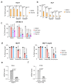
a, Crystal-violet stained 6-well dishes of SKHep1 cells that underwent therapy-induced senescence (etoposide, palbociclib or aurora kinase inhibitors) and were subsequently treated with ouabain or vehicle (DMSO). Timeline of the experiment is shown in Supplementary Fig 4a. Images are a representative experiment out of 3. b-d, Quantification of cell survival of SKHep1 cells (b), A549 cells (c) or HCT-116 cells (d) after treatment with the indicated drug combinations. Timeline of the experiment is shown in Supplementary Fig 4a (n = 4). Statistical significance was calculated using two-way ANOVA (Tukey’s test). e, Experimental design of the whole-body irradiation-induced senescence experiments. f, Representative immunofluorescence images of p16Ink4a (bottom) and SA-β-Galactosidase staining (top) in the lungs. Scale bar, 50 μm. g, Quantification of area positive for SA-β-Galactosidase staining and mRNA expression levels of p16Ink4a (n = 5 mock-irradiated control mice; n = 4 irradiated mice treated with ouabain; n = 4 irradiated mice treated with vehicle). h, Expression levels of ll1α and ll6 in lungs of non–irradiated mice (n = 5), irradiated mice treated with vehicle (n = 4) or irradiated mice treated with Ouabain (n = 4). Statistical significance was calculated using unpaired two-tailed Student's t-test. Data represent mean ± s.d; n represents independent experiments in b-d and number of mice in g-h.
Figure 6
Figure 6. Ouabain resets immune infiltration in old mice.
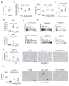
a, Experimental design for the ouabain treatment in old mice. T, time of enrolment. B, basal measures. O, ouabain treatment (3 consecutive days). b, Albumin levels of young (n = 7) and old mice, either treated with vehicle (n = 6) or ouabain (n = 8), were determined in whole-blood samples at the endpoint of the experiment. Data represent mean ± s.d; unpaired two-tailed Student's t-test. c, Motor coordination, balance and strength of old mice treated with vehicle (n = 6) or ouabain (n = 8) was assessed by performing the Rotarod test at 13 weeks after start of the experiment. Represented is the fold change versus the basal test. Data represent mean ± s.d; unpaired two-tailed Student's t-test. d, Expression levels of p16Ink4a in liver was determined by qRT-PCR following treatment with or vehicle (n = 5) or ouabain (n = 6). mRNA expression levels in young mice (n = 7) were used as reference. Data represent mean ± s.d; unpaired two-tailed Student's t-test. e, Left, quantification of SA-β-Galactosidase activity in the liver of young (n = 7) and old mice, either treated with vehicle (n = 6) or Ouabain (n = 8). Right, representative images of SA-β-Galactosidase activity in liver. Arrows indicate examples of SA-β-Galactosidase-positive cells. Scale bar, 50 μm. Data represent mean ± s.d; unpaired two-tailed Student's t-test. f, Left, experimental design for transcriptional profiling of livers from young or aged mice treated with ouabain or vehicle. Right, GSEA signature for oncogene-induced senescence and chemokines. g-h, Representative IHC images (g) and quantification (h) of the indicated immune cell markers in the liver of young (n = 6) and old mice, either treated with vehicle (n = 6) or ouabain (n = 8). Scale bar: 100µm. Data represent mean ± SEM; one-way ANOVA with Tukey’s post hoc comparison.

Comment in

  • Adding to the senolytic arsenal.
    Paiva SL. Paiva SL. Nat Rev Drug Discov. 2019 Nov;18(12):901. doi: 10.1038/d41573-019-00181-x. Nat Rev Drug Discov. 2019. PMID: 31780845 No abstract available.

References

    1. Kuilman T, Michaloglou C, Mooi WJ, Peeper DS. The essence of senescence. Genes Dev. 2010;24:2463–2479. - PMC - PubMed
    1. Herranz N, Gil J. Mechanisms and functions of cellular senescence. J Clin Invest. 2018;128:1238–1246. - PMC - PubMed
    1. Salama R, Sadaie M, Hoare M, Narita M. Cellular senescence and its effector programs. Genes Dev. 2014;28:99–114. - "V体育2025版" PMC - PubMed
    1. Coppe JP, Desprez PY, Krtolica A, Campisi J. The senescence-associated secretory phenotype: the dark side of tumor suppression. Annu Rev Pathol. 2010;5:99–118. - PMC - PubMed
    1. Kang TW, et al. Senescence surveillance of pre-malignant hepatocytes limits liver cancer development. Nature. 2011;479:547–551. - PubMed

Publication types

LinkOut - more resources