Skip to main page content
U.S. flag

An official website of the United States government

Dot gov

The . gov means it’s official VSports app下载. Federal government websites often end in . gov or . mil. Before sharing sensitive information, make sure you’re on a federal government site. .

Https

The site is secure. The https:// ensures that you are connecting to the official website and that any information you provide is encrypted and transmitted securely V体育官网. .

. 2019 Dec 2;51(5):575-586.e4.
doi: 10.1016/j.devcel.2019.10.007. Epub 2019 Nov 14.

V体育官网 - Prominin2 Drives Ferroptosis Resistance by Stimulating Iron Export

Affiliations

Prominin2 Drives Ferroptosis Resistance by Stimulating Iron Export

Caitlin W Brown et al. Dev Cell. .

Abstract

Ferroptosis, regulated cell death characterized by the iron-dependent accumulation of lethal lipid reactive oxygen species, contributes to tissue homeostasis and numerous pathologies, and it may be exploited for therapy. Cells differ in their sensitivity to ferroptosis, however, and a key challenge is to understand mechanisms that contribute to resistance. Using RNA-seq to identify genes that contribute to ferroptosis resistance, we discovered that pro-ferroptotic stimuli, including inhibition of the lipid hydroperoxidase GPX4 and detachment from the extracellular matrix, induce expression of prominin2, a pentaspanin protein implicated in regulation of lipid dynamics. Prominin2 facilitates ferroptosis resistance in mammary epithelial and breast carcinoma cells. Mechanistically, prominin2 promotes the formation of ferritin-containing multivesicular bodies (MVBs) and exosomes that transport iron out of the cell, inhibiting ferroptosis. These findings reveal that ferroptosis resistance can be driven by a prominin2-MVB-exosome-ferritin pathway and have broad implications for iron homeostasis, intracellular trafficking, and cancer VSports手机版. .

Keywords: GPX4; breast cancer; exosome; ferritin; ferroptosis; iron; mammary gland; multivesicular body; prominin2; therapy V体育安卓版. .


"VSports注册入口" Conflict of interest statement

Declaration of Interests: The authors declare no competing interests.

Figures

Figure 1.
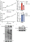
Figure 1.. Prominin2 is a ferroptosis stress response protein.
(A) MCF10A cells were detached from ECM for 30 min, 1 hr, 2 hrs or 3 hrs. Extracts from these cells were immunoblotted for prominin2 and actin. Immunoblot is representative of three independent experiments. (B) MCF10A and Hs578t cells were transfected with either control siRNA or prominin2 siRNA for 48 hrs and then maintained under adherent or ECM-detached conditions for 2 hrs. PROM2 mRNA expression was quantified by qPCR. Shown is representative experiment of three independent replicates. (C) siControl or siProminin2-treated MCF10A and Hs578t cells were maintained in ECM detached conditions for 24 hrs in the presence of either DMSO, ferrostatin-1 (2 μM), ZVAD-fmk (25 μM) or ferrostatin-1 and ZVAD-fmk and the number of viable cells was quantified. Shown is representative experiment of three independent replicates. (D) Adherent MCF10A, Hs578t and MDA-MB-231 cells were treated with either DMSO or RSL3 for 2 hrs and PROM2 mRNA expression was quantified by qPCR. Shown is representative experiment of three independent replicates. (E) Extracts from the cells in (D) were immunoblotted with antibodies specific for prominin2 and tubulin. The densitometric analysis of these immunoblots (normalized to 1.0 for each cell line) obtained from analysis of three independent experiments in shown below the blots. (F) Adherent MCF10A, Hs578t and MDA-MB-231 cells were treated with DMSO or RSL3 24 hrs and the percent of surviving cells was quantified. Absorbance was normalized to DMSO control. Shown is representative experiment of three independent replicates.
Figure 2.
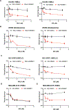
Figure 2.. Prominin2 increases resistance to ferroptosis.
(A) Prominin2 expression was diminished in adherent MCF10A cells for 48 hours using siRNA prior to treatment with RSL3, ML210, FIN56 or erastin for for 24 hrs. The percent of surviving cells was quantified and compared to cells transfected with an siRNA control. Absorbance was normalized to DMSO control. Shown is representative experiment of three independent replicates. See Supplemental Information for the IC50 values for these conditions. (B) Prominin2 expression was diminished in adherent Hs578t cells for 48 hours using siRNA prior to treatment with RSL3, ML210, FIN56, or erastin for 24 hrs. The percent of surviving cells was quantified and compared to cells transfected with an siRNA control. Absorbance was normalized to DMSO control. Shown is representative experiment of three independent replicates. See Supplemental Information for the IC50 values for these conditions. (C) Prominin2 expression was diminished in adherent MCF10A and Hs578t cells for 48 hours using siRNA. Cells were treated with either DMSO, RSL3 (5 μM) or RSL3 and ferrostatin-1 (2 μM) for 24 hrs and the percent of surviving cells was quantified. Shown is representative experiment of three independent replicates. (D) MDA-MB-231 cells were transfected with either a prominin2 expression construct or a vector control prior to treatment with either DMSO or RSL3 for 2 hrs. The percent of surviving cells was quantified. Absorbance was normalized to DMSO control. Shown is a representative experiment of three independent replicates.
Figure 3.
Figure 3.. Prominin2 promotes the formation of multivesicular bodies (MVBs) in response to GPX4 inhibition.
(A) Adherent MCF10A and Hs578t cells were treated with either DMSO or RSL3 for 2 hrs and immunostained using Abs specific for TSG101 (green) and prominin2 (red). Scale bar = 10 μm. For all immunofluorescence images, merged images are shown in the main figures and single channel images are provided in Supplemental Information. Also, for all experiments, image acquisition settings were the same for DMSO and RSL3-treated cells. Shown is representative image of three independent replicates. (B) Adherent Hs578t cells were treated with either DMSO or RSL3 for either 1, 1.5 or 2 hrs and immunostained using Abs specific for TSG101 (green) and prominin2 (red). Scale bar = 10 μm. Shown is representative image of three independent replicates. (C) MCF10A cells were transfected with either control siRNA or prominin2 siRNA for 48 hrs and then treated with either DMSO or RSL3 for 1.5 hrs. These cells were fixed and processed for transmission electron microscopy (TEM). The frequency of MVBs (white box) per section was quantified (right graph). Shown is representative image of five total cells quantified per group. (D) MCF10A cells and were transfected with either control siRNA or prominin2 siRNA for 48 hrs and then treated with either DMSO or RSL3 for 2 hrs. These cells were then immunostained using Abs specific for TSG101 (green) and prominin2 (red). Scale bar = 10 μm. Shown is representative image of three independent replicates. (E) Hs578t cells and were transfected with either control siRNA or prominin2 siRNA for 48 hrs and then treated with either DMSO or RSL3 for 2 hrs. These cells were then immunostained using Abs specific for TSG101 (green) and prominin2 (red). Scale bar = 10 μm. Shown is representative image of three independent replicates. (F) MDA-MB-231 cells were transfected with either a prominin2 expression construct or a vector control prior to treatment with either DMSO or RSL3 for 2 hrs. These cells were then immunostained using antibodies specific for TSG101 (green) and prominin2 (red). Scale bar = 10 μm. Shown is representative image of three independent replicates.
Figure 4.
Figure 4.. Multivesicular body formation is required for evasion of ferroptosis.
(A) TSG101 expression was diminished in adherent MCF10A cells for 48 hours using siRNA prior to treatment with DMSO or RSL3 for 24 hrs. The percent of surviving cells was quantified and compared to cells transfected with an siRNA control. Absorbance was normalized to DMSO control. Shown is representative experiment of three independent replicates. (B) TSG101 expression was diminished in adherent Hs578t cells for 48 hours using siRNA prior to treatment with DMSO or RSL3 for 24 hrs. The percent of surviving cells was quantified and compared to cells transfected with an siRNA control. Absorbance was normalized to DMSO control. Shown is representative experiment of three independent replicates. (C) TSG101 expression was diminished in adherent MCF10A cells for 48 hours using siRNA prior to treatment with DMSO, RSL3 or RSL3 and ferrostatin-1 for 24 hrs. The percent of surviving cells was quantified and compared to cells transfected with an siRNA control. Absorbance was normalized to DMSO control. Shown is representative experiment of three independent replicates. (D) Exosomes were isolated from cell culture medium of MCF10A and Hs578t cells treated for 24 hours with 1 μM RSL3 by ultracentrifugation. The exosome pellets were lysed and protein concentration was assessed. Exosomes from RSL3 treated MCF10A and Hs578t cells were immunoblotted for TSG101, prominin2, GM130 and CD63. See Supplemental Information for TEM of exosomes. Shown is representative immunoblot of three independent collections. (E) Prominin2 expression was diminished in MCF10A and Hs578t cells for 48 hours using siRNA prior to treatment with either DMSO, RSL3 (5 μM) or RSL3 and the exosome inhibitor GW4869 (20 μM) for 24 hrs and the percent of surviving cells was quantified. Shown is representative experiment of three independent replicates.
Figure 5.
Figure 5.. Prominin2 and ferritin contribute to the regulation of free iron.
(A) Adherent MCF10A and Hs578t cells were treated with either DMSO or RSL3 for 2 hrs and immunostained using Abs specific for ferritin (green) and prominin2 (red). Scale bar = 10 μm. Images are representative of three independent experiments with similar results. (B) Exosomes purified from RSL3-treated MCF10A and Hs578t cells as described in Fig. 3D were immunoblotted for prominin2, TSG101 and ferritin. (C) MDA-MB-231 cells were transfected with either a prominin2 expression construct or a vector control prior to treatment with either DMSO or RSL3 for 2 hrs. These cells were then immunostained using antibodies specific for ferritin heavy chain (green) and prominin2 (red). Scale bar = 10 μm. Shown are representative images of three independent experiments. (D) MCF10A were transfected with either control siRNA or ferritin heavy chain siRNA for 48hrs and then treated with increasing concentrations of RSL3 for 24 hrs, and the percent of surviving cells was quantified. Shown is representative experiment of three independent replicates. (E) Hs578t cells were transfected with either control siRNA or ferritin heavy chain siRNA for 48 hrs and then treated with increasing concentrations of RSL3 for 24 hrs, and the percent of surviving cells was quantified. Shown is representative experiment of three independent replicates. (F) MCF10A and Hs578t cells were transfected with either control siRNA or ferritin siRNA for 48 hrs and then treated with increasing concentrations of either DMSO, RSL3 and ferrostatin-1, or RSL3 and deferoxamine (DFO) for 24 hrs. The percent of surviving cells was then quantified. Shown is a representative experiment of three independent replicates.
Figure 6.
Figure 6.. GPX4 inhibition alters free iron concentration.
(A) Adherent MCF10A and Hs578t cells were transfected with either control siRNA or prominin2 siRNA for 48 hrs and then treated with 5 μM RSL3 for 2 hrs. Total free iron was quantified and reported in nmols. Shown is a representative experiment of three independent replicates. (B) Adherent MCF10A and Hs578t cells were transfected with either control siRNA or ferritin heavy chain siRNA for 48 hrs and then treated with RSL3 for 2hrs. Total free iron was quantified and reported in nmols. Shown is a representative experiment of three independent replicates. (C) Adherent MDA-MB-231 cells were transfected with either a prominin2 expression construct or a vector control prior to treatment with either DMSO or RSL3 for 2 hrs. Total free iron was quantified and reported as nmols. Shown is representative experiment of three independent replicates. (D) Adherent MCF10A and Hs578t cells were transfected with either control siRNA or TSG101 siRNA for 48 hrs and then treated with either DMSO or RSL3 for 2 hrs. Total free iron was quantified and reported as nanomoles. Shown is representative experiment of three independent replicates.
Figure 7.
Figure 7.. Detachment from the extracellular matrix induces prominin2-dependent MVB formation to rescue ferroptosis.
(A) Control and prominin2-depleted MCF10A cells were maintained in ECM-detached conditions for 24 hrs in the presence of either DMSO or deferoxamine and the number of viable cells was quantified. Shown is representative experiment of three independent replicates. (B) Control and prominin2-depleted MCF10A cells were maintained in ECM-detached conditions for the indicated times and total free iron was quantified. Shown is representative experiment of three independent replicates. (C) Control and prominin2-depleted MCF10A cells were maintained in ECM-detached conditions for 2 hrs and processed for TEM. The frequency of MVBs (white box) per section was quantified (right graph). Shown is representative image of five total cells quantified per group. (D) MCF10A and Hs578t cells were maintained in ECM-detached conditions for 2 hrs, fixed and immunostained for TSG101 (green) and prominin2 (red). Scale bar = 10 μm. Shown are representative images of three independent experiments.

Comment in

References

    1. Agmon E, Solon J, Bassereau P, and Stockwell BR (2018). Modeling the effects of lipid peroxidation during ferroptosis on membrane properties. Scientific reports 8, 5155. - PMC - PubMed
    1. Brown C, Amante JJ, Mercurio AM (2018). Cell clustering mediated by the adhesion protein PVRL4 is necessary for α6β4 integrin-promoted ferroptosis resistance in matrix-detached cells. The Journal of biological chemistry 293, 12741–12748. - PMC - PubMed
    1. Brown CW AJ, Goel HL, Mercurio AM (2017). The α6β4 integrin promotes resistance to ferroptosis. The Journal of cell biology 216, 4287–4297. - VSports注册入口 - PMC - PubMed
    1. Cella N C-UR, Montes GS, Hynes NE, Chammas R. (1996). The lysosomal-associated membrane protein LAMP-1 is a novel differentiation marker for HC11 mouse mammary epithelial cells. Differentiation 61, 113–120. - PubMed
    1. Dixon S.a.S., BR (2019). The Hallmarks of Ferroptosis. Annu Rev Cancer Biol 3, 35–54.

Publication types

MeSH terms

VSports手机版 - LinkOut - more resources