Skip to main page content
U.S. flag

An official website of the United States government

Dot gov

The . gov means it’s official. Federal government websites often end in VSports app下载. gov or . mil. Before sharing sensitive information, make sure you’re on a federal government site. .

Https

The site is secure V体育官网. The https:// ensures that you are connecting to the official website and that any information you provide is encrypted and transmitted securely. .

. 2020 Jan;30(1):21-33.
doi: 10.1038/s41422-019-0251-7. Epub 2019 Nov 15.

Guiding T lymphopoiesis from pluripotent stem cells by defined transcription factors

Affiliations

Guiding T lymphopoiesis from pluripotent stem cells by defined transcription factors

Rongqun Guo et al. Cell Res. 2020 Jan.

Abstract

Achievement of immunocompetent and therapeutic T lymphopoiesis from pluripotent stem cells (PSCs) is a central aim in T cell regenerative medicine. To date, preferentially reconstituting T lymphopoiesis in vivo from PSCs remains a practical challenge. Here we documented that synergistic and transient expression of Runx1 and Hoxa9 restricted in the time window of endothelial-to-hematopoietic transition and hematopoietic maturation stages in a PSC differentiation scheme (iR9-PSC) in vitro induced preferential generation of engraftable hematopoietic progenitors capable of homing to thymus and developing into mature T cells in primary and secondary immunodeficient recipients. Single-cell transcriptome and functional analyses illustrated the cellular trajectory of T lineage induction from PSCs, unveiling the T-lineage specification determined at as early as hemogenic endothelial cell stage and identifying the bona fide pre-thymic progenitors. The induced T cells distributed normally in central and peripheral lymphoid organs and exhibited abundant TCRαβ repertoire VSports手机版. The regenerative T lymphopoiesis restored immune surveillance in immunodeficient mice. Furthermore, gene-edited iR9-PSCs produced tumor-specific T cells in vivo that effectively eradicated tumor cells. This study provides insight into universal generation of functional and therapeutic T cells from the unlimited and editable PSC source. .


"VSports在线直播" Conflict of interest statement

The authors declare no competing interests.

"V体育官网" Figures
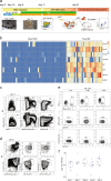
Fig. 1
Fig. 1
T cell regeneration in vivo from iRunx1-p2a-Hoxa9-edited embryonic stem cells. a The strategy of stepwise T lineage induction by defined transcription factors. iRunx1-ESC, and iRunx1-Xi-ESC lines (C57BL/6 background, CD45.2 strain) were used for T cell lineage induction. Xi means one of the eight transcription factors Hoxa5, Hoxa7, Hoxa9, Hoxa10, Hlf, Ikzf1, Nkx2-3, Setbp1. b Heatmaps of the eight transcription factors abundantly expressed in embryonic pre-HSCs but rarely expressed in iRunx1-ESC-derived iHECs. The expression value (TPM) of each gene was converted by log2 and illustrated by pheatmap (R package). One column represents one cell repeat. (iRunx1-iHEC, n = 50 single cells, T1-pre-HSC, n = 28 single cells). c Sorting gates of iHEC population at day 11 derived from iRunx1-Hoxa9-ES line (iR9-ESC). Two representative plots from five independent experiments are shown. d Immuno-phenotypes of pre-thymic progenitors in induced hematopoietic progenitor cells from iHECs after ten-day maturation. Two representative plots from five independent experiments are shown. Lin was defined as CD2CD3CD4CD8CD11bGr1Ter119CD19NK1.1TCRγδ. Pre-thymic progenitors were defined as Linc-kit+CD127+/CD135+. e Pluripotent stem cell-derived T cells in peripheral blood (PB) of B-NDG mice were analyzed by flow cytometry 4 weeks after transplantation. One million iHEC-derived hematopoietic cells were transplanted into individual B-NDG mice (CD45.1+) irradiated by X-ray (2.25 Gy). Three representative mice from five independent experiments were analyzed. f Summary of pluripotent stem cell-derived T cells in PB of individual B-NDG mice from five independent experiments. Forty B-NDG mice transplanted with ESC-derived iHPCs were analyzed. The box plot shows the percentage of the CD3+ iT cells in PB, the percentage values were illustrated by ggplot2 (R package). A base-10 logarithmic scale was used for the Y-axis. One point represents one mouse
Fig. 2
Fig. 2
Tissue distributions, transcriptome characterization, and TCRα/β diversities of ESC-derived T Cells. a Flow cytometry analysis of mature iT cells in spleen (SP), lymph node (LN), and PB of B-NDG mice transplanted with ESC-derived hematopoietic cells. Each B-NDG mouse was transplanted with one million iHPCs collected at day 21. Representative mouse was sacrificed and analyzed at 5 and 6 weeks after transplantation. Data from two representative mice are shown. b Flow cytometry analysis of iDN cells in the thymus of B-NDG mice transplanted with ESC-derived hematopoietic cells. Each B-NDG mouse was transplanted with one million iHPCs at day 21. Representative mouse was sacrificed and analyzed at 4 and 5 weeks after transplantation. Data from four representative mice of two independent experiments are shown. Lin was defined as Ter119CD11bGr1CD19B220NK1.1TCRγδ. c Flow cytometry analysis of iHPC derivatives in bone marrow (BM). Each B-NDG mouse was transplanted with one million iHPCs collected at day 10 in the presence of OP9-DL1 feeder cells. Representative mouse was sacrificed and analyzed 5 weeks and 6 weeks after transplantation. The BM-derived iHPCs (CD45.2+Linc-kitmidSca1+) were sorted for the 2nd transplantation. Data from two mice are shown. d Flow cytometry analysis of iT and iNK cells in PB, spleen (SP) and bone marrow (BM) 6 weeks after the 2nd transplantation. Five hundred LSK cells from primary iT mice were used as input for secondary transplantation. The secondary recipients were sacrificed and analyzed 6 weeks after transplantation. Data from one mouse are shown. e Characterization of surface markers on CD4SP and CD8SP iT cells. CD4SP and CD8SP iT cells were sorted from the spleens of B-NDG mice transplanted with ESC-derived hematopoietic cells at week 5. One biological replicate per column. Myeloid cells (n = 2 sample repeats): Ter119CD3CD19CD11b+; B cells (n = 4 sample repeats): Ter119CD11bCD3CD19+; CD4+ cells (n = 3 sample repeats): Ter119CD19CD11bCD4+; CD8+ cells (n = 3 sample repeats): Ter119CD19CD11bCD8+; iCD4+ cells (n = 3 sample repeats): CD45.2+Ter119CD19CD11bCD4+; iCD8+ cells (n = 3 sample repeats): CD45.2+Ter119CD19CD11bCD8+. f Characterization of transcription factors in CD4SP and CD8SP iT cells. g Chord diagram of TCRα diversity in thymus iT cells. h Chord diagram of TCRβ diversity in thymus iT cells. i Chord diagram of TCRα diversity in spleen iT cells. j Chord diagram of TCRβ diversity in spleen iT cells. Aliquots of sorted 15,000 naïve CD4SP and CD8SP iT cells from either thymus or spleen of iT-B-NDG mice were used as cell inputs for TCRαβ sequencing
Fig. 3
Fig. 3
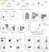
Assessment of T potential of single iHECs from iR9-ESC. a The strategy of T cell induction from iR9-ESC-derived single iHECs. Single iHECs were sorted into individual wells (24-well plates) pre-seeded with OP9-DL1 feeder cells (10,000 cells/well) 12 h prior maturation in EM medium with doxycycline (1 μg/mL). Doxycycline was sustained for 10 days during the maturation step. After maturation, the bulk blood cells were assessed for T lineage generation potential. For in vivo T cell regeneration, the single iHEC-derived bulk hematopoietic cells (day 10) were transplanted into individual B-NDG recipients. For in vitro T cell induction, the medium was changed to T cell induction medium (TIM, α-MEM, 20% DFBS, and 1% GlutaMAX) supplemented with 2% conditioned medium derived from supernatants of AFT024-hFlt3L and AFT024-hIL7 cell culture for sustaining 12 days. b Single iHECs efficiently gave rise to T cells. Three hundred and eighty-four single iHECs at day 11 were sorted into individual wells (24 well plates). Thirty single-iHEC-formed blood colonies were induced for T cell generation in vitro. Cell collections of Twenty-five single-iHEC-formed blood colonies were transplanted into 25 individual B-NDG mice for the assessment of T lymphopoiesis in vivo. c Flow cytometry analysis of induced T cells from in vitro induction of single iHECs. iT cells from single iHEC culture product (day 22) were analyzed. Plots of iT cells induced from one representative colony are shown. d Single iHEC-derived hematopoietic cells gave rise to mature iT cells in PB of B-NDG recipient mice 4 weeks after transplantation. Plots of one representative mouse are shown
Fig. 4
Fig. 4
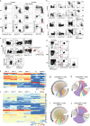
Single-cell transcriptomic characterization of iHECs and iHPCs. a Principal component analysis (PCA) of iHECs and developmental E11 AGM-derived ECs, T1 pre-HSCs, T2 pre-HSCs, E12 HSCs, E14 HSCs, and adult HSCs. TPM values of genes in iHECs (n = 70), natural E11 AGM-derived ECs (n = 17), T1 pre-HSCs (n = 28), T2 pre-HSCs (n = 32), E12 HSCs (n = 21), E14 HSCs (n = 32) and adult HSCs (n = 47) from single-cell RNA-Seq data were calculated with Stringtie package. b The expression of the top 100 genes contributing most to PC2 (50 genes for each direction). The expression value (TPM) of each gene was converted by log2 and illustrated by pheatmap (R package). One column represents one cell repeat. c Violin plots show the expression profile of selected artery (A)- and vein (V)-related genes (A: Nrp1, Efnb2, and Hey1; V: Nrp2, Nr2f2, and Ephb4) in single iHECs. The expression value (TPM) of each gene was converted by log2 and illustrated by ggplot2 (R package). One point represents one cell. d Violin plots show the expression profile of selected surface markers (Cdh5, Esam, Tek, Procr, Cd47, and Cd63) in single iHECs. The expression value (TPM) of each gene was converted by log2 and illustrated by ggplot2 (R package). One point represents one cell. e Violin plots show the expression profile of selected transcription factors (Fli1, Erg1, Lmo2, Lyl1, Tal1, Sox7, Runx1, Mycn, Gata2, Bcl11a, Hoxa9, and Hoxb5) related to hematopoietic development in single iHECs. The expression value (TPM) of each gene was converted by log2 and illustrated by ggplot2 (R package). One point represents one cell. f Two-dimensional t-SNE analysis of iHEC and iHPC single-cell RNA-Seq. For single-cell RNA-Seq, the iHECs were collected at day 11, and the iHPCs were collected at day 14, 17 and 21. Each dot represents one cell. The TPM values of genes in iHECs (n = 65), iHPCs at day 14 (n = 21), day 17 (n = 18) and day 21 (n = 56) from single-cell RNA-Seq data were calculated with Stringtie package. Cell types were defined as: (1) iHEC, CD31+CD41lowCD45c-kit+CD201high; (2) day-14 and day-17 iHPC, CD45+Lin(Ter119/Gr1/F4-80/CD2/CD3/CD4/CD8/CD19/FcεRIα); (3) day-21 iHPC, Ter119CD45+c-kit+CD127+. g t-SNE analysis of the expression pattern of selected endothelia-related transcription factors (Sox7, Sox18, and Ets1) in iHECs and iHPCs. h t-SNE analysis of the expression pattern of selected hematopoiesis-related transcription factors (Lyl1, Etv6, Prdm5, Myb, Sfpi1, and Meis1) in iHECs and iHPCs. i t-SNE analysis of the expression pattern of selected T cell development-related transcription factors (Lmo2, Bcl11a, Ikzf1, Myc, Gata3, and Tcf7) in iHECs and iHPCs at day 14, day 17, and day 21. j t-SNE analysis of the expression pattern of selected lymphopoiesis-related surface protein-coding genes (Kit, Flt3, Cd7, Ccr9, Ccr7, and Cxcr4) in iHECs and iHPCs at day 14, day 17, and day 21
Fig. 5
Fig. 5
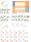
Adoptively transferred iT cells reject allogeneic skin in Rag1/ mice. a The images of allogeneic skin grafts. Representative images of rejected allogeneic skin tissues on ESC-iT-Rag1/ (day 9) mice (n = 2) and grafted skin tissue on control Rag1/ mice (day 30) were shown. b Flow cytometry analysis of the adoptively transferred ESC-iT cells in peripheral blood (PB) of Rag1/ recipients 9 days after the allogeneic skin was grafted. Plots of two representative mice are shown. c Flow cytometry analysis of the activation status of the ESC-iT cells in the rejected allogeneic skin tissues. The rejected allogeneic skin tissues were from the adoptively ESC-iT cell-transferred Rag1/ recipients 9 days after the allogeneic skin grafted. The activated ESC-iT cells were defined as CD4+/CD8+CD44highCD69+. Rejected skin tissues from two representative ESC-iT cell-transferred Rag1/ mice were analyzed. d Flow cytometry analysis of the intracellular cytokines IFNγ and IL-17 secreted by the CD4+ or CD8+ ESC-iT cells in rejected allogeneic skin tissues. The 1st allogeneic skin grafts were analyzed at day 9 and the 2nd allogeneic skin grafts were analyzed at day 6 after skin transplantation. Data from the primary and secondary rejected skin tissues from one representative ESC-iT cell-transferred Rag1/ mouse are shown
Fig. 6
Fig. 6
OT1-iT cell therapy suppresses the solid tumor growth in mice transplanted with E.G7-OVA cells. a Schematic diagram of the generation of OT1-engineered iT cells for anti-tumor therapy. Mouse MEFs were isolated from CD45.2+ C57BL/6 mouse and reprogrammed into iPSCs with retroviruses expressing Oct4, Klf4, and Sox2. Then an rtTA-TRE-Runx1-Hoxa9-HygroR DNA cassette was inserted into the Rosa26 locus. Next, a CAG-OT1-IRES-GFP-PuroR expression element was inserted into the Hipp11 locus of iR9-iPSC. OT1-iR9-iPSC results in the production of CD8+ T cells carrying TCRVα2 and TCRVβ5 (MHC class I-restricted, ovalbumin-specific TCR). OT1-iR9-iPSC-derived iHECs were induced into iHPCs (OT1-iHPCs) as described in the materials and methods section. The iHPCs were injected into irradiated (4.5 Gy) Rag1/ recipient mice (3 million/mouse, 8–10-week-old, C57BL/6 background). E.G7-OVA tumor cell line (C57BL/6 background) were transplanted into the groin of the Rag1/ (n = 8) or OT1-iT-Rag1/ (n = 8) mice by subcutaneous injection (0.2 million/mouse) 6 weeks after OT1-iHPC transplantation. b TCRVα2 and TCRVβ5 expression in OT1-iR9-iPSCs measured by intracellular staining. The iR9-iPSC was used as negative control. c Sorting gates of the OT1-iR9-iPSC-derived iHEC population at day 11. The cells were enriched by streptavidin-beads recognizing biotin-CD31 before sorting. Representative plots from three independent experiments are shown. d Immuno-phenotypes of pre-thymic progenitors in iHPCs induced from OT1-iR9-iPSC-derived iHECs after ten-day maturation. Representative plots from three independent experiments are shown. Lin was defined as CD2CD3CD4CD8CD11bGr1Ter119CD19NK1.1TCRγδ. pre-thymic progenitors were defined as Linc-kit+CD127+/CD135+. e TCRVα2 and TCRVβ5 expression of iT cells in PB of Rag1/ mice 6 weeks after transplantation of OT1-iR9-iPSC-derived iHPCs. Three representative mice from three independent experiments were analyzed. f Tumor growth in Rag1/ and OT1-iT-Rag1/ mice. E.G7-OVA cells were transplanted into the groin of the Rag1/ (n = 8) or OT1-iT-Rag1/ mice (n = 8) by subcutaneous injection (0.2 million/mouse). The length and width of the tumors were measured every other day by a caliper, and each tumor size was calculated as length × width (mm2). Mice with tumor size larger than 20 mm at the longest axis were euthanized for ethical consideration. ***P < 0.001 (independent t-test, two-tailed). g Characterization of the OT1-iT cells in tumors. Tumors were isolated at day 19 after injection and disaggregated by collagenase IV to single cell suspensions. The effector iT cells were defined as CD44+CD62L. The memory iT cells were defined as CD44+CD62L+. IFNγ secreted by CD8+ OT1-iT cells in tumors were intracellular stained. Representative plots from two tumors are shown

"VSports手机版" References

    1. Schmitt TM, Zuniga-Pflucker JC. Induction of T cell development from hematopoietic progenitor cells by delta-like-1 in vitro. Immunity. 2002;17:749–756. - PubMed
    1. Mohtashami M, et al. Direct comparison of Dll1- and Dll4-mediated Notch activation levels shows differential lymphomyeloid lineage commitment outcomes. J. Immunol. 2010;185:867–876. - PubMed
    1. Montel-Hagen A, et al. Organoid-Induced Differentiation of Conventional T Cells from Human Pluripotent Stem Cells. Cell Stem Cell. 2019;24:376–389 e378. - PMC - PubMed
    1. Awong G, et al. Human proT-cells generated in vitro facilitate hematopoietic stem cell-derived T-lymphopoiesis in vivo and restore thymic architecture. Blood. 2013;122:4210–4219. - PMC - PubMed
    1. Shukla S, et al. Progenitor T-cell differentiation from hematopoietic stem cells using Delta-like-4 and VCAM-1. Nat. Methods. 2017;14:531–538. - "V体育官网入口" PubMed

Publication types

MeSH terms

V体育平台登录 - Substances