Skip to main page content
U.S. flag

An official website of the United States government

Dot gov

The . gov means it’s official. Federal government websites often end in . gov or . mil. Before sharing sensitive information, make sure you’re on a federal government site. VSports app下载.

Https

The site is secure. The https:// ensures that you are connecting to the official website and that any information you provide is encrypted and transmitted securely V体育官网. .

. 2019 Nov 6;15(11):e1008096.
doi: 10.1371/journal.ppat.1008096. eCollection 2019 Nov.

Candida albicans triggers NADPH oxidase-independent neutrophil extracellular traps through dectin-2

Affiliations

V体育安卓版 - Candida albicans triggers NADPH oxidase-independent neutrophil extracellular traps through dectin-2

Sheng-Yang Wu et al. PLoS Pathog. .

Abstract

Candida albicans is one of the top leading causes of healthcare-associated bloodstream infection. Neutrophil extracellular traps (NET) are known to capture and kill pathogens. It is reported that opsonized C. albicans-triggered NETosis is NADPH oxidase-dependent. We discovered a NADPH oxidase-independent NETosis pathway in neutrophil response to unopsonized C. albicans. While CR3 engagement with opsonized C. albicans triggered NET, dectin-2 recognized unopsonized C. albicans and mediated NET formation. Engagement of dectin-2 activated the downstream Syk-Ca2+-PKCδ-protein arginine deiminase 4 (PAD4) signaling pathway which modulated nuclear translocation of neutrophil elastase (NE), histone citrullination and NETosis. In a C. albicans peritonitis model we observed Ki67+Ly6G+ NETotic cells in the peritoneal exudate and mesenteric tissues within 3 h of infection. Treatment with PAD4 inhibitor GSK484 or dectin-2 deficiency reduced % Ki67+Ly6G+ cells and the intensity of Ki67 in peritoneal neutrophils. Employing DNA digestion enzyme micrococcal nuclease, GSK484 as well as dectin-2-deficient mice, we further showed that dectin-2-mediated PAD4-dependent NET formation in vivo restrained the spread of C. albicans from the peritoneal cavity to kidney. Taken together, this study reveals that unopsonized C VSports手机版. albicans evokes NADPH oxidase-independent NETosis through dectin-2 and its downstream signaling pathway and dectin-2-mediated NET helps restrain fungal dissemination. .


Conflict of interest statement

The authors have declared that no competing interests exist.

"VSports在线直播" Figures
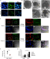
Fig 1
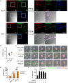
Fig 1. Both opsonized and unopsonized C. albicans induce NETosis.
Neutrophils were stimulated or not (Ctrl) with opsonized (Ops.) and unopsonized (Unops.) C. albicans (Ca) at MOI of 2. (A) Cells were stained with anti-histone H3 antibody and Hoechst 33342. Immunofluorescence images were viewed under confocal microscope at 3 h of stimulation. (B) Transmission electron microscopy of unstimulated neutrophils (HBSS 0 h) or neutrophils stimulated with unopsonized C. albicans for 1 h (Unops.Ca 1 h) or 2 h (Unops.Ca 2 h) or with opsonized C. albicans for 1 h (Ops.Ca 1 h). Yellow arrows point to disintegrated nuclear envelop, whereas the red arrow points to disrupted cell membrane where cytosolic materials are being released. (C) Cells were stained with anti-histone H3 antibody (green), cell-impermeable DNA dye SYTOX Orange (red), and cell-permeable DNA dye Hoechst 33342 (blue). Immunofluorescence images were viewed under fluorescence microscope at 3 h of stimulation. DIC, differential interference contrast image. Images in the boxed areas are enlarged and shown on the right. (D) % NETotic cells = the number of cells that had NET morphology (SYTOX Orange+, web-like structure) after stimulation with opsonized (Ops) or unopsonized (Unops.) C. albicans divided by the total number of cells (blue) counted in images as prepared in (C). (E) C. albicans strains HLC 54 (yeast-locked) and SC 5314 (germination-competent) were incubated in RPMI medium for 4 h to allow competent cells to germinate. Neutrophils were then stimulated with opsonized and unopsonized HLC 54 (Yeast) and SC 5314 (Hyphae). Ctrl for the unops. group was cells incubated in HBSS only. Ctrl for the ops. group was cells incubated in HBSS containing 5% mouse serum. Extracellular DNA was quantified by Quant-iT PicoGreen dsDNA assay (n = 3). *, p < 0.05; ***, p < 0.005; n.s., not significant, as analyzed by Student’s t test.
Fig 2
Fig 2. Unopsonized C. albicans-induced NET formation is independent of NCF-1- and mitochondrial ROS.
Ncf-1+/+ and Ncf-1-/- neutrophils were stimulated or not (Ctrl) with opsonized (Ops.) or unopsonized (Unops.) C. albicans at MOI of 2. (A) Cells were stimulated with unopsonized C. albicans for 3 h and stained with anti-histone H3 antibody (green), cell-impermeable DNA dye SYTOX Orange (red), and cell-permeable DNA dye Hoechst 33342 (blue). Immunofluorescence images were viewed under fluorescence microscope. DIC, differential interference contrast image. Images in the boxed areas are enlarged and shown on the right. (B) % NETotic cells = the number of cells that had NET morphology (SYTOX Orange+, web-like structure) divided by the total number of cells (blue) counted in images as prepared in (A). (C) Live cells were stained with cell-permeable DNA dye Draq5 (blue) and cell-impermeable DNA dye SYTOX Orange (red) before stimulation with GFP-expressing C. albicans strain OG1 (green). NETosis in response to opsonized and unopsonized pre-germinated C. albicans was observed over 180 min after addition of C. albicans. Zeiss LSM 780 confocal microscope was employed for time-lapse imaging (Images were obtained from S1, S2 and S3 Videos separately). (D) Cells were stimulated with opsonized and unopsonized C. albicans for 3 h. Cell supernatants were collected for Quant-iT PicoGreen dsDNA assay. (n = 3) (E) Cells were pretreated with 5, 10, 20 μM of mitochondria ROS inhibitor, MitoTEMPO, for 30 min before stimulation with unopsonized C. albicans. Cell supernatants were collected for Quant-iT PicoGreen dsDNA assay. (n = 3). All experiments were performed three times. Data from one representative experiment are presented as mean ± standard deviation (SD). **, p < 0.01; n.s., not significant, as analyzed by Student’s t test.
Fig 3
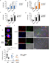
Fig 3. Neutrophil killing of unopsonized C. albicans requires dectin-2-mediated NET formation.
WT, Itgam-/- (A), clec4n-/- (B), clec7a-/- (C), and MyD88-/- (D) neutrophils were stimulated or not (Ctrl) with opsonized (Ops. Ca) or unopsonized (Unops. Ca) C. albicans at MOI of 2 for 3 h. Extracellular DNA was quantified by Quant-iT PicoGreen dsDNA assay. (n = 3). All experiments were performed three times. Data from one representative experiment are shown and presented as mean ± SD. *, p < 0.05; **, p < 0.01; n.s., not significant, as analyzed by Student’s t test. (E) Neutrophils were stimulated with pre-germinated (Unops. Hyphae) or ungerminated (Unops. Yeast) GFP-expressing C. albicans OG1 (green). Cells were cytospun, permeabilized and stained for dectin-2 (red) and nucleus (blue). Arrows point to where dectin-2 contacts either the yeast or hyphal form of the fungus. (F) Clec4n+/+ and clec4n-/- neutrophils were stimulated with unopsonized C. albicans at MOI of 2 for 3 h and stained with anti-histone H3 antibody (green), cell-impermeable DNA dye SYTOX Orange (red), cell-permeable DNA dye Hoechst 33258 (blue). Immunofluorescence images were viewed under fluorescence microscope. DIC, differential interference contrast image. Yellow arrows point to cells that undergo NETosis. (G) WT, Itgam-/-, and clec4n-/- neutrophils were incubated with unopsonized C. albicans at MOI of 2 in HBSS supplemented with 10 U/ml of MNase or heat-inactivated MNase (h.i. MNase). Wells containing C. albicans only without neutrophils were used as control. Controls were incubated in medium containing h.i. MNase or MNase. Three hours after incubation, medium was collected and cold H2O (pH = 11) was added to lyse cells. C. albicans was detached by mini cell scraper and vigorous pipetting. The number of viable fungi was determined by plating the supernatant on yeast-peptone-dextrose agar plate. Colony counts (CFU) were enumerated 2–3 days later. The ability of neutrophils to kill Candida is presented as % killing of C. albicans which was calculated by dividing the difference of CFU counts between the control group (without neutrophils) and neutrophil-added groups with MNase or h.i. MNase treatment by the counts of respective control. WT, n = 9; Itgam-/- and clec4n-/-, n = 5 each. Each n represents neutrophils collected from one mouse. Data were pooled from 3 independent experiments and presented as mean ± SD. *, p < 0.05; n.s., not significant, as analyzed by Student’s t test by comparing the 2 groups linked by a bracket.
Fig 4
Fig 4. Unopsonized C. albicans induces NETosis through dectin-2-Syk-Ca2+-PKCδ pathway.
WT (A-H) and Clec4n-/- (H) neutrophils were stimulated with unopsonized C. albicans. (A-E) Cells were pre-treated (+) or not (-) with Syk inhibitor (Syki, 10 μM of SykI) (A), Ca2+ chelator (10 μM of BAPTA-AM) (B), PKC inhibitor (PKCi, 10 μM of Ro318220) (C), and inhibitor to PKC isoforms (250 μM of inhibitor to PKCα+β1, 2.5 μM of inhibitor to PKCβ, and 0.8, 4, 20 μM of inhibitor to PKCδ) (D) for 30 min before stimulation with (+) or without (-) unopsonized C. albicans at MOI of 2 for 3 h. Extracellular DNA was quantified by Quant-iT PicoGreen dsDNA assay. Relative inhibition (%) was calculated by dividing the value of inhibitor-treated group by that of the untreated (A-C). (E) Cells were first loaded with Ca2+ indicator and then pre-treated (Syki) or not (DMSO) with 10 μM of SykI for 30 min. After treatment, cells were stimulated (DMSO and Syki) or not (Ctrl) with unopsonized C. albicans at MOI of 4. Intracellular Ca2+ content was analyzed by flow cytometry from before stimulation until 450 sec after. Arrow points to the time when C. albicans was added. Maximal % of Ca2+-positive cells in unopsonized C. albicans-stimulated group was taken as 100% intensity (Max). The Ca2+ response of other time points was normalized against the maximal response and is shown as relative Ca2+-positive cells (%) (calculated under the kinetic mode of FlowJo software). Line graphs showing the kinetics of Ca2+ responses in the three groups were analyzed and overlaid by FlowJo software. Bar graph on the right shows % Ca2+-positive cells at maximal response (20–50 sec, gating strategy is shown in S1 Fig). (F, G) Cells were pre-treated with Syk inhibitor SykI (F) or Ca2+ chelator BAPTA-AM (G) before stimulation by pre-germinated unopsonized C. albicans at MOI of 2. At 30 min of stimulation, cell lysates were collected and subject to Western blot analysis for phosphorylated-Syk and -PKCδ. β-actin was used as loading control. Relative intensities of p-Syk and p-PKCδ are quantified by ImageJ and shown as bar graphs next to the blot. (H) WT and clec4n-/- neutrophils were stimulated with pre-germinated unopsonized C. albicans at MOI of 2 for 15 and 30 min. Cell lysates were collected and subject to Western blot analysis for p-Syk and p-PKCδ as described in (G). Relative intensities of p-Syk and p-PKCδ in stimulated cells were normalized against their respective unstimulated controls and shown as relative fold induction. All Western blot experiments were performed three times. Data from one representative experiment are shown. Data are presented as mean ± standard error of the mean (SEM). *, p < 0.05; **, p < 0.01, as analyzed by Student’s t test comparing the 2 groups linked by a bracket.
Fig 5
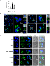
Fig 5. NE nuclear translocation is involved in NCF-1-independent NETosis through Syk-Ca2+-PKCδ.
(A) Neutrophils were pre-treated (+) or not (-) with neutrophil elastase inhibitor (NEi, 10 μM of sivelestat) for 30 min before stimulation (+) or not (-) with unopsonized C. albicans at MOI of 2 for 3 h. Extracellular DNA was quantified by Quant-iT PicoGreen dsDNA assay. *, p < 0.05 as analyzed by Student’s t test comparing the groups treated with and without inhibitor. (B) Neutrophils were seeded on coverslips and stimulated with unopsonized C. albicans at MOI of 2. At indicated time after stimulation, cells were stained with anti-neutrophil elastase antibody (green) and cell-permeable DNA dye Hoechst 33258 (blue). Immunofluorescence images were viewed under confocal microscope. DIC, differential interference contrast image. Red arrow point to nuclear translocation of NE. (C) Cells were pre-treated with Syk inhibitor (10 μM of SykI, Syki), Ca2+ chelator (10 μM of BAPTA-AM), PKCδ inhibitor (20 μM of Rottlerin, PKCδi), and NE inhibitor (10 μM of sivelestat, NEi) before stimulation. Ctrl., cells incubated in HBSS containing 0.5% DMSO. At 2 h of stimulation, cells were stained with anti-neutrophil elastase antibody (green) and cell-permeable DNA dye Hoechst 33258 (blue). Immunofluorescence images were viewed under confocal microscope. DIC, differential interference contrast image.
Fig 6
Fig 6. Unopsonized C. albicans-triggered NETosis is dependent on PAD4.
(A) Neutrophils were pre-treated(+) or not (-) with PAD1-4 (10 μM of CC-Cl-amidine) or PAD4 (10 μM of GSK484) inhibitor for 30 min before stimulation (+) or not (-) with unopsonized C. albicans at MOI of 2 for 3 h. Extracellular DNA was quantified by Quant-iT PicoGreen dsDNA assay. (B) Cells were pre-treated with PAD4 inhibitor (PAD4i, 10 μM of GSK484) for 30 min before stimulation. At 2 h of stimulation, cells were stained with anti-neutrophil elastase antibody (green) and cell-permeable DNA dye Hoechst 33258 (blue). Immunofluorescence images were viewed under confocal microscope. DIC, differential interference contrast image. Ctrl., cells incubated in HBSS containing 0.1% DMSO. (C) Neutrophils pretreated or not (Ctrl) with PAD4 inhibitor (10 μM of GSK484, PAD4i) were stimulated with unopsonized C. albicans at MOI of 2 for 3 h. Cells were stained with anti-histone H3 antibody (green), cell-impermeable DNA dye SYTOX Orange (red), cell-permeable DNA dye Hoechst 33258 (blue). Immunofluorescence images were viewed under fluorescence microscope. DIC, differential interference contrast image. Arrows point to H3-containing web-like structure. (D) Cells were pre-treated with PKCδ inhibitor (PKCδI, 20 μM of Rottlerin) before stimulation with C. albicans. At 30 min of stimulation, cell lysates were collected and subject to Western blot analysis for citrullinated-histone H3 (cit.H3). GAPDH was used as a loading control. The experiment was performed 3 times. Data from one representative experiment are shown. Relative intensities of cit.H3 against GAPDH are shown below the blot. Data are presented as mean ± standard error of the mean (SEM).*, p < 0.05; **, p < 0.01, as analyzed by Student’s t test comparing the two groups treated with and without inhibitor (A, D).
Fig 7
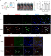
Fig 7. C. albicans infection triggers NETosis in mice.
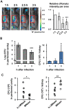
(A) Peritoneal exudates were harvested from naïve mice and from mice at 4 h after receiving two peritoneal injections of 9% casein (18 h apart (18 h → 4 h). Cells were stained with anti-Ly6G and anti-F4/80 antibodies and subject to flow cytometric analysis. Contour plots show the % of Ly6G+F4/80- neutrophils (blue population) and Ly6G-F4/80+ macrophages (green population) among total cells. (B-D) WT mice were given peritoneal injections of casein as described above. At 4 h after the second casein injection, mice received 1 × 10⁸ of C. albicans intraperitoneally. (B) Mice were injected with SYTOX Orange intraperitoneally at the same time when C. albicans strain SC5314 was administered. Mice were imaged on the side by IVIS (Ex/Em = 570/620) to record SYTOX Orange signals for 3 h starting at the time when C. albicans was administered. Photons in user-specified region of interest (ROI, gated area) was measured by Living Image 3.2 software. Relative intensity of total photons in ROI at each time point was calculated based on the intensity at 1 h after infection. n = 10. Data are presented as mean ± SD. ***, p < 0.005, as analyzed by Student’s t test comparing the intensity at each time point to that at 1 h after infection. (C) Three hours after C. albicans infection, peritoneal exudates were collected and seeded on coverslips. Cells on the coverslips were permeabilized and stained for Ki67 (orange), histone H3 (red), Ly6G (green) and nucleus (blue) and viewed under fluorescence microscope. Arrows point to Ly6G+Ki67+ cells. (D) Three hours after C. albicans infection, mesenteric tissues were collected and embedded in O.C.T. Cryosections were stained for Ki67 (red), Ly6G (green) and nucleus (blue) and viewed under confocal microscope. Arrows point to Ly6G+Ki67+ cells.
Fig 8
Fig 8. NET formation reduces C. albicans spread from peritoneal cavity to kidney.
WT mice were injected with 9% casein intraperitoneally as described above. (A) At 4 h after the second injection, mice were injected with 3 × 10⁸ of dTomato-expressing C. albicans strain CAF2 intraperitoneally and imaged on the side by IVIS (Ex/Em = 570/620). dTomato signals were recorded for 3 h starting 1 h after C. albicans administration. Photons in ROI (gated area) was measured by Living Image 3.2 software. Relative intensity of total photons in ROI was calculated based on the intensity at 1 h after infection. n = 5. Data are presented as mean ± SD. (B) Mice were injected with 1 × 10⁸ of C. albicans strain SC 5314 intraperitoneally at 4 h after second casein injection. At indicated times after infection, peritoneal fluid and kidneys were collected. Fungal burdens were determined by plating. n = 4. (C) Mice were injected with 100 U of MNase or heat-inactivated MNase (h.i. MNase, in otherwise equivalent amount) intraperitoneally at the time of infection with 1×10⁸ of C. albicans. At 3 h after infection, peritoneal fluid and kidneys were collected. Fungal burdens were determined by plating. n = 6. *, p < 0.05; **, p < 0.01; ***, p < 0.005, as analyzed by Student’s t test comparing indicated time points to 1 h after infection (A, B) or the two groups linked by a bracket (C).
Fig 9
Fig 9. GSK treatment impedes NET formation and promotes C. albicans spread from peritoneal cavity to kidney.
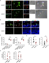
(A) WT mice were given two injections of 9% casein as described above. At 4 h after the second injection, mice were injected with GSK484 (20 mg/Kg) or HBSS containing 10% of DMSO intraperitoneally (Ctrl.) at the time of challenge with 1 × 10⁸ C. albicans. At 3 h after infection, peritoneal exudates, mesenteric tissues and kidneys were collected. (A) Peritoneal exudates were seeded on coverslips and incubated for 1 h. Cells were permeabilized and stained for Ki67 (orange), histone H3 (red), Ly6G (green) and nucleus (blue) and viewed under fluorescence microscope. DIC, differential interference contrast image. Arrows point to Ly6G+Ki67+ cells. (B) Mesenteric tissues were collected and embedded in O.C.T. Cryosections were stained for Ki67 (red), Ly6G (green) and nucleus (blue) and viewed under fluorescence microscope. (C) Peritoneal exudates were stained with anti-CD11b, -Ly6G and -Ki67antibodies and subject to flow cytometric analysis. The percentages of Ki67+ cells among total neutrophils (CD11b+Ly6G+) population are shown as % Ki67+ of neutrophils. The mean fluorescence intensity (MFI) of Ki67 represents the level of Ki67 expression in Ki67+ cells. Gating strategy for Ki67 was illustrated in S4B Fig. Data were pooled from two independent experiments. (D) Fungal counts in total peritoneal fluid and kidney homogenates were determined by plating. Fungal colonies were counted 2–3 days later. Data were pooled from 4 independent experiments. *, p < 0.05; ***, p < 0.005, as analyzed by Student’s t test.
Fig 10
Fig 10. Dectin-2 deficiency reduces C. albicans-induced NETosis and increases fungal spread from peritoneal cavity to kidney.
(A-C) Clec4n+/+ and clec4n-/- mice were given two injections of 9% casein as described above. At 4 h after the second injection, mice were injected with or without 1 × 10⁸ of C. albicans intraperitoneally. At 3 h after infection, peritoneal exudates, mesenteric tissues and kidneys were collected. (A) Peritoneal exudates were seeded on coverslips and incubated for 1 h. Cells were permeabilized and stained for Ki67 (orange), histone H3 (red), Ly6G (green) and nucleus (blue) and viewed under fluorescence microscope. Arrows point to Ly6G+Ki67+ cells. (B) Mesenteric tissues were collected and embedded in O.C.T. Cryosections were stained for Ki67 (red), Ly6G (green) and nucleus (blue) and viewed under fluorescence microscope. DIC, differential interference contrast image. (C) Peritoneal exudates were stained with anti-CD11b, -Ly6G and -Ki67 antibodies and subject to flow cytometric analysis. The percentages of Ki67+ cells among total neutrophils (CD11b+Ly6G+) population are shown as % Ki67+ of neutrophils. The mean fluorescence intensity (MFI) of Ki67 represents the level of Ki67 expression in Ki67+ cells. Data were pooled from 2 independent experiments. Gating strategy for Ki67 was illustrated in S4B Fig. Naïve mice are uninfected mice receiving casein injection only. (D) Fungal counts in total peritoneal fluid and kidney homogenates were determined by plating. Fungal colonies were counted 2–3 days later. (Clec4n+/+ n = 13; Clec4n-/- n = 9). Data were pooled from four independent experiments. ***, p < 0.005, as analyzed by Student’s t test. (E) Clec4n-/- mice were given two injections of 9% casein as described above. Four hours after the second injection, mice were injected with 100 U of MNase or heat-inactivated MNase (h.i. MNase, in otherwise equivalent amount) intraperitoneally at the time when 1×10⁸ of C. albicans was administered. At 3 h after infection, peritoneal exudates and kidneys were harvest. Fungal counts in total peritoneal fluid and kidney homogenates were determined by plating. Fungal colonies were counted 2–3 days later. (n = 7–8). Data were pooled from 2 independent experiments. n.s., not significant, as analyzed by Student’s t test.

References

    1. Kullberg BJ, Arendrup MC. Invasive Candidiasis. N Engl J Med. 2015;373(15):1445–56. 10.1056/NEJMra1315399 . - DOI - PubMed
    1. Chen PY, Chuang YC, Wang JT, Sheng WH, Yu CJ, Chu CC, et al. Comparison of epidemiology and treatment outcome of patients with candidemia at a teaching hospital in Northern Taiwan, in 2002 and 2010. J Microbiol Immunol Infect. 2014;47(2):95–103. 10.1016/j.jmii.2012.08.025 . - DOI - PubMed
    1. Brown GD, Denning DW, Gow NA, Levitz SM, Netea MG, White TC. Hidden killers: human fungal infections. Sci Transl Med. 2012;4(165):165rv13 10.1126/scitranslmed.3004404 . - "VSports" DOI - PubMed
    1. Yang YL, Wang AH, Wang CW, Cheng WT, Li SY, Lo HJ, et al. Susceptibilities to amphotericin B and fluconazole of Candida species in Taiwan Surveillance of Antimicrobial Resistance of Yeasts 2006. Diagnostic microbiology and infectious disease. 2008;61(2):175–80. 10.1016/j.diagmicrobio.2008.01.011 . - V体育2025版 - DOI - PubMed
    1. Gullo A. Invasive fungal infections: the challenge continues. Drugs. 2009;69 Suppl 1:65–73. 10.2165/11315530-000000000-00000 . - DOI - PubMed

VSports注册入口 - Publication types

MeSH terms

LinkOut - more resources