V体育官网入口 - Skip to main page content
U.S. flag

An official website of the United States government

Dot gov

The . gov means it’s official. Federal government websites often end in . gov or VSports app下载. mil. Before sharing sensitive information, make sure you’re on a federal government site. .

Https

The site is secure. The https:// ensures that you are connecting to the official website and that any information you provide is encrypted and transmitted securely. V体育官网.

. 2019 Nov;23(11):7200-7209.
doi: 10.1111/jcmm.14389. Epub 2019 Sep 26.

Long non-coding RNA TUG1 promotes airway remodelling by suppressing the miR-145-5p/DUSP6 axis in cigarette smoke-induced COPD

Affiliations

Long non-coding RNA TUG1 promotes airway remodelling by suppressing the miR-145-5p/DUSP6 axis in cigarette smoke-induced COPD

VSports - Wenchao Gu et al. J Cell Mol Med. 2019 Nov.

Abstract

Chronic obstructive pulmonary disease (COPD) is a progressive lung disease that is primarily caused by cigarette smoke (CS)-induced chronic inflammation. In this study, we investigated the function and mechanism of action of the long non-coding RNA (lncRNA) taurine-up-regulated gene 1 (TUG1) in CS-induced COPD VSports手机版. We found that the expression of TUG1 was significantly higher in the sputum cells and lung tissues of patients with COPD as compared to that in non-smokers, and negatively correlated with the percentage of predicted forced expiratory volume in 1 second. In addition, up-regulation of TUG1 was observed in CS-exposed mice, and knockdown of TUG1 attenuated inflammation and airway remodelling in a mouse model. Moreover, TUG1 expression was higher in CS extract (CSE)-treated human bronchial epithelial cells and lung fibroblasts, whereas inhibition of TUG1 reversed CSE-induced inflammation and collagen deposition in vitro. Mechanistically, TUG1 promoted the expression of dual-specificity phosphatase 6 (DUSP6) by sponging miR-145-5p. DUSP6 overexpression reversed TUG1 knockdown-mediated inhibition of inflammation and airway remodelling. These findings suggested an important role of TUG1 in the pathological alterations associated with CS-mediated airway remodelling in COPD. Thus, TUG1 may be a promising therapeutic target in CS-induced airway inflammation and fibroblast activation. .

Keywords: COPD; DUSP6; TUG1; airway inflammation; airway remodelling; miR-145-5p. V体育安卓版.


Conflict of interest statement

The authors confirm that there are no conflict of interest.

Figures

Figure 1
Figure 1
TUG1 expression was higher in patients with COPD. A, Sputum cells were obtained from non‐smokers (n = 15), smokers (n = 20) and patients with COPD (n = 30). The expression of TUG1 in sputum cells was measured by qRT‐PCR. B, Spearman’s correlation analysis between TUG1 expression and FEV1%. C, Lung tissue samples were obtained from non‐smokers (n = 5), smokers (n = 5) and patients with COPD (n = 10). The expression of TUG1 in lung tissue samples was measured by qRT‐PCR. D, Spearman’s correlation analysis between TUG1 expression and FEV1%. *P < 0.05 as compared to non‐smoker group
Figure 2
Figure 2
Knockdown of TUG1 inhibited CS‐induced airway remodelling and airway inflammation in vivo. A, TUG1 expression was assessed by qRT‐PCR in the lung tissues of air‐ or CS‐exposed mice transduced or not transduced with the TUG1 knockdown construct. B‐D, Counting of total bronchoalveolar lavage fluid (BALF) cells and neutrophils, and histological analysis of the lung sections were performed by haematoxylin and eosin staining to visualize inflammatory cell recruitment. E and F, Representative Masson and α‐SMA staining of lung sections from air‐ or CS‐exposed mice transduced or not transduced with the TUG1 knockdown construct. G, The expression of the inflammatory cytokines IL‐6 and TGF‐β1 in the lung tissues was quantified by ELISA. *P < 0.05 versus Air/PBS group; & P < 0.05 as compared to CS/PBS group
Figure 3
Figure 3
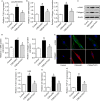
Knockdown of TUG1 inhibited CSE‐induced airway remodelling and airway inflammation in vitro. A, The expression of TUG1 was analysed in CSE‐treated parenchymal fibroblasts. B‐D, The mRNA and protein expression levels of α‐SMA and collagen I were determined by qRT‐PCR and Western blot analysis, respectively, in these groups. E, α‐SMA and collagen I expression was detected by immunofluorescent staining. F, Expression of TUG1 was analysed in HBE cells treated with 5% CSE for 48 h and transfected with shTUG1. G and H, Expression levels of IL‐6 and TGF‐β1 were assessed by qRT‐PCR. *P < 0.05 as compared to control group; & P < 0.05 as compared to CSE/shNC group
Figure 4
Figure 4
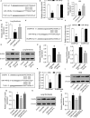
TUG1 positively regulated DUSP6 expression by sponging miR‐145‐5p. A, The putative miR‐145‐5p–binding sequence of TUG1 mRNA. B, Luciferase activity was measured. C and D, Expression of miR‐145‐5p was analysed in CSE‐treated HBE cells and lung fibroblasts transfected with the TUG1 knockdown construct. E, wt or mut miR‐145‐5p target sequences of the 3′UTR from DUSP6. F, Luciferase activity was measured. G‐I, DUSP6 expression was detected by Western blotting in CSE‐treated HBE cells and lung fibroblasts transfected with miR‐145‐5p mimic. J, The putative miR‐145‐5p–binding sequence of TUG1 and DUSP6. K, Luciferase activity was measured. L‐O, The protein expression of DUSP6 was measured in different groups. *P < 0.05 as compared to control group; & P < 0.05 as compared to CSE/shNC group or CSE/miR‐NC group; # P < 0.05 as compared to CSE/shTUG1 group
Figure 5
Figure 5
Overexpression of DUSP6 reverses the inhibition of airway inflammation and fibrosis mediated by HBE cells and lung fibroblasts after TUG1 knockdown. A and B, Expression of DUSP6 was determined in CSE‐treated HBE cells and lung fibroblasts transfected with shTUG1 or DUSP6 alone or co‐transfected with shTUG1 and DUSP6. C, Protein expression levels of α‐SMA and collagen I were measured in lung fibroblasts. D, IL‐6 and TGF‐β1 levels were measured in HBE cells. *P < 0.05 as compared to NC group; & P < 0.05 as compared to CSE/shTUG1 group

"V体育ios版" References

    1. Vestbo J, Hurd SS, Agustí AG, et al. Global strategy for the diagnosis, management, and prevention of chronic obstructive pulmonary disease: GOLD executive summary. Am J Respir Crit Care Med. 2013;187:347‐365. 10.1164/rccm.201204-0596PP. - DOI - PubMed
    1. Tam A, Wadsworth S, Dorscheid D, Man SF, Sin DD. The airway epithelium: more than just a structural barrier. Ther Adv Respir Dis. 2011;5:255‐273. 10.1177/1753465810396539. - DOI - PubMed
    1. Togo S, Holz O, Liu X, et al. Lung fibroblast repair functions in patients with chronic obstructive pulmonary disease are altered by multiple mechanisms. Am J Respir Crit Care Med. 2008;178:248‐260. 10.1164/rccm.200706-929OC. - DOI - PMC - PubMed
    1. Liu Q, Li H, Wang Q, et al. Increased expression of TROP2 in airway basal cells potentially contributes to airway remodeling in chronic obstructive pulmonary disease. Respir Res. 2016;17:159 10.1186/s12931-016-0463-z. - DOI - PMC - PubMed
    1. Shi X, Sun M, Liu H, Yao Y, Song Y. Long non‐coding RNAs: a new frontier in the study of human diseases. Cancer Lett. 2013;339:159‐166. 10.1016/j.canlet.2013.06.013. - "VSports手机版" DOI - PubMed

Publication types

"VSports在线直播" MeSH terms