Skip to main page content
U.S. flag

An official website of the United States government

Dot gov

The . gov means it’s official. Federal government websites often end in VSports app下载. gov or . mil. Before sharing sensitive information, make sure you’re on a federal government site. .

Https

The site is secure. The https:// ensures that you are connecting to the official website and that any information you provide is encrypted and transmitted securely. V体育官网.

. 2019 Sep 3;28(10):2501-2508.e4.
doi: 10.1016/j.celrep.2019.07.107.

The Hippo Pathway Effector TAZ Regulates Ferroptosis in Renal Cell Carcinoma (V体育安卓版)

Affiliations

The Hippo Pathway Effector TAZ Regulates Ferroptosis in Renal Cell Carcinoma

"VSports最新版本" Wen-Hsuan Yang et al. Cell Rep. .

Abstract

Despite recent advances, the poor outcomes in renal cell carcinoma (RCC) suggest novel therapeutics are needed. Ferroptosis is a form of regulated cell death, which may have therapeutic potential toward RCC; however, much remains unknown about the determinants of ferroptosis susceptibility VSports手机版. We found that ferroptosis susceptibility is highly influenced by cell density and confluency. Because cell density regulates the Hippo-YAP/TAZ pathway, we investigated the roles of the Hippo pathway effectors in ferroptosis. TAZ is abundantly expressed in RCC and undergoes density-dependent nuclear or cytosolic translocation. TAZ removal confers ferroptosis resistance, whereas overexpression of TAZS89A sensitizes cells to ferroptosis. Furthermore, TAZ regulates the expression of Epithelial Membrane Protein 1 (EMP1), which, in turn, induces the expression of nicotinamide adenine dinucleotide phosphate (NADPH) Oxidase 4 (NOX4), a renal-enriched reactive oxygen species (ROS)-generating enzyme essential for ferroptosis. These findings reveal that cell density-regulated ferroptosis is mediated by TAZ through the regulation of EMP1-NOX4, suggesting its therapeutic potential for RCC and other TAZ-activated tumors. .

Keywords: EMP1; Epithelial Membrane Protein 1; Hippo pathway; NADPH Oxidase 4; NOX4; TAZ; WW Domain Containing Transcription Regulator 1; cell density; erastin; ferroptosis; renal cell carcinoma V体育安卓版. .


Conflict of interest statement

Declaration of Interests

The authors declare no competing interests.

"VSports在线直播" Figures
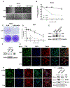
Figure 1.
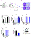
Figure 1.. Cell density regulates the sensitivity of RCC to the erastin-induced ferroptosis
(A) Bright field images of RCC4 cells cultured in low/high densities when treated with the indicated concentrations of erastin. Scale bar, 200 μm. (B) Cell viability assay by CelltiterGlo after RCC4 cells of low/high cell densities treated with indicated concentrations of erastin for 24 h. Data are represented as mean ± SEM from triplicate of one of four independent experiments (***p < 0.001). (C) Crystal violet staining of RCC4 cultured in low/high densities treated with vehicle or 1μM erastin. (D) Cell viability assay by CelltiterGlo after 293T cells grown at low vs. high (2500/10,000 cells per 96 well) cell densities treated with indicated concentrations of erastin. Data are represented as mean ± SEM from triplicate of one of two independent experiments (***p < 0.001). (E) Western blot analysis of YAP and TAZ in renal carcinoma cell lines, RCC4 and 786-O cells, as well as in the breast cancer cell line, MDA-MB-231. Representative data of at least three independent experiments. (F) Western blot analysis of YAP and TAZ in RCC4 cells grown at low vs. high density. Representative data of at least two independent experiments. (G-H) Confocal immunofluorescence images of TAZ in RCC4 cells (G) or RCC PDX, 13-789, tumor cells (H) grown at low vs. high density. DAPI staining of cell nuclei and rhodamine-phalloidin staining for F actin cytoskeleton to mark cell boundaries. Merged images with nuclear stain, DAPI, are also shown. Scale bars, 50 μm. (I) Western blot measuring YAP and TAZ abundance and phospho-TAZ (Ser89) in RCC PDX 13-789 cells of cytosolic/nucleic extractions. Histone H3 serves as a nuclear marker; β-actin serves as a cytosolic marker. See also Figure S1.
Figure 2.
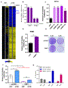
Figure 2.. TAZ regulates sensitivity to erastin-induced ferroptosis
(A-F) Silencing of TAZ reduces erastin-induced ferroptosis. RCC4 cells were treated with siRNA control, siNT, or siRNA targeting TAZ, siTAZ, for two days. (A) Cells were harvested for western blot analysis to check knockdown efficiency. (B) Cell viability was determined by CelltiterGlo after 24 hours of indicated dosage of erastin treatment. Data are represented as mean±SEM, n=3 (*p < 0.05; **p<0.01; ***p < 0.001). (C) Crystal violet staining of RCC4 treated with 1μM erastin for 2 days after incubating with siRNA control, siNT, or silencing of TAZ, siTAZ for 1 day. Representative data from one of two independent studies. (D) RCC4 cells were treated with siRNA control, siNT, or siRNA targeting TAZ, siTAZ for 24 h. Later, cell viability was determined by CelltiterGlo after 24 h treatment of 1μM erastin. Data are represented as mean±SEM, n=3 by comparing to the DMSO controls of each group (*p < 0.05; ***p < 0.001, n.s : not significant). (E-G) Treatment of multiple individual siRNAs targeting TAZ (#1 to #5), siTAZ (pooled), or siRNA control, non-targeting (siNT) for two days in renal carcinoma cells, RCC4 cells (E), or 786O cells (F), or RCC PDX 13-789 cells (G). Cell viability was determined by CelltiterGlo after 24 h treatment of 2.5μM, 1μM and 16μM of erastin, respectively. Data are represented as mean±SEM, n=3 by comparing to the DMSO controls of each group (*p < 0.05; **p<0.01; ***p < 0.001). (H-I) Overexpression of TAZ sensitizes RCC4 cells to erastin-induced ferroptosis. (H) RCC4 cells were transfected with pLX304 control vector or TAZS89A plasmid and then treated with 4μM erastin for 24 hours. Cell viability was then determined by CelltiterGo. Data are represented as mean±SEM, n=3 by comparing to the DMSO controls. (***p < 0.001) (I) Cells were also harvested for western blot analysis of TAZ. See also Figure S2.
Figure 3.
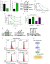
Figure 3.. EMP1 is a direct target gene of TAZ that regulates erastin-induced ferroptosis sensitivity
(A) Gene expression profiles were analyzed by U133A2.0 microarray in RCC4 cells after treating with 1μM erastin for 7 hours following silencing TAZ for two days. (B) RT-qPCR validates that EMP1 is downregulated when TAZ is silenced by siRNAs. Data are represented as mean±SEM, n=3 after normalized to the DMSO controls (mock). (C-D) siRNAs targeting EMP1 reduces erastin-induced ferroptosis. Treatment of siRNA control, non-targeting (siNT), multiple individual siRNAs targeting EMP1 (#1 to #4), or siEMP1 (pooled), for two days in renal carcinoma cells, RCC4 cells (C), or 786O cells (D). Cell viability was determined by CelltiterGlo after 24 hours of 2μM and 1μM erastin, respectively. Data are represented as mean±SEM, n=3 by comparing to the DMSO controls. (E) Overexpression EMP1 sensitizes RCC4 cells to ferroptosis. Crystal violet staining of RCC4 cells that are stably overexpress control vector, pLX304, or pLX304-EMP1-V5 (EMP1) after treating with 3μM erastin for 1 day. (F) Genetic interaction between TAZ and EMP1. After stably overexpressing with EMP1, RCC4 cells were treated with siRNAs targeting TAZ. Later, cell viabilities were determined by CelltiterGlo after 24 hours of 2 μM erastin treatment. Data are represented as mean±SEM, n=3 after normalized to the DMSO controls (*p < 0.05; **p<0.01; ***p < 0.001). (G) EMP1 is the direct target of TAZ. From RCC4 lysates, ChIP-qPCR with TAZ antibody (CST #70148) validates that TAZ binds to EMP1 promoter. ChIP-qPCR of the CTGF promoter serves as a positive control, while Ch14 serves as a negative control. Data from three technical replicates, mean±SEM, from one representative experiment out of four are shown. See also Figure S3.
Figure 4.
Figure 4.. EMP1 regulates ferroptosis through NOX4
(A) siRNA-mediated knockdown of EMP1 gene reduces the mRNA level of NOX4. Data are represented as mean ± SEM from triplicate of one of three independent experiments (*p < 0.05). (B) Overexpression of EMP1 increases NOX4 mRNA level. Data are represented as mean ± SEM from triplicate of one of three independent experiments (***p < 0.001). (C) Higher amount of NOX4 protein correlates with EMP1 overexpression. V5 antibody indicated EMP1-V5 overexpression. The western blots of NOX4 were quantified, normalized to the β-tubulin, expressed relative to the control vector, and showed as below numbers. Representative data from one of at least three independent studies. (D-E) Inhibition of NOX4 blocks erastin-induced ferroptosis. Cell viability was determined by CelltiterGlo after 24 hours of indicated concentrations of erastin with (D) NOX4 inhibitor, 10 μM GKT136901, mean±SEM, n=3, (E) silencing NOX4 by two individual siRNAs. mean±SEM, n=7, Data are represented by comparing to the DMSO controls (**p<0.01; ***p < 0.001). (F) Genetic interaction between EMP1 and NOX4. Cell viabilities were determined by CelltiterGlo after RCC4 cells were treated with EMP1 overexpression for 1 day following 20 μM NOX4 inhibitor, GKT136901 and 8 μM erastin for 1 day. Data are represented as mean±SEM, n=3 after normalized to the DMSO controls. (G-H) Western blot analysis of phospho-p38 (Thr180/Tyr182), total p38, and control β-tubulin upon overexpression of EMP1 (G) or siRNA-mediated silencing of EMP1 (H). (I) Western blot analysis of NOX4 upon overexpression of EMP1 with treatment of p38 inhibitor, SB203580. (J) Inhibition of TAZ, EMP1, or NOX4 abolishes elevated lipid-ROS by erastin treatment. Lipid ROS in RCC4 cells treated with knockdown TAZ or EMP1 as well as inhibition of NOX4 was assessed by flow cytometry using C11-BODIPY. Representative data from one of five independent studies. (K) A schematic model illustrating the erastin-induced ferroptosis regulated by TAZ-EMP1-NOX4 axis. See also Figure S4.

References (V体育官网入口)

    1. Angeli JPF, Schneider M, Proneth B, Tyurina YY, Tyurin VA, Hammond VJ, Herbach N, Aichler M, Walch A, and Eggenhofer E (2014). Inactivation of the ferroptosis regulator Gpx4 triggers acute renal failure in mice. Nature cell biology 16, 1180. - V体育官网 - PMC - PubMed
    1. Borbély G, Szabadkai I, Horváth Z, Markó P, Varga Z, Breza N, Baska F, Vántus T, Huszár M, Geiszt M, et al. (2010). Small-Molecule Inhibitors of NADPH Oxidase 4. Journal of Medicinal Chemistry 53, 6758–6762. - PubMed
    1. Cao JJ, Zhao XM, Wang DL, Chen KH, Sheng X, Li WB, Li MC, Liu WJ, and He J (2014). YAP is overexpressed in clear cell renal cell carcinoma and its knockdown reduces cell proliferation and induces cell cycle arrest and apoptosis. Oncol Rep 32, 1594–1600. - PubMed
    1. Chen PH, Chi JT, and Boyce M (2018). Functional crosstalk among oxidative stress and O-GlcNAc signaling pathways: Abstract. Glycobiology. - PMC (VSports在线直播) - PubMed
    1. Chen PH, Smith TJ, Wu J, Siesser PF, Bisnett BJ, Khan F, Hogue M, Soderblom E, Tang F, Marks JR, et al. (2017). Glycosylation of KEAP1 links nutrient sensing to redox stress signaling. EMBO J 36, 2233–2250. - PMC - PubMed

MeSH terms