Skip to main page content
U.S. flag

An official website of the United States government

Dot gov

The . gov means it’s official. Federal government websites often end in . gov or . mil. Before sharing sensitive information, make sure you’re on a federal government site VSports app下载. .

Https

The site is secure. The https:// ensures that you are connecting to the official website and that any information you provide is encrypted and transmitted securely V体育官网. .

. 2019 Jul 23;5(16):e130621.
doi: 10.1172/jci.insight.130621.

"V体育安卓版" Self-reactive B cells in the GALT are actively curtailed to prevent gut inflammation

Self-reactive B cells in the GALT are actively curtailed to prevent gut inflammation

Ashima Shukla et al. JCI Insight. .

Abstract

Immune homeostasis in the gut associated lymphoid tissues (GALT) is critical to prevent the development of inadvertent pathologies. B cells as the producers of antibodies and cytokines plays an important role in maintaining the GALT homeostasis. However, the mechanism by which B cells specifically direct their responses towards non-self-antigens and become ignorant to self-antigens in the GALT is not known. Therefore, we developed a novel mouse model by expressing Duck Egg Lysozyme (DEL) in gut epithelial cells in presence of HEL reactive B cells. Notably, we observed a transient activation and rapid deletion of self-reactive B cells in Peyers Patches and Mesenteric lymph nodes upon self-antigen exposure. The survival of self-reactive B cells upon exposure to their self-antigen was partially rescued by blocking receptor editing but could be completely rescued by stronger survival signal like ectopic expression of BCL2. Importantly, rescuing the self-reactive B cells promoted production of auto-antibodies and gut inflammation. Mechanistically, we identify a specific activation of TGFβ signaling in self-reactive B cells in the gut and a critical role of this pathway in maintaining peripheral tolerance. Collectively, our studies describe functional consequences and fate of self-reactive B cells in GALT and provide novel mechanistic insights governing self-tolerance of B cells in the gut VSports手机版. .

Keywords: B cells; Immunology; Inflammation; Tolerance. V体育安卓版.


Conflict of interest statement

Conflict of interest: The authors have declared that no conflict of interest exists.

Figures

Figure 1
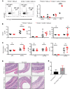
Figure 1. Self-reactive B cells diminish upon gut-associated antigen encounter.
(A) Breeding scheme to obtain Villincre+mDELloxpSWHEL mice and the gain of DEL expression and loss of Thy 1.1 upon Villincre recombination, as measured by flow cytometry. (B) HEL-specific cell population (gated on B220+HEL+) in spleens, pLNs, mLNs, and PPs of Villincre+mDELloxpSWHEL and Villincre+SWHEL mice. (C) B cells frequencies (gated on live cells, B220+) as analyzed by flow cytometry in spleens, pLNs, mLNs, and PPs of Villincre+mDELloxpSWHEL and Villincre+SWHEL mice at the age of 6–8 weeks. Data were analyzed using 2-way ANOVA; the experiment was repeated more than 3 times. (D) Schematic of reconstitution experimental (RVillincre+ and RVillincre+mDELloxp mice). (E) B cell percentages in spleens, pLNs, mLNs, and PPs and HEL-specific cell percentages in spleens, pLNs, mLNs, and PPs and absolute numbers of HEL-specific B cells in PPs isolated from RVillincre+ and RVillincre+mDELloxp mice after 8 weeks after reconstitution. Data are representative of 3 mice per group; the experiment was repeated more than 3 times; and data were analyzed using 2-way ANOVA and grouped multiple t test. ****P < 0.0001, **P < 0.005, *** P < 0.001, * P < 0.05.
Figure 2
Figure 2. The pool of self-reactive B cells is renewed in gut by bone marrow.
Mice were kept on BrDU-containing water for 8 weeks after reconstitution. Newly formed HEL-specific B cell (gated on lymphocytes, B220+, HEL+, anti-BrDU+) frequencies were measured by flow cytometry in spleens, pLNs, mLNs, and PPs of (A) Villincre+SWHEL and Villincre+mDELloxpSWHEL and (B) RVillincre+ and RVillincre+mDELloxp mice. Data were analyzed by 2-way ANOVA and grouped multiple t test. (C) Frequencies of BrDU+ non-HEL-specific cells in spleens, pLNs, mLNs, and PPs and frequencies of BrDU+ HEL-specific transitional stage 1 B cells (T1), marginal zone B cells (MZ), follicular B cells (Fo), and transitional stage 2 B cells (T2) in spleens. (D) Histograms displaying the MFI of eFluor670 to determine proliferation of adoptively transferred HEL-specific B cells (gated on lymphocytes, single cells, eFluor670+, B220+, HEL+) in spleens, pLNs, mLNs, and PPs into Villincre+mDELwt and Villincre+mDELloxp mice. Data are representative of 2 mice per group sacrificed on 1, 3, and 5 days after transfer. Grouped statistical analysis was performed by multiple t test. **P < 0.005.
Figure 3
Figure 3. Self-reactive B cells are activated upon antigen encounter in gut.
HEL-specific cells were isolated from spleens, pLNs, mLNs, and PPs of RVillincre+ and RVillincre+mDELloxp mice. BCR density, as measured by MFI of (A) HEL-binding histograms of HEL-specific cells, with (E) the MFI of HEL binding of HEL-specific cells, and(B) anti-IgM histograms of HEL-specific cells, with (E) the MFI of IgM of HEL-specific cells from 8 mice per group. Activation of HEL-specific cells was measured. (C) CD69 histograms of HEL binding, with (E) the MFI of CD69. (D) CD86 histograms of HEL binding, with (E) the MFI of CD86 in HEL-specific cells from 8 mice per group. (F) Gated on B220+, dot plots of HEL-specific cells (HEL+) and germinal center cells (GL7+) in spleens, pLNs, mLNs, and PPs. (G) GL7+HEL+ cells in spleens, pLNs, mLNs, and PPs of 5 mice per group. Grouped statistical analysis was performed by multiple t test. ****P < 0.0001, **P < 0.005, ***P < 0.001, *P < 0.05.
Figure 4
Figure 4. Self-reactive B cells undergo Bim-mediated apoptosis and Rag-mediated receptor editing.
(A) Gated on B220+HEL+ cells, dot plot displaying gain of Bim expression in HEL-specific cells upon antigen encounter in PPs and graph displaying the percentage of Bim+ HEL-specific cells in spleens, pLNs, mLNs, and PPs from Villincre+SWHEL and Villincre+mDELloxpSWHEL mice. *P = 0.0442, ****P < 0.0001. Data are representative of more than 3 experimental repeats and 7 mice per group. (B) Immunohistochemistry staining of HEL binding (cy3) and Bim (cy5) in PPs from Villincre+SWHEL and Villincre+mDELloxpSWHEL mice (original magnification, ×20). Villincre+ and Villincre+mDELloxp mice were reconstituted with the bone marrow from SWHELRag2KO-transgenic mice (C) The frequency of B220+ cells in the spleens, pLNs, mLNs, and PPs and absolute numbers of HEL-specific B cells from PPs of RVillincre+(Rag2KO) and RVillincre+mDELloxp(Rag2KO) mice. (D) Gated on B220+ cells, graph displaying the frequency of HEL-specific cells in the spleens, pLNs, mLNs, and PPs of RVillincre+(Rag2KO), RVillincre+mDELloxp(Rag2KO), RVillincre+(Rag2KO+μMT), and RVillincre+mDELloxp(Rag2KO+μMT) mice. (E) Absolute numbers of HEL-specific cells in PPs of RVillincre+(Rag2KO+μMT) and RVillincre+mDELloxp(Rag2KO+μMT) mice. (F) Log2 fold change in the numbers of HEL-specific cells from PPs of reconstituted Villincre+ and Villincre+mDELloxp mice by SWHEL, SWHEL(Rag2KO) and SWHEL(Rag2KO+μMT) bone marrow. (G) Activation of HEL-specific cells from spleens, pLNs, mLNs, and PPs of RVillincre+(Rag2KO) and RVillincre+mDELloxp(Rag2KO) mice by HEL binding MFI and MHC-II MFI. ****P < 0.0001, **P < 0.005, *P = 0.0253. Data are represented of 4 mice group; experiments were performed 3 times for Rag2KO rescue. Grouped statistical analysis was performed by multiple t test and 2-way ANOVA in Prism.
Figure 5
Figure 5. Ectopic expression of Bcl2 rescues the survival of self-reactive B cells in gut.
(A) Schematic for experimental design to reconstitute Villincre+ and Villincre+mDELloxp mice with SWHEL-Bcl2–transgenic mice bone marrow. (B) Gated on lymphocyte population, graph displaying the frequency of B220+ cells in spleens, pLNs, mLNs, and PPs of RVillincre+ (Bcl2) and RVillincre+mDELloxp (Bcl2) mice. (C) Gated on B220+ population, graph displaying HEL-binding cell frequencies in the spleens, pLNs, mLNs, and PPs of RVillincre+ (Bcl2) and RVillincre+mDELloxp (Bcl2) mice. MFI of (D) HEL binding, (E) MHC-II, and (F) CD86 on HEL-specific cells in the spleens, pLNs, mLNs, and PPs of RVillincre+ (Bcl2) and RVillincre+mDELloxp (Bcl2) mice. (G) Gated on B220+Hel+ cells, graph displaying the frequency of Bim+ cells. ***P < 0.001, **P < 0.01, ****P < 0.0001, *P < 0.05; n = 9 mice per group. Grouped statistical analysis was performed by multiple t test.
Figure 6
Figure 6. Deregulation of B cell tolerance in gut leads to IBD-like disease.
(A) Flow contour plot showing the IgG1- and IgA-switched HEL-specific cells in the PPs and graph showing the frequency of HEL-specific IgA-switched cells in the spleens, pLNs, mLNs, and PPs of RVillincre+(Bcl2) and RVillincre+mDELloxp (Bcl2) mice. The MFI of HEL-specific anti-IgA, anti-IgG1, and IgG2c (B) in serum and (C) in fecal extracts of RVillincre+(Bcl2) and RVillincre+mDELloxp (Bcl2) mice. (D) H&E staining of small and large intestine showing infiltration and inflammation in RVillincre+mDELloxp (Bcl2) mice (original magnification, ×5 [muscularis]; ×20 [inflammation and villus]). (E) The gut histological scoring of n = 6 and n = 7, respectively, per group of RVillincre+ (Bcl2) and RVillincre+mDELloxp (Bcl2) mice.
Figure 7
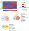
Figure 7. TGF-β signaling is enriched in self-reactive B cells upon antigen encounter in PPs.
(A) Heatmap of supervised hierarchical clustering of differentially expressed genes (DEGs) in self-reactive B cells (HEL+ve) or normal B cells (HEL–ve) in spleens, mLNs, and PPs isolated from Villincre+mDELloxpSWHEL mice. (B) The number of genes significantly (P = 0.01, 1.5-fold) over- or underexpressed in self-reactive B cells from spleen, mLNs, and PPs. Significant DEGs were run on MetaCore pathway analysis. (C) Venn diagram of common or unique molecular pathways significantly (P < 0.05) enriched in non-HEL-specific cells isolated from spleens, mLNs, and PPs. (D) Venn diagram of common or unique molecular pathways enriched in HEL-specific cells isolated from spleens, mLNs, and PPs (P < 0.05). Among the unique pathways identified in C and D in PPs (E) are common or unique molecular pathways and a list of unique molecular pathways enriched in non-HEL and HEL-specific cells in PPs.
Figure 8
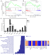
Figure 8. BCL2 induces an IgA immune network in self-reactive B cells.
Gene set enrichment of TGF-β signature genes (GSE7460) (A) consisting of DEGs in HEL-specific cells from PPs versus spleen and (B) consisting of DEGs in HEL-specific PPs versus non-HEL-specific PPs. (C) Fold expression of 9 genes in HEL-specific cells from PPs of RVillincre+ and RVillincre+mDELloxp mice (these genes were picked from TGF-β signature genes; GSE7460). (D) List of pathways enriched in HEL-specific cells isolated from PPs of RVillincre+mDELloxp (Bcl2) mice verus HEL-specific cells from isolated from PPs from Villincre+mDELloxpSWHEL mice. (E) List of genes significantly overexpressed in log2 scale in PPs from RVillincre+mDELloxp (Bcl2) verus HEL-specific cells from isolated from PPs from Villincre+mDELloxpSWHEL mice from intestinal immune network for IgA production (P < 0.05). n = 3 HEL-specific samples, n = 2 for non-HEL-specific samples, and n = 3 for RVillincre+mDELloxp (Bcl2).
Figure 9
Figure 9. Inhibition of TGF-β signaling rescues self-reactive B cells upon antigen encounter in gut.
(A) Histogram and graph displaying TGFβRI expression and MFI of TGFβR1 staining in HEL-specific cells from spleens, pLNs, mLNs, and PPs of RVillincre+ (Bcl2) and RVillincre+mDELloxp (Bcl2) mice. (B) Experimental design schematic for study of the role of TGF-β signaling in self-reactive B cells. Gated on B220+, the left column shows the percentage of CFSE+ (non-HEL-specific cells) and right column shows the percentage of eFLuor670+ cells in spleens, pLNs, mLNs, and PPs. (C) Histogram and graph displaying the p-Smad2/3 expression and MFI of p-Smad2/3 staining, as measured by flow cytometry in DMSO- and TGFβI/II R inhibitor–treated HEL-specific cells from spleen, pLNs, mLNs and PPs Villincre+ and Villincre+mDELloxp mice 72 hours after adoptive transfer. Graph of HEL-specific cells treated with DMSO or TGFβI/II R inhibitor in spleens, pLNs, mLNs, and PPs (D) of CD69 MFI and S1PR1 MFI. The statistical analysis was done by paired t test in Prism. ****P < 0.0001, ***P = 0.0002, **P = 0.0077, *P = 0.0105, n = 4 and experiment was repeated 3 times.

References

    1. Eckburg PB, et al. Diversity of the human intestinal microbial flora. Science. 2005;308(5728):1635–1638. doi: 10.1126/science.1110591. - VSports app下载 - DOI - PMC - PubMed
    1. Macdonald TT, Monteleone G. Immunity, inflammation, and allergy in the gut. Science. 2005;307(5717):1920–1925. doi: 10.1126/science.1106442. - DOI - PubMed
    1. Fasano A. Leaky gut and autoimmune diseases. Clin Rev Allergy Immunol. 2012;42(1):71–78. doi: 10.1007/s12016-011-8291-x. - DOI - PubMed
    1. Macpherson AJ, Uhr T. Induction of protective IgA by intestinal dendritic cells carrying commensal bacteria. Science. 2004;303(5664):1662–1665. doi: 10.1126/science.1091334. - DOI - PubMed
    1. Artis D. Epithelial-cell recognition of commensal bacteria and maintenance of immune homeostasis in the gut. Nat Rev Immunol. 2008;8(6):411–420. doi: 10.1038/nri2316. - DOI (V体育官网入口) - PubMed

Publication types

MeSH terms