Skip to main page content
U.S. flag

An official website of the United States government

Dot gov

The . gov means it’s official. Federal government websites often end in . gov or . mil. Before sharing sensitive information, make sure you’re on a federal government site. VSports app下载.

Https

The site is secure. The https:// ensures that you are connecting to the official website and that any information you provide is encrypted and transmitted securely V体育官网. .

. 2020 Feb;27(2):758-772.
doi: 10.1038/s41418-019-0385-7. Epub 2019 Jul 8.

FBXO45-MYCBP2 regulates mitotic cell fate by targeting FBXW7 for degradation

Affiliations

FBXO45-MYCBP2 regulates mitotic cell fate by targeting FBXW7 for degradation

Kai T Richter et al. Cell Death Differ. 2020 Feb.

Abstract

Cell fate decision upon prolonged mitotic arrest induced by microtubule-targeting agents depends on the activity of the tumor suppressor and F-box protein FBXW7. FBXW7 promotes mitotic cell death and prevents premature escape from mitosis through mitotic slippage. Mitotic slippage is a process that can cause chemoresistance and tumor relapse. Therefore, understanding the mechanisms that regulate the balance between mitotic cell death and mitotic slippage is an important task. Here we report that FBXW7 protein levels markedly decline during extended mitotic arrest. FBXO45 binds to a conserved acidic N-terminal motif of FBXW7 specifically under a prolonged delay in mitosis, leading to ubiquitylation and subsequent proteasomal degradation of FBXW7 by the FBXO45-MYCBP2 E3 ubiquitin ligase. Moreover, we find that FBXO45-MYCBP2 counteracts FBXW7 in that it promotes mitotic slippage and prevents cell death in mitosis. Targeting this interaction represents a promising strategy to prevent chemotherapy resistance VSports手机版. .


Conflict of interest statement

The authors declare that they have no conflict of interest.

Figures

Fig. 1
Fig. 1
FBXW7 protein levels are reduced under prolonged mitotic arrest. a FBXW7α protein levels decrease during prolonged mitotic arrest. HeLa cells were treated with 250 ng/ml nocodazole for 2 h. Mitotic cells were collected by mitotic shake-off (time point 0) and further incubated with 250 ng/ml nocodazole. The cells were harvested at different time points by a mitotic shake-off. (Top) Cell extracts were prepared and analyzed by western blotting. (Bottom, left) Relative FBXW7α signal intensities were quantified. Average signal intensities and standard deviations from n = 4 experiments were calculated. Statistical significance (difference to time point 0) was analyzed by a two-tailed, unpaired t-test with unequal variance. *p < 0.05; **p < 0.01. (Bottom, right) Mitotic arrest was confirmed for two time points (0 h and 8 h) by FACS analysis. Asynchronous (asy.) cells served as a control. b Decrease in FBXW7α protein levels during prolonged mitotic arrest depends on proteasomal activity. (Top) HeLa cells were treated with 250 ng/ml nocodazole for 2 h. Mitotic cells were collected by mitotic shake-off (time point 0) and further incubated with 250 ng/ml nocodazole. MG132 was added in order to inhibit the proteasome. Cells were harvested at different time points by a mitotic shake-off. Cell extracts were prepared and analyzed by western blotting. (Bottom) Relative FBXW7α, Cyclin B1, and MCL-1 signal intensities were quantified. Average signal intensities and standard deviations from n = 4 experiments were calculated. Statistical significance (difference to time point 0) was analyzed by a two-tailed, unpaired t-test with unequal variance. *p < 0.05; **p < 0.01; n.s. not significant. c Network of known and putative FBXW7 interaction partners. Immunoprecipitation of Flag-FBXW7α from HEK-293T cells treated with the proteasomal inhibitor MG132 for 4 h and subsequent mass spectrometry analysis identified several known SCF components and the SCF regulators NEDD8 and ARIH1 as well as FBXW7 substrates. Moreover, FBXO45 and MYCBP2 were identified as FBXW7 interaction partners and could act as putative FBXW7 regulators
Fig. 2
Fig. 2
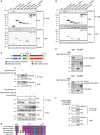
FBXO45-MYCBP2 binds to a conserved N-terminal motif of FBXW7α a, b FBXO45 and FBXW7α specifically interact in reciprocal immunoprecipitations. Several Flag-tagged F-box and DCAF proteins were overexpressed in HEK-293T cells for 24 h. Cell extracts were used for an immunoprecipitation directed against the Flag tag. c (Top) Schematic representation of Δ106-126 deletion in FBXW7α used in immunoprecipitation experiments. Positions of N-terminal domain (blue), dimerization domain (green), F-box domain (yellow), and WD40 domain (red) are indicated. (Bottom) FBXO45 and MYCBP2 interact with the N-terminal domain of FBXW7α. Flag-FBXW7α FL or Flag-FBXW7αΔ106-126 were overexpressed in HEK-293T cells for 24 h. Cell extracts were used for α-Flag immunoprecipitations. d Sequence alignment of amino acid residues 106–126 from human FBXW7α with homologs from Macaca mulatta, Mus musculus, Xenopus laevis, and Danio rerio. e Either MBP alone or MBP-FBXW7α-N167 were incubated with in vitro translated and [35S]-methionine containing FBXO45 or MYCBP2(1951-2950). MBP was pulled down with amylose beads. Pull-down samples were analyzed by SDS-PAGE and Colloidal Coomassie staining. In vitro translated proteins were detected by autoradiography. f FBXW7α forms a complex with MYCBP2 and FBXO45. Flag-MYCBP2(1951-2950) and HA-FBXO45 were co-expressed in HEK-293T cells for 24 h. Cell extracts were used for an immunoprecipitation with α-Flag agarose beads. Immunoprecipitated proteins were eluted by competition with Flag peptide. Eluates were used for a second immunoprecipitation directed against the HA tag. Immunoprecipitates obtained after sequential immunoprecipitation were analyzed by western blotting
Fig. 3
Fig. 3
FBXO45 and FBXW7α interact during mitotic arrest. a FBXW7α is a nuclear protein, whereas FBXO45 is localized in the cytoplasm. Cytoplasmic and nuclear extracts were prepared from HeLa cells and analyzed by western blotting. b FBXO45 and FBXW7α interact predominantly in mitosis. Flag-FBXO45 was overexpressed in HeLa cells. Asynchronous or mitotic cells that had been treated with nocodazole for 17 h were harvested. Cytoplasmic extracts were used for an immunoprecipitation directed against the Flag tag
Fig. 4
Fig. 4
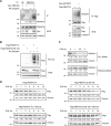
FBXO45 and MYCBP2 downregulate FBXW7α protein levels during mitotic arrest. a HeLa cells were transfected with 30 nM FBXO45 or FBXW7 siRNAs for 72 h. GL2 siRNA was used as a control. The cells were arrested in mitosis by nocodazole (noco.) treatment for 17 h and collected by a mitotic shake-off. Mitotic cells were compared with an asynchronous (asy.) cell population. (Top) Cell extracts were analyzed by western blotting. Crossreacting band in FBXO45 immunoblot is marked with an asterisk. As the samples were analyzed in three western blots, three vinculin immunoblots are shown as loading controls. (Bottom) Quantification of relative FBXW7α and FBXO45 signal intensities are shown. Relative FBXW7α and FBXO45 signals in the GL2 controls were set to 1. Average signal intensities and standard deviations from n = 4 experiments were calculated. Statistical significance was analyzed by a two-tailed, unpaired t-test with unequal variance. *p < 0.05; n.s. not significant. b Expression of Flag-MYCBP2(1951-2950) was induced in a stable U2OS Flp-In-T-Rex cell line by doxycycline treatment. The cells were arrested in mitosis by nocodazole treatment (noco.). Alternatively, they were treated with thymidine (thym.) or left untreated (asy.). U2OS cells that do not express the MYCBP2 fragment served as controls. (Top) Cell extracts were analyzed by western blotting. (Bottom) Quantification of relative FBXW7α signal intensity was performed as described in (A)
Fig. 5
Fig. 5
FBXO45 and MYCBP2 promote the ubiquitylation and destabilization of FBXW7α. a Myc-FBXO45 and HA-Ubiquitin or Myc-MYCBP2 and Flag-FBXW7α were overexpressed in HEK-293T cells for 48 h. Cells were arrested in mitosis by nocodazole treatment for 17 h. At 4 h before harvesting, cells were treated with MG132. Cell extracts were used for immunoprecipitations directed against endogenous FBXW7α or against the Flag tag. b Flag-FBXW7α FL or Flag-FBXW7αΔ106-126 were co-expressed with His-Ubiquitin in HEK-293T cells. The cells were arrested in mitosis by nocodazole treatment. At 4 h before harvesting, cells were treated with MG132. Cell extracts were prepared and α-His pull-downs were performed. c HeLa cells were transfected with 30 nM of GL2 or FBXO45 siRNA for 72 h. At 17 h before harvesting, the cells were treated with nocodazole. Mitotic cells were collected by mitotic shake-off and further incubated with nocodazole and cycloheximide (CHX). The cells were harvested at different time points after the addition of cycloheximide by mitotic shake-off. Cell extracts were analyzed by western blotting. Samples were analyzed on a single gel. Relative FBXW7α signal intensities were quantified. d Flag-FBXW7α FL or Flag-FBXW7αΔ106-126 were overexpressed in U2OS Flp-In-T-Rex cells by treatment with doxycycline. Cells were arrested in mitosis by nocodazole treatment. Cycloheximide (CHX) was added and cells were harvested at different time points after the addition of cycloheximide. MG132 was added in order to inhibit the proteasome. Cell extracts were analyzed by western blotting. Relative Flag signal intensities were quantified
Fig. 6
Fig. 6
FBXO45 and MYCBP2 cause mitotic slippage. a U2OS cells were treated with 250 ng/mL nocodazole. At 4 h after nocodazole addition, cells were analyzed by live-cell imaging. Representative images from live-cell imaging are shown. Time point 0 marks induction of mitotic cell death or mitotic slippage. Scale bars: 20 µm. b U2OS cells were transfected with 30 nM of GL2 or different FBXO45 siRNAs for 48 h and further incubated with 250 ng/mL nocodazole. Percentages of cells undergoing mitotic cell death were quantified. Cells from n = 3 independent experiments were analyzed. In each experiment, 50 cells were quantified. c U2OS cells were cotransfected with 40 nM of GL2 or FBXO45 siRNA and an empty vector (EV) or an siRNA-resistant version of Flag-FBXO45. At 48 h after transfection, the cells were treated with nocodazole and mitotic cell fate was analyzed by live-cell imaging. Cells from n = 4 independent experiments were quantified. d Expression of Flag-MYCBP2(1951-2950) was induced in U2OS cells by treatment with doxycycline for 48 h. Cells were arrested in mitosis by treatment with nocodazole and mitotic cell fate was analyzed by live-cell imaging. Cells from n = 3 independent experiments were analyzed. e U2OS cells were transfected with 30 nM of GL2, FBXO45, or FBXW7 siRNA or cotransfected with FBXO45 and FBXW7 siRNA. At 48 h after transfection, the cells were treated with nocodazole and mitotic cell fate was analyzed by live-cell imaging. Cells from n = 3 independent experiments were quantified. For statistical analysis in be, significance was analyzed by a two-tailed, unpaired t-test. *p < 0.05, **p < 0.01, n.s. not significant

VSports - References

    1. Hershko A, Ciechanover A. The ubiquitin system. Annu Rev Biochem. 1998;67:425–79. - PubMed
    1. Davis RJ, Welcker M, Clurman BE. Tumor suppression by the Fbw7 ubiquitin ligase: mechanisms and opportunities. Cancer Cell. 2014;26:455–64. - "V体育官网入口" PMC - PubMed
    1. Nateri AS, Riera-Sans L, Da Costa C, Behrens A. The ubiquitin ligase SCFFbw7 antagonizes apoptotic JNK signaling. Science. 2004;303:1374–8. - PubMed
    1. Wei W, Jin J, Schlisio S, Harper JW, Kaelin WG., Jr The v-Jun point mutation allows c-Jun to escape GSK3-dependent recognition and destruction by the Fbw7 ubiquitin ligase. Cancer Cell. 2005;8:25–33. - PubMed
    1. Welcker M, Orian A, Jin J, Grim JE, Harper JW, Eisenman RN, et al. The Fbw7 tumor suppressor regulates glycogen synthase kinase 3 phosphorylation-dependent c-Myc protein degradation. Proc Natl Acad Sci USA. 2004;101:9085–90. - V体育ios版 - PMC - PubMed

Publication types

MeSH terms

Substances