Skip to main page content
U.S. flag

An official website of the United States government

Dot gov

The VSports app下载. gov means it’s official. Federal government websites often end in . gov or . mil. Before sharing sensitive information, make sure you’re on a federal government site. .

Https

The site is secure. The https:// ensures that you are connecting to the official website and that any information you provide is encrypted and transmitted securely V体育官网. .

. 2019 Aug 21;87(9):e00872-18.
doi: 10.1128/IAI.00872-18. Print 2019 Sep.

Salmonella enterica Effectors SifA, SpvB, SseF, SseJ, and SteA Contribute to Type III Secretion System 1-Independent Inflammation in a Streptomycin-Pretreated Mouse Model of Colitis

Affiliations

Salmonella enterica Effectors SifA, SpvB, SseF, SseJ, and SteA Contribute to Type III Secretion System 1-Independent Inflammation in a Streptomycin-Pretreated Mouse Model of Colitis

Shigeki Matsuda et al. Infect Immun. .

Abstract

Salmonella enterica serovar Typhimurium (S. Typhimurium) induces inflammatory changes in the ceca of streptomycin-pretreated mice. In this mouse model of colitis, the type III secretion system 1 (T3SS-1) has been shown to induce rapid inflammatory change in the cecum at early points, 10 to 24 h after infection. Five proteins, SipA, SopA, SopB, SopD, and SopE2, have been identified as effectors involved in eliciting intestinal inflammation within this time range. In contrast, a T3SS-1-deficient strain was shown to exhibit inflammatory changes in the cecum at 72 to 120 h postinfection. However, the effectors eliciting T3SS-1-independent inflammation remain to be clarified. In this study, we focused on two T3SS-2 phenotypes, macrophage proliferation and cytotoxicity, to identify the T3SS-2 effectors involved in T3SS-1-independent inflammation. We identified a mutant strain that could not induce cytotoxicity in a macrophage-like cell line and that reduced intestinal inflammation in streptomycin-pretreated mice. We also identified five T3SS-2 effectors, SifA, SpvB, SseF, SseJ, and SteA, associated with T3SS-1-independent macrophage cytotoxicity. We then constructed a strain lacking T3SS-1 and all the five T3SS-2 effectors, termed T1S5 VSports手机版. The S. Typhimurium T1S5 strain significantly reduced cytotoxicity in macrophages in the same manner as a mutant invA spiB strain (T1T2). Finally, the T1S5 strain elicited no inflammatory changes in the ceca of streptomycin-pretreated mice. We conclude that these five T3SS-2 effectors contribute to T3SS-1-independent inflammation. .

Keywords: Salmonella; cytotoxicity; inflammation; type III effectors. V体育安卓版.


Figures

FIG 1
FIG 1
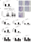
The proliferation of S. Typhimurium in macrophages is not related to intestinal inflammation in streptomycin-pretreated mice. (A) Proliferation rates of the indicated S. Typhimurium mutant strains in RAW cells. The proliferation rate of the S. Typhimurium invA (T1) strain in RAW cells was taken as 100. (B and C) Histopathology of the murine cecum 5 days after infection with the indicated S. Typhimurium mutant strains. Representative images of hematoxylin and eosin-stained cecal sections are shown. Scale bars, 100 μm (B). Histopathology scores were determined by blinded examination of cecal sections. Each bar represents an individual mouse (C). (D) Transcript levels of Ifng, Il6, Tnfa, Kc, and Mip2 in the ceca of mice infected with the indicated S. Typhimurium strains at 5 days after infection measured by qPCR. (E) Numbers of bacteria recovered from the ceca, the mesenteric lymph nodes, and the spleens of mice 5 days after infection. A sample t test (A) and the two-tailed Mann-Whitney U test (B, D, and E) were performed for statistical analysis. Individual data are shown as a scatter plot, and bars indicate the means. Asterisks indicate differences that were statistically significant (P < 0.05). NS, not significant.
FIG 2
FIG 2
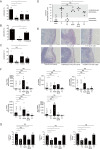
The S. Typhimurium strains, T1SpvB, T1SifA, and T1Fl, that reduce the cytotoxicity of macrophages elicit T3SS-1-independent inflammation in mice. (A to C) LDH release from RAW cells infected with the indicated S. Typhimurium mutant strains at 20 h after infection. (D and E) Histopathology of the murine cecum 5 days after infection with the indicated S. Typhimurium mutant strains. The histopathology score was determined by the blinded examination of cecal sections. Each plot represents an individual mouse (D). Representative images of hematoxylin and eosin-stained cecal sections are shown. Scale bars, 100 μm (E). (F) Transcript levels of Ifng, Il6, Tnfa, Kc, and Mip2 in the ceca of mice infected with the indicated S. Typhimurium strains at 5 days after infection, as measured by qPCR. (G) Numbers of bacteria recovered from the ceca, mesenteric lymph nodes and spleen of mice 5 days after infection. The combined histopathological score, the expression levels of genes, and bacterial numbers in the tissue of mice infected with invA (T1) or invA spiB (T1T2) were determined as shown in Fig. 1B, D, and E, respectively. The two-tailed Student t test (A to C) and the Mann-Whitney U test (E to G) were performed for statistical analysis. Individual data are shown as a scatter plot, and bars indicate the means. Asterisks indicate differences that were statistically significant (P < 0.05). NS, not significant.
FIG 3
FIG 3
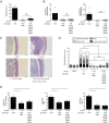
Effect of different combinations of the sifA, spvB, fliC, and fljB mutations on T3SS-1-independent inflammation in mice. (A) LDH release from RAW cells infected with the indicated S. Typhimurium mutant strains at 20 h after infection. (B) The levels of Tnfa and Mip2 in the ceca of mice infected with the indicated S. Typhimurium strains at 5 days after infection. (C and D) Histopathology of the murine cecum 5 days after infection with the indicated S. Typhimurium mutant strains. Representative images of hematoxylin and eosin-stained cecal sections are shown. Scale bars, 100 μm (C). The histopathology score was determined by blinded examination of the cecal sections. Each bar represents an individual mouse (D). A two-tailed Student t test (A) and the Mann-Whitney U test (B and D) were performed for statistical analysis. Individual data are shown as a scatter plot, and bars indicate the means. Asterisks indicate differences that were statistically significant (P < 0.05). NS, not significant.
FIG 4
FIG 4
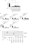
Screening of 29 T3SS-2 effectors for cytotoxicity of RAW cells. (A) LDH release from RAW cells infected with the indicated S. Typhimurium mutant strains at 20 h after infection. The cytotoxicities of RAW cells infected with the invA ΔsifA, invA ΔspvB, or invA ΔsseL strain were determined as shown in Fig. 1A, 2A, or 2C. Asterisks indicate that the cytotoxicities of RAW cells infected with the T3SS-1-deficient and T3SS-2 effector mutants (invA Δeffector) were significantly lower than those of cells infected with the T3SS-1-deficient strain (invA). (B) LDH release from RAW cells infected with the indicated S. Typhimurium mutant strains at 20 h after infection. NT, not tested. (C) Numbers of bacteria recovered from RAW cells infected with the indicated S. Typhimurium mutant strains at 2 and 20 h after infection. A two-tailed Student t test (A to C) was performed for statistical analysis. Individual data are shown as a scatter plot, and bars indicate the means. Asterisks indicate differences that were statistically significant (P < 0.05). NS, not significant.
FIG 5
FIG 5
Effect of different combinations of the sifA, spvB, sseF, sseJ, and steA mutations on T3SS-1-independent inflammation in mice (A) LDH release from RAW cells infected with the indicated S. Typhimurium mutant strains at 20 h after infection. (B) Levels of Tnfa and Mip2 in the ceca of mice infected with the indicated S. Typhimurium strains at 5 days after infection. (C and D) Histopathology of the murine cecum 5 days after infection with the indicated S. Typhimurium mutant strains. Representative images of hematoxylin and eosin-stained cecal sections are shown. Scale bars, 100 μm. (C) Histopathological scoring of cecal sections. (D) Each bar represents an individual mouse. (E) Numbers of bacteria recovered from colon contents, mesenteric lymph nodes (MLN), and spleens of mice. A two-tailed Student t test (A) and Mann-Whitney U test (B, D, and E) were performed for statistical analysis. Individual data are shown as a scatter plot, and bars indicate the means (A, B, and E). Asterisks indicate differences that were statistically significant (P < 0.05). NS, not significant.
FIG 6
FIG 6
Effect of deletions of four of the five effectors, sifA, spvB, sseF, sseJ, and steA, on T3SS-1-independent inflammation in mice. (A) LDH release from RAW cells infected with the indicated S. Typhimurium mutant strains at 20 h after infection. (B) Transcript levels of Ifng, Il6, Tnfa, Kc, and Mip2 in the ceca of mice infected with the indicated S. Typhimurium strains at 5 days after infection, as measured by qPCR. (C) Combined histopathological scoring of cecal sections. A two-tailed Student t test (A) and the Mann-Whitney U test (B and C) were performed for statistical analysis. Individual data are shown as a scatter plot, and bars indicate the means (A and B). Each plot represents an individual mouse (C). Asterisks indicate differences that were statistically significant (P < 0.05). NS, not significant.
FIG 7
FIG 7
Effect of different combinations of three of the four mutations—spvB, sseF, sseJ, and steA—in addition to the sifA mutation on T3SS-1-independent inflammation in mice. (A) Levels of Tnfa and Mip2 in the ceca of mice infected with the indicated S. Typhimurium strains at 5 days after infection, as measured by qPCR. Individual data are shown as a scatter plot, and bars indicate the means. (B) Combined histopathological scoring of cecal sections. Each bar represents an individual mouse. A two-tailed Mann-Whitney U test was performed for statistical analysis. Asterisks indicate differences that were statistically significant (P < 0.05). NS, not significant.

"V体育安卓版" References

    1. Tsolis RM, Adams LG, Ficht TA, Bäumler AJ. 1999. Contribution of Salmonella typhimurium virulence factors to diarrheal disease in calves. Infect Immun 67:4879–4885. - PMC - PubMed
    1. Coombes BK, Coburn BA, Potter AA, Gomis S, Mirakhur K, Li Y, Finlay BB. 2005. Analysis of the contribution of Salmonella pathogenicity islands 1 and 2 to enteric disease progression using a novel bovine ileal loop model and a murine model of infectious enterocolitis. Infect Immun 73:7161–7169. doi:10.1128/IAI.73.11.7161-7169.2005. - DOI - PMC - PubMed
    1. Hapfelmeier S, Müller AJ, Stecher B, Kaiser P, Barthel M, Endt K, Eberhard M, Robbiani R, Jacobi CA, Heikenwalder M, Kirschning C, Jung S, Stallmach T, Kremer M, Hardt WD. 2008. Microbe sampling by mucosal dendritic cells is a discrete, MyD88-independent step in ΔinvG S. Typhimurium colitis. J Exp Med 205:437–450. doi:10.1084/jem.20070633. - DOI - PMC - PubMed
    1. Zhang S, Santos RL, Tsolis RM, Stender S, Hardt WD, Bäumler AJ, Adams LG. 2002. The Salmonella enterica serotype Typhimurium effector proteins SipA, SopA, SopB, SopD, and SopE2 act in concert to induce diarrhea in calves. Infect Immun 70:3843–3855. doi:10.1128/iai.70.7.3843-3855.2002. - V体育2025版 - DOI - PMC - PubMed
    1. Marijke Keestra A, Winter MG, Klein-Douwel D, Xavier MN, Winter SE, Kim A, Tsolis RM, Bäumler AJ. 2011. A Salmonella virulence factor activates the NOD1/NOD2 signaling pathway. mBio 2:e00266-11. doi:10.1128/mBio.00266-11. - DOI - PMC - PubMed

Publication types (VSports在线直播)

MeSH terms (V体育ios版)