Skip to main page content
U.S. flag

An official website of the United States government

Dot gov

The . gov means it’s official. Federal government websites often end in . gov or . mil. Before sharing sensitive information, make sure you’re on a federal government site VSports app下载. .

Https

The site is secure. The https:// ensures that you are connecting to the official website and that any information you provide is encrypted and transmitted securely. V体育官网.

. 2019 Jun 19;10(7):485.
doi: 10.1038/s41419-019-1723-x.

"VSports在线直播" Docosahexaenoic acid inhibits both NLRP3 inflammasome assembly and JNK-mediated mature IL-1β secretion in 5-fluorouracil-treated MDSC: implication in cancer treatment

Affiliations

Docosahexaenoic acid inhibits both NLRP3 inflammasome assembly and JNK-mediated mature IL-1β secretion in 5-fluorouracil-treated MDSC: implication in cancer treatment

Adélie Dumont et al. Cell Death Dis. .

Abstract

Limitation of 5-fluorouracil (5-FU) anticancer efficacy is due to IL-1β secretion by myeloid-derived suppressor cells (MDSC), according to a previous pre-clinical report VSports手机版. Release of mature IL-1β is a consequence of 5-FU-mediated NLRP3 activation and subsequent caspase-1 activity in MDSC. IL-1β sustains tumor growth recovery in 5-FU-treated mice. Docosahexaenoic acid (DHA) belongs to omega-3 fatty acid family and harbors both anticancer and anti-inflammatory properties, which could improve 5-FU chemotherapy. Here, we demonstrate that DHA inhibits 5-FU-induced IL-1β secretion and caspase-1 activity in a MDSC cell line (MSC-2). Accordingly, we showed that DHA-enriched diet reduces circulating IL-1β concentration and tumor recurrence in 5-FU-treated tumor-bearing mice. Treatment with 5-FU led to JNK activation through ROS production in MDSC. JNK inhibitor SP600125 as well as DHA-mediated JNK inactivation decreased IL-1β secretion. The repression of 5-FU-induced caspase-1 activity by DHA supplementation is partially due to β-arrestin-2-dependent inhibition of NLRP3 inflammasome activity but was independent of JNK pathway. Interestingly, we showed that DHA, through β-arrestin-2-mediated inhibition of JNK pathway, reduces V5-tagged mature IL-1β release induced by 5-FU, in MDSC stably overexpressing a V5-tagged mature IL-1β form. Finally, we found a negative correlation between DHA content in plasma and the induction of caspase-1 activity in HLA-DR- CD33+ CD15+ MDSC of patients treated with 5-FU-based chemotherapy, strongly suggesting that our data are clinical relevant. Together, these data provide new insights on the regulation of IL-1β secretion by DHA and on its potential benefit in 5-FU-based chemotherapy. .


Conflict of interest statement

The authors declare that they have no conflict of interest.

Figures
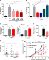
Fig. 1
Fig. 1. IL-1β secretion is reduced by DHA in 5-FU-treated MDSC.
a MSC-2 were treated by 5-FU (1 µM) for 24 h ±DHA (20 to 60 µM) and cell culture media were collected for analysis of IL-1β concentration by ELISA. Error bars represent mean ± SD from four independent experiments. b Analysis of IL-1β secretion by ELISA in MSC-2 treated with 5-FU (1 µM) for 24 h ±DHA, eicosapentaenoic (EPA), oleic (OA), and linoleic (LA) acids at 60 µM. Error bars represent mean ± SD from 3 to 6 independent experiments. c Analysis of IL-1β mRNA expression, by RT-qPCR, in MSC-2 treated for 12 and 24 h with 5-FU ±DHA (60 µM). Error bars represent mean ± SD from three independent experiments. d Expression of IL-1β mRNA, by RT-qPCR, in MSC-2 treated for 12 h with 5-FU ±Lipopolysaccharide (LPS). Error bars represent mean ± SD from three independent experiments. e Determination of plasma IL-1β content in EL4 tumor-bearing mice fed control (ctrl) or DHA-enriched diet, initiated 7 days prior to a single intraperitoneal injection of 5-FU (50 mg/kg). Plasma was collected 48 h after the injection of 5-FU. f Monitoring of tumor growth in mice fed a ctrl diet (n = 8, black) or DHA-enriched diet (n = 9, purple) and in mice treated with a single 5-FU injection (50 mg/kg; blue arrow) and fed a ctrl diet (n = 9, gray) or DHA-enriched diet (n = 9, red). Error bars represent mean ± SEM. *p < 0.05; **p < 0.01; ***p < 0.001; ****p < 0.0001; ns nonsignificant
Fig. 2
Fig. 2. DHA treatment decreases 5-FU-induced JNK activation in MDSC.
a Representative immunoblotting (n = 3) showing expression of phospho-JNK (p-JNK) and total JNK in MSC-2 treated with 5-FU (1 µM) for the indicated times. Quantification of p-JNK and total JNK bands was determined by densitometry. Data are expressed as ratio between p-JNK and total JNK for the different time points and compared to time point (0). b Analysis of IL-1β secretion, by ELISA, in MSC-2 treated with 5-FU (1 µM) ±inhibitor of JNK (SP600125; SP) for 24 h. Error bars represent mean ± SD of three independent experiments. c Representative immunoblotting (n = 4) with anti-IL-1β and anti-β-actin antibodies showing expression of pro-IL-1β in cell lysate and mature IL-1β in supernatant (SN) of MSC-2 treated as in b. d Representative immunoblotting (n = 4) showing phospho-JNK (p-JNK) and total JNK in MSC-2 treated with 5-FU (1 µM) for 12 h ±DHA (20–60 µM) and oleic acid (OA; 60 µM). Relative ratio between p-JNK and total JNK was analyzed by densitometry and compared to the nontreated sample (ctrl). e JNK phosphorylation was assessed by flow cytometry in MDSC (CD11b+ Gr-1+) obtained from spleen of EL4 tumor-bearing mice. Mice were fed a control (ctrl) or DHA-enriched diet initiated 7 days prior to a single intraperitoneal injection of 5-FU (50 mg/kg). MDSC were collected and labeled 24 h after the single 5-FU injection. Error bars represent mean ± SEM. f Representative immunoblotting (n = 3) showing expression of phospho-JNK and total JNK in 5-FU-treated MSC-2 and co-treated with bafilomycin A (Baf.), chloroquin (CQ), N-acetyl-cysteine (NAC), Tempol (Tpol), and inhibitor of pan-caspases (ZVAD) for 12 h. Ratio between p-JNK and total JNK was determined by densitometry and compared to the nontreated sample (ctrl). g Analysis of ROS content. MSC-2 were treated for 8 h with 5-FU (1 µM) ±DHA (60 µM) or anti-oxidant N-acetyl-cysteine (NAC). Cells were loaded with CM-H2DCFDA and analyzed by flow cytometry to determine intracellular ROS content. Error bars represent mean ± SD from three independent experiments. h IL-1β secretion by ELISA in MSC-2 treated with 5-FU (1 µM) ±anti-oxidants N-acetyl-cysteine (NAC) and Tempol (Tpol) for 24 h. Error bars represent mean ± SD from three independent experiments. *p < 0.05; **p < 0.01; ***p < 0.001; ****p < 0.0001; ns nonsignificant
Fig. 3
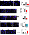
Fig. 3. Inhibition of NLRP3 inflammasome assembly and activation by DHA.
a Analysis of ASC speck formation. MSC-2 were treated for 12 h with 5-FU (1 µM) ±DHA (60 µM) and stained with anti-ASC antibody and DAPI. Arrows show ASC specks. Bar graphs are the mean ± SD of ASC speck-positive cells from three independent replicates. b, c In situ Proximity Ligation Assay for analysis NLRP3 and caspase-1 b or NLRP3 and ASC c interaction. MSC-2 were treated for 12 h with 5-FU (1 µM) ±DHA (60 µM). In one experiment, cells (80–100) were analyzed for a positive PLA signal (at least one red dot in a cell). Error bars represent mean ± SD of at least three independent experiments. d Analysis of ASC speck formation in MSC-2 treated with 5-FU in combination with JNK inhibitor. Cells were exposed to 5-FU (1 µM) ±SP600125 (4 µM; SP) for 12 h and stained with anti-ASC antibody and DAPI. Arrows show ASC specks. Bar graphs are the mean ± SD of ASC speck-positive cells from two independent experiments. e Study NLRP3 and β-arrestin-2 interaction by in situ proximity ligation assay. MSC-2 were treated for 12 h with 5-FU (1 µM) ±DHA (60 µM). As previously, cells were analyzed for a positive-PLA signal (at least one red dot in a cell). Error bars represent mean ± SD of at least three independent experiments. **p < 0.01; ***p < 0.001; ****p < 0.0001; ns nonsignificant. Scale bar = 10 µm
Fig. 4
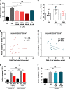
Fig. 4. Repression of caspase-1 activity in 5-FU-treated MDSC mediated by DHA exposure.
a Analysis of 5-FU-induced caspase-1 activity by FLICA in MSC-2 treated with 5-FU (1 µM) for 12 h ±DHA (20–60 µM) or oleic acid (OA). Error bars represent mean ± SD from three independent experiments. b Measure of caspase-1 activity by FLICA in MDSC (CD11b+ Gr-1+) purified 48 h after a single intraperitoneal injection of 5-FU (50 mg/kg) to EL4 tumor-bearing mice fed control (ctrl) or DHA-enriched diet. Error bars represent mean ± SEM. c, d Correlation between plasma DHA content before 5-FU therapy and ratio of caspase-1 activity (after (D1) and before (D0) 5-FU chemotherapy) in HLA-DR CD33+ CD14+ cells c or in HLA-DR CD33+ CD15+ cells d (n = 26 patients). e Effect of DHA on 5-FU induced caspase-1 activity in β-arrestin-2-silenced MDSC. MSC-2 with stable knockdown of β-arrestin-2 was obtained by transduction with β-arrestin-2 (ARRB2) shRNA lentiviral vector and compared to ctrl shRNA vector. Cells were treated with 5-FU (1 µM) ±DHA (60 µM) for 12 h and harvested for active caspase-1 analysis by FLICA. Error bars represent mean ± SD from three independent experiments. f Caspase-1 activity analyzed by FLICA in 5-FU-treated MSC-2 for 12 h ±JNK inhibitor SP600125 (4 and 10 µM). Error bars represent mean ± SD from four independent experiments. *p < 0.05; **p < 0.01; ***p < 0.001; ****p < 0.0001; ns nonsignificant
Fig. 5
Fig. 5. DHA decreases 5-FU-mediated mature IL-1β release. MSC-2 cells were stably transduced with a pMSCV plasmid containing V5-tagged mature IL-1β.
a Analysis by western-blotting of JNK phosphorylation (p-JNK), total JNK (JNK) and mature V5-tagged IL-1β content with anti-p-JNK, anti-JNK and anti-V5 antibodies in cell lysate and supernatant (SN) of MSC-2. Cells were treated by 5-FU (1 µM) at indicated times. Data represent three replicates. b Mature IL-1β level, determined by western-blotting in supernatant (SN) and cell lysates with anti-V5 and anti-β-actin antibodies. MSC-2 were treated for 24 h with 5-FU (1 µM) ±SP600125 (4 µM). Image is representative of four independent experiments. c Mature IL-1β level was determined as in b after 24 h of treatment with 5-FU (1 µM) ±DHA (60 µM). Data are representative of four independent experiments. d Relative changes in p-JNK/JNK ratio were quantified by western-blotting in MSC-2 stably expressing β-arrestin-2 (ARRB2) or ctrl shRNA. MSC-2 were treated with 5-FU (1 µM) ±DHA (60 µM) and were harvested 12 h after the treatment for western-blotting analysis with anti-p-JNK, anti-JNK, anti-β-arrestin-2, and anti-β-actin antibodies. Ratio p-JNK to total JNK is determined by densitometry and the comparison is relative to the 5-FU condition (ctrl). Data are representative of three replicates. e Mature IL-1β secretion was evaluated in β-arrestin-2 (ARRB2)-silenced and ctrl shRNA MSC-2 stably expressing V5-tagged mature IL-1β treated for 24 h with 5-FU (1 µM) ±DHA (60 µM). Protein levels from supernatants (SN) and cell lysates were analyzed by western-blotting with anti-V5, anti-β-arrestin-2, and anti-β-actin antibodies. Relative change of V5-tagged mature IL-1β secretion was determined by densitometry in MSC-2 treated with 5-FU ±DHA compared to ctrl MSC-2. Image is representative of three independent experiments
Fig. 6
Fig. 6
Graphical abstract. DHA limits 5-FU-induced IL-1β release in MDSC through inhibition of NLRP3 inflammasome assembly and JNK-dependent mature IL-1β secretion

References

    1. Longley DB, Harkin DP, Johnston PG. 5-Fluorouracil: mechanisms of action and clinical strategies. Nat. Rev. Cancer. 2003;3:330–338. doi: 10.1038/nrc1074. - DOI - PubMed
    1. Prenen H, Vecchione L, Van Cutsem E. Role of targeted agents in metastatic colorectal cancer. Target. Oncol. 2013;8:83–96. doi: 10.1007/s11523-013-0281-x. - DOI (VSports在线直播) - PubMed
    1. Wang W, McLeod HL, Cassidy J, Collie-Duguid ES. Mechanisms of acquired chemoresistance to 5-fluorouracil and tomudex: thymidylate synthase dependent and independent networks. Cancer Chemother. Pharmacol. 2007;59:839–845. doi: 10.1007/s00280-006-0384-5. - DOI - PubMed
    1. Cotte AK, et al. Lysophosphatidylcholine acyltransferase 2-mediated lipid droplet production supports colorectal cancer chemoresistance. Nat. Commun. 2018;9:322. doi: 10.1038/s41467-017-02732-5. - DOI - PMC - PubMed
    1. Bronte V, et al. Recommendations for myeloid-derived suppressor cell nomenclature and characterization standards. Nat. Commun. 2016;7:12150. doi: 10.1038/ncomms12150. - DOI (VSports在线直播) - PMC - PubMed

V体育官网入口 - Publication types

MeSH terms (V体育官网入口)

LinkOut - more resources