Skip to main page content
U.S. flag

An official website of the United States government

Dot gov

The . gov means it’s official. Federal government websites often end in . gov or VSports app下载. mil. Before sharing sensitive information, make sure you’re on a federal government site. .

Https

The site is secure. The https:// ensures that you are connecting to the official website and that any information you provide is encrypted and transmitted securely. V体育官网.

. 2019 Oct;71(4):742-752.
doi: 10.1016/j.jhep.2019.05.027. Epub 2019 Jun 11.

VSports最新版本 - Loss of Fbxw7 synergizes with activated Akt signaling to promote c-Myc dependent cholangiocarcinogenesis

Affiliations

Loss of Fbxw7 synergizes with activated Akt signaling to promote c-Myc dependent cholangiocarcinogenesis

Jingxiao Wang et al. J Hepatol. 2019 Oct.

Abstract

Background & aims: The ubiquitin ligase F-box and WD repeat domain-containing 7 (FBXW7) is recognized as a tumor suppressor in many cancer types due to its ability to promote the degradation of numerous oncogenic target proteins. Herein, we aimed to elucidate its role in intrahepatic cholangiocarcinoma (iCCA). VSports手机版.

Methods: Herein, we first confirmed that FBXW7 gene expression was reduced in human iCCA specimens V体育安卓版. To identify the molecular mechanisms by which FBXW7 dysfunction promotes cholangiocarcinogenesis, we generated a mouse model by hydrodynamic tail vein injection of Fbxw7ΔF, a dominant negative form of Fbxw7, either alone or in association with an activated/myristylated form of AKT (myr-AKT). We then confirmed the role of c-MYC in human iCCA cell lines and its relationship to FBXW7 expression in human iCCA specimens. .

Results: FBXW7 mRNA expression is almost ubiquitously downregulated in human iCCA specimens V体育ios版. While forced overexpression of Fbxw7ΔF alone did not induce any appreciable abnormality in the mouse liver, co-expression with AKT triggered cholangiocarcinogenesis and mice had to be euthanized by 15 weeks post-injection. At the molecular level, a strong induction of Fbxw7 canonical targets, including Yap, Notch2, and c-Myc oncoproteins, was detected. However, only c-MYC was consistently confirmed as a FBXW7 target in human CCA cell lines. Most importantly, selected ablation of c-Myc completely impaired iCCA formation in AKT/Fbxw7ΔF mice, whereas deletion of either Yap or Notch2 only delayed tumorigenesis in the same model. In human iCCA specimens, an inverse correlation between the expression levels of FBXW7 and c-MYC transcriptional activity was observed. .

Conclusions: Downregulation of FBXW7 is ubiquitous in human iCCA and cooperates with AKT to induce cholangiocarcinogenesis in mice via c-Myc-dependent mechanisms VSports最新版本. Targeting c-MYC might represent an innovative therapy against iCCA exhibiting low FBXW7 expression. .

Lay summary: There is mounting evidence that FBXW7 functions as a tumor suppressor in many cancer types, including intrahepatic cholangiocarcinoma, through its ability to promote the degradation of numerous oncoproteins. Herein, we have shown that the low expression of FBXW7 is ubiquitous in human cholangiocarcinoma specimens V体育平台登录. This low expression is correlated with increased c-MYC activity, leading to tumorigenesis. Our findings suggest that targeting c-MYC might be an effective treatment for intrahepatic cholangiocarcinoma. .

Keywords: Cholangiocarcinogenesis; Cholangiocarcinoma murine model; FBXW7; Notch2; Yap; c-Myc VSports注册入口. .


Conflict of interest statement

Conflict of interest: The authors declare no conflict of interest.

Figures

Figure 1.
Figure 1.. Downregulation of the FBXW7 gene in human intrahepatic cholangiocarcinoma (iCCA) specimens.
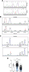
(A) Chromatogram showing the wild-type sequence (upper panel) and the R465H mutant form (lower panel) of FBXW7 exon 8. The red arrow indicates the mutated nucleotide. (B, C) Examples of loss of heterozygosity (LOH) at the D4S2998 (B) or D4S2934 (C) locus encompassing the FBXW7 gene. The two alleles are indicated by asterisks (*), and red arrows indicate the presence of LOH at one allele. (D) Quantitative real-time RT-PCR analysis of FBXW7 mRNA levels in normal livers (n=8), iCCA (n=82), and corresponding non-tumorous surrounding liver tissues (ST; n=82). Abbreviations: ns, not significant; *** p<0.0001 when compared to NL and ST.
Figure 2.
Figure 2.. Co-expression of AKT and a dominant negative form of Fbxw7 (Fbxw7ΔF) leads to intrahepatic cholangiocarcinoma development in mice.
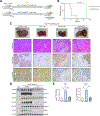
(A) Timeline of tumor development in AKT/Fbxw7ΔF mice (upper panel); macroscopy of AKT/Fbxw7ΔF livers (second panel); histopathologic features of the lesions (third panel); Ck19 immunostaining (fourth panel). (B) HA-tag immunohistochemistry showing cytoplasmic and nuclear staining for AKT and Fbxw7ΔF. The lesions are highly proliferative, as indicated by Ki67 nuclear immunoreactivity. (C) Western blotting of pathways activated in AKT/Fbxw7ΔF cholangiocellular lesions. Gapdh was used as a loading control. (D) mRNA levels of canonical Yap (Ctgf, Cyr61; upper panel) and Notch (Hey1, Nrarp; lower panel) target genes. (E) mRNA levels of c-Myc targets (Cdk4, Ccnd2, Skp2, Tert, Ldha, Ldhc, and Odc1). Scale bar: 200μm for 10×; 100μm for 20×. Abbreviations: H&E, hematoxylin and eosin staining; NL, normal liver; T, tumor; ns, not significant. * p <0.05; ** p <0.01; *** p <0.001.
Figure 3.
Figure 3.. Deletion of Yap delays, without suppressing, cholangiocarcinogenesis in AKT/Fbxw7ΔF mice.
(A) Study design. Yapflox/flox conditional knockout mice were subjected to HTVi of either AKT/Fbxw7ΔF/pCMV (control, n=9) or AKT/Fbxw7ΔF/Cre (n=10) plasmids. (B) Survival curve showing the delay of intrahepatic cholangiocarcinoma development following deletion of Yap. (C) Delay of tumor development in AKT/Fbxw7ΔF mice, as revealed by macroscopic examination of the livers (upper panel), histopathology of the lesions, Ck19 immunoreactivity (as a marker of biliary tumors) and Yap immunoreactivity. (D) Western blotting of AKT/Fbxw7ΔF mouse livers from control and Yap deleted (Cre) mice. Gapdh was used as a loading control. Scale bar: 200μm for 10×; 100μm for 20×. Abbreviations: H&E, hematoxylin and eosin staining; NL, normal liver; SB, sleeping beauty transposase.
Figure 4.
Figure 4.. Deletion of Notch2 retards, without impairing, cholangiocarcinogenesis in AKT/Fbxw7ΔF mice.
(A) Study design. Notch2lox/flox conditional knockout mice were subjected to HTVi of either AKT/Fbxw7ΔF/pCMV (control, n=8) or AKT/Fbxw7ΔF/Cre (n=10) plasmids. (B) Delay of tumor development in AKT/Fbxw7ΔF mice, as revealed by macroscopic examination of the livers and histopathology of the lesions, is accompanied by reduction of the Ck19 biliary marker and increase of the Hnf-4α hepatocellular marker. (C) Survival curve showing the delay of intrahepatic cholangiocarcinoma development following deletion of Notch2. (D) Analysis of Ck19-positive areas in control (pCMV) and Notch2-depleted (Cre) livers. (E) Western blotting of AKT/Fbxw7ΔF livers from control and Notch2 deleted mice. Scale bar: 200μm. Abbreviations: H&E, hematoxylin and eosin staining; NL, normal liver. *** p <0.001.
Figure 5.
Figure 5.. Inactivation of c-Myc completely abolishes cholangiocarcinogenesis in AKT/Fbxw7ΔF mice.
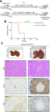
(A) Study design. FVB/N mice were subjected to HTVi of either AKT/Fbxw7ΔF/pT3 (control, n=6) or AKT/Fbxw7ΔF/MadMyc (n=8) plasmids. MadMyc is a dominant negative form of c-Myc. (B) Survival curve of AKT/Fbxw7ΔF/pT3 and AKT/Fbxw7ΔF/MadMyc mice. (C) Inhibition of tumor development in AKT/Fbxw7ΔF/MadMyc mice, as revealed by macroscopic examination of the livers and histopathology of the lesions, is accompanied by immunoreactivity for the Ck19 biliary marker limited to normal biliary epithelial cells (indicated by arrows) and low/absent immunoreactivity for the proliferation marker Ki67. Scale bar: 100μm. Abbreviation: H&E, hematoxylin and eosin staining.
Figure 6.
Figure 6.
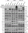
Western blot analysis showing c-MYC as a consistent target of FBXW7 in cholangiocarcinoma (CCA) cell lines. GAPDH was used as a loading control.
Figure 7.
Figure 7.. Expression levels of the FBXW7 gene are inversely correlated with c-MYC activity in human intrahepatic cholangiocarcinoma (iCCA).
(A) Absence of significant correlation between FBXW7 and c-MYC mRNA levels, as assessed by linear regression analysis. (B) A significant, negative correlation was found between mRNA levels of FBXW7 and c-MYC activity using the same statistical approach.
Figure 8.
Figure 8.. Immunohistochemical patterns of FBXW7 and c-Myc in human intrahepatic cholangiocarcinoma specimens.
Three representative cases (iCCA 1–3) are shown. Specifically, iCCA 1 (upper panel) recapitulates the most frequent pattern observed in the human iCCA collection, consisting of tumors with low levels of FBXW7 and strong immunoreactivity for c-MYC. iCCA 2 (middle panel) shows instead the example of an iCCA with high immunoreactivity for FBXW7 and weak/punctate staining of c-MYC protein. Finally, iCCA 3 (lower panel), consisting of a tumor displaying concomitant, elevated immunolabeling for both FBXW7 and c-MYC. CK19 staining was used as a biliary differentiation marker of the tumors. Scale bar: 100μm. Abbreviation: H&E, hematoxylin and eosin staining.

References

    1. Siegel RL, Miller KD, Jemal A. Cancer statistics, 2018. CA Cancer J Clin 2018;68:7–30. - PubMed
    1. Sia D, Villanueva A, Friedman SL, Llovet JM. Liver Cancer Cell of Origin, Molecular Class, and Effects on Patient Prognosis. Gastroenterology 2017;152:745–761. - PubMed
    1. Lee SY, Cherqui D. Operative management of cholangiocarcinoma. Semin Liver Dis 2013;33:248–261. - PubMed
    1. Khan SA, Emadossadaty S, Ladep NG, Thomas HC, Elliott P, Taylor-Robinson SD, et al. Rising trends in cholangiocarcinoma: is the ICD classification system misleading us? J Hepatol 2012;56:848–854. - "V体育平台登录" PubMed
    1. Yamamoto M, Takasaki K, Otsubo T, Katsuragawa H, Katagiri S. Recurrence after surgical resection of intrahepatic cholangiocarcinoma. J Hepatobiliary Pancreat Surg 2001;8:154–157. - PubMed

"VSports最新版本" Publication types

MeSH terms