Skip to main page content
U.S. flag

An official website of the United States government

Dot gov

The . gov means it’s official. Federal government websites often end in . gov or . mil VSports app下载. Before sharing sensitive information, make sure you’re on a federal government site. .

Https

The site is secure V体育官网. The https:// ensures that you are connecting to the official website and that any information you provide is encrypted and transmitted securely. .

. 2019 Jun 12;25(6):777-788.e8.
doi: 10.1016/j.chom.2019.04.004. Epub 2019 May 14.

"V体育官网入口" Resistin-like Molecule α Provides Vitamin-A-Dependent Antimicrobial Protection in the Skin

Affiliations

Resistin-like Molecule α Provides Vitamin-A-Dependent Antimicrobial Protection in the Skin

Tamia A Harris et al. Cell Host Microbe. .

Abstract

Vitamin A deficiency increases susceptibility to skin infection. However, the mechanisms by which vitamin A regulates skin immunity remain unclear. Here, we show that resistin-like molecule α (RELMα), a small secreted cysteine-rich protein, is expressed by epidermal keratinocytes and sebocytes and serves as an antimicrobial protein that is required for vitamin-A-dependent resistance to skin infection. RELMα was induced by microbiota colonization of the murine skin, was bactericidal in vitro, and was protected against bacterial infection of the skin in vivo. RELMα expression required dietary vitamin A and was induced by the therapeutic vitamin A analog isotretinoin, which protected against skin infection in a RELMα-dependent manner. The RELM family member Resistin was expressed in human skin, was induced by vitamin A analogs, and killed skin bacteria, indicating a conserved function for RELM proteins in skin innate immunity. Our findings provide insight into how vitamin A promotes resistance to skin infection VSports手机版. .

Keywords: antimicrobial protein; bacteria; innate immunity; microbiome; skin; skin infection; vitamin A V体育安卓版. .


Conflict of interest statement

Declaration of Interests

The authors declare no competing interests. Correspondence and requests for materials should be addressed to L. V. H (Lora.Hooper@qiuluzeuv.cn) or T. A VSports最新版本. H (Tamia.Harris-Tryon@qiuluzeuv.cn).

Figures
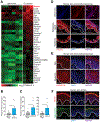
Figure 1:
Figure 1:. RELMα is expressed in the skin and expression is induced by the microbiota.
(A) Heatmap comparing transcript abundances in the skin of germ-free mice (n=6) and germ-free mice after topical exposure to Staphylococcus aureus (n=3). Transcript abundance was determined by RNAseq. The heatmap shows expression levels (log10(FPKMs+0.1)) ordered by transcript abundance. Retnla is highlighted in red. (B) qRT-PCR analysis of skin Retnla expression in germ-free mice and germ-free mice after exposure to S. aureus for 3 days. (C) qRT-PCR analysis of skin Retnla expression in germ-free mice, germ-free mice exposed to a conventional microbiota for 4 days (conv-D), or mice from a conventional facility (conv-L). (D) Immunofluorescence detection of RELMα in mouse skin. Epidermis (arrow, above dashed line) and sebaceous gland (arrowhead, inside dashed line) are indicated. (E) Immunofluorescence detection of RETN in human skin. (F) Fluorescence in situ hybridization (FISH) detection of RETN in human skin. Defa5 staining as negative control. Nuclei are stained with DAPI. Scale bars, 25 μm. Epidermis above dashed line. SG= sebaceous gland. Epi=Epidermis. Means±SEM are plotted; *P<0.05 as determined by one-tailed Welch’s t-test. See also Figure S1, Figure S2, and Table S1.
Figure 2:
Figure 2:. RELMα kills bacteria by disrupting their membranes.
(A) Purified recombinant mRELMα or hRETN was added to mid-logarithmic phase Streptococcus pyogenes for 2 hours and surviving bacteria were quantified by dilution plating. Colony forming units (CFUs) are expressed as a percentage of untreated bacteria. (B) 2.5 μM of mRELMα or hRETN was added to mid-logarithmic phase bacteria for 2 hours and surviving bacteria were quantified by dilution plating. Means±SEM are plotted. (C) Antibacterial efficacy of mRELMα and hRETN against wild-type S. aureus and an isogenic S. aureus mutant that lacks staphyloxanthin (ΔCRTM). Means±SEM are plotted. *P<0.05 **P<0.01 by paired Student’s t-test. (D) Carboxyfluorescein (CF)-loaded liposomes were exposed to 5 μM mouse RELMα. Dye efflux was measured over time and is expressed as a percentage of maximal efflux in the presence of the detergent octylglucoside (OG). (E) Percentage of dye release for varying doses of RELMα at the 500s time point. (F) Propidium iodide uptake by S. pyogenes in the presence of increasing concentrations of mRELMα. Assays were performed in triplicate. All results are representative of at least two independent experiments. See also Figure S3 and S4.
Figure 3:
Figure 3:. Mice lacking RELMα have an altered skin microbiota.
(A) Retnla−/− and wild-type littermates were separated at weaning into separate cages for at least 8 weeks, and skin samples were taken from the dorsal surface. Skin microbiota compositions were determined by 16S rRNA analysis and are plotted on a principle coordinate analysis (PCoA) plot. (B) Relative abundances of bacterial genera in wild-type and Retnla−/− mice respectively. (C) Relative abundance of coagulase negative Staphylococcus and Streptococcus species on the dorsal flank of wild type compared to Retnla−/− mice. Means±SEM are plotted *P<0.05 by Welch’s t-test. See also Figure S4 and S5.
Figure 4:
Figure 4:. Mice lacking RELMα are more susceptible to bacterial infection.
(A,B) S. pyogenes (A) or wild-type or staphyloxanthin-deficient (ΔCRTM) Staphylococcus strains (B) were grown to logarithmic phase and applied on a gauze rectangle to the dorsal skin of Retnla−/− or wild-type mice under occlusion for 2 days. CFU were determined in sections of inoculated skin. (C) S. pyogenes was injected intradermally into the skin of Retnla−/− or wild-type mice. Skin abscesses were removed and CFU were determined. Each symbol represents one mouse. Means±SEM are plotted *P<0.05; ***P<0.001; ns=not significant by Welch’s t-test (A,B) or unpaired t-test (C). See also Figure S4.
Figure 5:
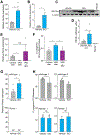
Figure 5:. Vitamin A is required for RELMα expression.
(A) RAR binding to the human RETN promoter was measured by chromatin immunoprecipitation (ChIP) with an anti-RAR antibody. Predicted retinoic acid response elements (RAREs) were identified by in silico analysis using NUBIScan (Podvinec et al., 2002) and are indicated by an asterisk (*). Predicted RARE sequences are shown in Figure S6. Data are representative of two independent experiments. (B) qRT-PCR analysis of human RETN expression in the human sebocyte cell line SZ95. Cells were treated with retinol, IL-1β, the pan-RAR inhibitor BMS493, or a combination. (C) Mice on a Vitamin A deprived diet express less Retnla transcript. qRT-PCR analysis of Retnla expression in the skin of mice on a control or vitamin A-deficient (VAD) diet administered for 9 weeks or 13 weeks. (D,E) Mice on a Vitamin A deprived diet express less RELMα protein. Western blot detection of RELMα in skin of mice fed a control or VAD diet (D), with quantification (E). (F) FISH detection of Retnla in mouse skin shows decreased Retnla transcripts in sebaceous glands (dashed line) of VAD diet-fed mice. Scale bar, 25 μm. Data are representative of at least two experiments. *P<0.05, **P<0.01,***P<0.001 by One-way ANOVA (B, C); unpaired t-test (E). See also Figure S6, Table S1, and Table S2.
Figure 6:
Figure 6:. RELMα provides vitamin A-dependent antimicrobial protection of the skin.
(A,B) Treatment of mice with the synthetic retinoid isotretinoin (ISO) for 3 days increases the abundance of transcripts encoding RELMα (A) and a known target of retinoids, retinoic acid receptor β (B). (C,D) Western blot of mouse RELMα expression in the skin of wild-type mice treated orally with the synthetic retinoid isotretinoin (ISO) or vehicle (C) with quantification in (D). (E) qRT-PCR analysis of mouse Retnla expression in the skin of mice on a control diet, a VAD diet, or a VAD diet supplemented with ISO. (F) Mid-logarithmic phase S. pyogenes was injected intradermally into the skin of mice on a control diet, VAD diet, or VAD diet rescued with ISO. Skin abscesses were removed and CFU were determined. (G) qRT-PCR analysis of mouse Retnla expression in the skin of wildtype or Retnla−/− mice treated orally by gavage with ISO or vehicle. nd, not detected. (H) Mid-logarithmic phase S. pyogenes was injected intradermally into the skin of wild-type or Retnla−/− mice treated by gavage with ISO or vehicle. Each symbol represents one mouse. Means±SEM are plotted. (A-G) Data are representative of at least two experiments. (H) Each graph represents an independent experiment. One-way ANOVA (G); Welch’s t-test (C); unpaired t-test (A,B,H); Kruskal-Wallis test (E,F). *P<0.05, **P<0.01, ***P<0.001, ns=not significant. See also Figure S4 and Table S1.

Comment in

"VSports手机版" References

    1. Albahrani AA, and Greaves RF (2016). Fat-soluble vitamins: clinical indications and current challenges for chromatographic measurement. Clin. Biochem. Rev 37, 27–47. - PMC - PubMed
    1. Banerjee RR, and Lazar MA (2001). Dimerization of resistin and resistin-like molecules is determined by a single cysteine. J. Biol. Chem 276, 25970–25973. - PubMed
    1. Belkaid Y, and Segre JA (2014). Dialogue between skin microbiota and immunity. Science 346, 954–959. - PubMed
    1. Beylot C, Auffret N, Poli F, Claudel JP, Leccia MT, Del Giudice P, and Dreno B (2014). Propionibacterium acnes: an update on its role in the pathogenesis of acne. J. Eur. Acad. Dermatol. Venereol 28, 271–278. - PubMed
    1. Braff MH, Di Nardo A, and Gallo RL (2005). Keratinocytes store the antimicrobial peptide cathelicidin in lamellar bodies. J. Investig. Dermatol 124, 394–400. - PubMed

V体育官网 - MeSH terms

Substances