Skip to main page content
U.S. flag

An official website of the United States government

Dot gov

The VSports app下载. gov means it’s official. Federal government websites often end in . gov or . mil. Before sharing sensitive information, make sure you’re on a federal government site. .

Https

The site is secure V体育官网. The https:// ensures that you are connecting to the official website and that any information you provide is encrypted and transmitted securely. .

. 2019 May 9;5(1):14.
doi: 10.1038/s41522-019-0087-4. eCollection 2019.

A microbiota-generated bile salt induces biofilm formation in Clostridium difficile (V体育2025版)

Affiliations

A microbiota-generated bile salt induces biofilm formation in Clostridium difficile

"VSports在线直播" Thomas Dubois et al. NPJ Biofilms Microbiomes. .

Abstract

Clostridium difficile is a major cause of nosocomial infections. Bacterial persistence in the gut is responsible for infection relapse; sporulation and other unidentified mechanisms contribute to this process. Intestinal bile salts cholate and deoxycholate stimulate spore germination, while deoxycholate kills vegetative cells VSports手机版. Here, we report that sub-lethal concentrations of deoxycholate stimulate biofilm formation, which protects C. difficile from antimicrobial compounds. The biofilm matrix is composed of extracellular DNA and proteinaceous factors that promote biofilm stability. Transcriptomic analysis indicates that deoxycholate induces metabolic pathways and cell envelope reorganization, and represses toxin and spore production. In support of the transcriptomic analysis, we show that global metabolic regulators and an uncharacterized lipoprotein contribute to deoxycholate-induced biofilm formation. Finally, Clostridium scindens enhances biofilm formation of C. difficile by converting cholate into deoxycholate. Together, our results suggest that deoxycholate is an intestinal signal that induces C. difficile persistence and may increase the risk of relapse. .

Keywords: Bacteriology; Biofilms; Pathogens V体育安卓版. .


Conflict of interest statement

The authors declare no competing interests.

Figures
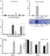
Fig. 1
Fig. 1
Effects of bile salts and deoxycholate on biofilm formation by C. difficile strain 630Δerm. a Bacteria were grown in BHISG supplemented with the indicated concentrations of bile salt extract or b deoxycholate. a, b Biofilm formation was evaluated at 72 h. In (b), representative image of a CV stained biofilm produced by cells cultured in the presence of increasing DOC concentrations. c Biofilm formation was evaluated after 48 h growth in the presence of 240 µM bile acids [cholic acid (CA), deoxycholic acid (DCA), lithocholic acid (LCA), chenodexycholic acid (CDCA)] or bile salts [cholate (CHO), deoxycholate (DOC) or chenodeoxycholate (CDOC)]. Asterisks indicate statistical significance determined with a Kruskal–Wallis test followed by an uncorrected Dunn’s test (*p ≤ 0.05, **p ≤ 0.001, ***p ≤ 0.001 vs BHISG with 0 mg/mL bile salts or 0 µM DOC). d Biofilm formation kinetics in the presence or absence of 240 µM DOC. Biofilm formation (ad) was measured using a crystal violet assay that included two PBS washing before staining (see the Methods section). e Kinetics of CFU grown in the presence or absence of 240 µM DOC. The CFU counts were performed with unwashed biofilms (see the Methods section). Asterisks indicate statistical significance determined with a two-way ANOVA followed by a Fisher LSD test (***p ≤ 0.001, ****p ≤ 0.0001 vs 24 h). Data shown indicate the mean and the error bars represent the standard error of the mean of at least five experiments performed on different days
Fig. 2
Fig. 2
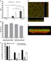
Characterization of the biofilm architecture and population in the presence of DOC. a CLSM images of C. difficile biofilms formed in the absence or presence of 240 µM DOC stained with Syto 9 (green/living) and propidium iodide (red/dead). b CLSM images of C. difficile grown in the absence or presence of 240 µM DOC and stained with FM4-64. a, b The CLSM analyses were performed with unwashed biofilms. c Cell length (µm) distribution within a population grown in BHISG or BHIG with 240 µM DOC. Asterisks indicate statistical significance determined with a two-tail Mann–Whitney test (****p ≤ 0.0001 vs BHISG without DOC)
Fig. 3
Fig. 3
Analysis of the biofilm matrix composition in the absence and presence of DOC. a CLSM analysis of the biofilm stained with ConA-Cy3 (red/α-D-glucosyl or α-D-mannosyl residues), WGA-Cy3 (red/N-acetylglucosamine or sialic acid residues), calcofluor white (blue/β-1,3 or β-1,4 polysaccharides), BOBO-3 (red/eDNA), or Sypro Ruby (red/proteins). The CLSM observations were performed with unwashed biofilms. b Dispersion of 48 h biofilms with proteinase K, DNase I, and NaIO4. c Inhibition of biofilm formation by proteinase K and DNase I. d CFU of biofilms treated with proteinase K and DNase I. The viability assays were performed with unwashed biofilms. e Biofilm formation in BHISG with 240 µM DOC by the parental strain, the cwp19 inactivated strain and cwp19 complemented strain. Asterisks indicate statistical significance determined with a Kruskal–Wallis test followed by an uncorrected Dunn’s test (*p ≤ 0.05, **p ≤ 0.01 vs the control or 630∆erm.) Biofilm formation (b, c, e) was measured using a crystal violet assay that included two PBS washing before staining. Images are representative fields acquired from 3 different biological replicates. Each bar represents the mean of at least 5 biological replicates performed on different days. The error bars represent the standard error of the mean
Fig. 4
Fig. 4
DOC represses both sporulation and toxin production of C. difficile. a CFU of viable cells (black) and heat-resistant spores (gray) in the absence or presence of 240 µM CHO or 240 µM DOC. The sporulation assays performed with unwashed biofilms. b Relative expression levels of tcdA and tcdB measured by qRT-PCR in cells grown in the absence or presence of 240 µM DOC or 240 µM CHO. Relative expression levels (∆∆Ct method) are the ratio of mRNA level in the presence of bile salts to the mRNA level in the absence of bile salts. Reactions of qRT-PCR were normalized using DNA polIII (CD1305), rpoA (CD0098), pgi (CD3285), and tpi (CD3172). c ELISA-based quantification of TcdA production by cells grown in the absence or presence of 240 µM DOC or 240 µM CHO. Concentrations were standardized to the amount of protein as measured by the Bradford method. d ELISA-based quantification of TcdA release into the supernatant by cells grown in the absence or presence of 240 µM DOC or 240 µM CHO. Asterisks indicate statistical significance determined with a two-way ANOVA followed by a Fisher LSD test (***p ≤ 0.001, ****p ≤ 0.0001 vs BHIS). Each bar represents the mean of at least 6 biological replicates performed on different days and error bars represent the standard error of the mean
Fig. 5
Fig. 5
Biofilm formation in the presence of DOC requires the CD1687 and is regulated by CcpA, CodY, and Spo0A, but not by SigB. a Biofilm formation by the parental strain (630∆erm), CD1687 mutants, a CD1687 mutant with an empty vector (pRPF185) and a CD1687 complemented strain (pRPF185::CD1687) grown in BHISG with 240 µM DOC. The inducer Atc (100 ng/mL) is added at the beginning of the experiment and every 24 h until the end of the experiment. b CFU of the parental strain (630∆erm) and the CD1687 mutant. c Localization of the CD1687 lipoprotein in the presence (I) or absence (NI) of the inducer; cell wall fraction (CW); membrane fraction (MB), cytoplasm fraction (CY). Samples are derived from the same experiments and processed in parallel. d Biofilm production by the parental strain (630∆erm), codY, ccpA, and sigB mutants grown in BHISG with 240 µM DOC. e Biofilm production by the parental strain (630∆erm), spo0A, sigE, and sigF mutants grown in the BHISG with 240 µM DOC. f CFU by the parental strain (630∆erm), codY, ccpA, and spo0A mutants grown in the BHISG with 240 µM DOC. Asterisks indicate statistical significance determined with a Kruskal–Wallis test followed by an uncorrected Dunn’s test (*p ≤ 0.05, **p ≤ 0.01, ****p ≤ 0.0001 vs 630∆erm). Biofilm formation (a, d, e) was measured using a crystal violet assay that included two PBS washes before staining. The viability assays (b, f) were performed with unwashed biofilms. Each bar represents the mean and the error bars represent the standard error of the mean of at least 5 biological replicates performed on different days
Fig. 6
Fig. 6
C. difficile biofilm formation enhanced by C. scindens. a Single-species and dual-species biofilm formation by C. difficile (Cd) and C. scindens (Cs) grown in the presence of 240 µM CHO or 240 µM DOC. Biofilm formation was measured using a crystal violet assay that included two PBS washing before staining. b CFU of C. difficile and C. scindens in single- and dual-species biofilms grown in the presence of 240 µM CHO in BHISG. The CFU counts were performed on unwashed biofilms. c Conversion of CHO to DOC by C. difficile and C. scindens in single- and dual-species cultures. d CLSM images and e 3D-reconstruction of a dual-species biofilm stained with Syto-61 (Red). Green cells are auto-fluorescent C. difficile. The CLSM analyses were performed with unwashed biofilms. Asterisks indicate statistical significance determined with a two-way ANOVA followed by a Fisher LSD test (****p ≤ 0.0001 vs CD + CS in BHISG + CHO). The error bars represent the standard error of the mean. Each bar represents the mean of at least 4 biological replicates performed on different days

References (V体育官网)

    1. Freeman J, et al. The changing epidemiology of Clostridium difficile infections. Clin. Microbiol. Rev. 2010;23:529–549. doi: 10.1128/CMR.00082-09. - DOI - PMC - PubMed
    1. Gupta A, Khanna S. Community-acquired Clostridium difficile infection: an increasing public health threat. Infect. Drug Resist. 2014;7:63–72. - PMC - PubMed
    1. Figueroa I, et al. Relapse versus reinfection: recurrent Clostridium difficile infection following treatment with fidaxomicin or vancomycin. Clin. Infect. Dis. 2012;55:S104–S109. doi: 10.1093/cid/cis357. - "V体育平台登录" DOI - PMC - PubMed
    1. Janoir C. Virulence factors of Clostridium difficile and their role during infection. Anaerobe. 2016;37:13–24. doi: 10.1016/j.anaerobe.2015.10.009. - V体育平台登录 - DOI - PubMed
    1. Sorg JA, Sonenshein AL. Bile salts and glycine as cogerminants for Clostridium difficile spores. J. Bacteriol. 2008;190:2505–2512. doi: 10.1128/JB.01765-07. - DOI (VSports在线直播) - PMC - PubMed

"VSports app下载" Publication types

MeSH terms (VSports app下载)