Skip to main page content (V体育安卓版)
U.S. flag

An official website of the United States government

Dot gov

The . gov means it’s official. Federal government websites often end in VSports app下载. gov or . mil. Before sharing sensitive information, make sure you’re on a federal government site. .

Https

The site is secure V体育官网. The https:// ensures that you are connecting to the official website and that any information you provide is encrypted and transmitted securely. .

. 2019 Jul;20(7):865-878.
doi: 10.1038/s41590-019-0388-z. Epub 2019 May 13.

The IRE1 endoplasmic reticulum stress sensor activates natural killer cell immunity in part by regulating c-Myc

Affiliations

The IRE1 endoplasmic reticulum stress sensor activates natural killer cell immunity in part by regulating c-Myc

Han Dong et al. Nat Immunol. 2019 Jul.

"V体育ios版" Abstract

Natural killer (NK) cells are critical mediators of host immunity to pathogens. Here, we demonstrate that the endoplasmic reticulum stress sensor inositol-requiring enzyme 1 (IRE1α) and its substrate transcription factor X-box-binding protein 1 (XBP1) drive NK cell responses against viral infection and tumors in vivo. IRE1α-XBP1 were essential for expansion of activated mouse and human NK cells and are situated downstream of the mammalian target of rapamycin signaling pathway. Transcriptome and chromatin immunoprecipitation analysis revealed c-Myc as a new and direct downstream target of XBP1 for regulation of NK cell proliferation. Genetic ablation or pharmaceutical blockade of IRE1α downregulated c-Myc, and NK cells with c-Myc haploinsufficency phenocopied IRE1α-XBP1 deficiency. c-Myc overexpression largely rescued the proliferation defect in IRE1α-/- NK cells. Like c-Myc, IRE1α-XBP1 also promotes oxidative phosphorylation in NK cells. Overall, our study identifies a IRE1α-XBP1-cMyc axis in NK cell immunity, providing insight into host protection against infection and cancer VSports手机版. .


Conflict of interest statement

Competing interests

L. H. G V体育安卓版. is a former Director of Bristol-Myers Squibb and is currently on the board of directors of and holds equity in GlaxoSmithKline Pharmaceuticals and the Waters Corporation. She chairs the scientific advisory board, is a co-founder of and holds equity in Quentis Therapeutics. She also serves on the scientific advisory boards of Repare Therapeutics, Abro and Kaleido Therapeutics.

Figures
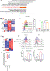
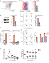
Figure 1.
Figure 1.. Induction of IRE1α-XBP1 UPR in mouse and human activated NK cells in vitro and in vivo.
(a) Gene ontology (GO) analysis of the top 500 differentially expressed genes in Ly49H+ splenic NK cells harvested from either naïve or MCMV-infected mice day 1.5 post infection (PI). Top 10 GO clusters with the key word ‘stimulus’ were shown. n = 3 mice/group. (b) RNA-seq analysis showing the expression of canonical IRE1α-XBP1 target genes during a time course PI. (c) Flow cytometric analysis of Venus reporter expression in splenic NK cells from ERAI transgenic mice at day 2 PI. (d) RNA-seq analysis showing the expression of canonical IRE1α-XBP1 target genes in splenic NK cells harvested from WT mice and cultured either in the presence or absence of IL-12 and IL-18 for 16 hr (GSE106138). (e) Flow cytometric analysis of Venus reporter expression in sorting-purified splenic and BM NK cells from ERAI transgenic mice following 16 hr culture in the presence or absence of IL-12 and IL-18, or IL-15. Each column (b, d) or symbol (c, e) represents an individual mouse. (f) Quantitative real-time PCR analysis of XBP1 splicing activity in sorted-purified human primary NK cells from PBMCs after 16 hrs culture in the presence or absence of IL-12 and IL-18, or IL-15. β-ACTIN was used as reference, and data are shown as fold change normalized to the unstimulated levels. n = 6 PBMC donors. Error bars represent mean with s.d. (c, e) or with minimal to maximal (f). Data were analyzed by one-way analysis of variance (ANOVA) with the Tukey post-test (c, e), or two-sided one sample t-test (f). *p<0.05, ***p<0.001, ****p<0.0001, ns = not significant. Data are representative of 3 (c, e) or 2 (f) independent experiments.
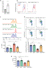
Figure 2.
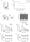
Figure 2.. IRE1α is required for optimal protective antiviral NK cell responses.
(a, b) IRE1NK and littermate control mice were infected with a lethal dose of MCMV. (a) Viral titers in the blood at day 4 PI. n = 8 mice/group. (b) Survival curve. n as indicated in the key. (c) Schematic of co-transfer experiments in d-h: Equal numbers of Ly49H+ NK cells from WT (CD45.1) and knockout (KO; CD45.2) donors were co-transferred into recipient Ly49H-deficient mice 1 day before infection with MCMV. (d) Quantification of the percentage of transferred WT and IRE1NK Ly49H+ NK cells in peripheral blood at specified time points PI. Lines showed expansion kinetics by connecting mean values of adjacent time points in ggroup. (e) As in d, except showing the relative percentage within the transferred Ly49H+ NK cells. (f) Relative percentages of transferred WT and IRE1NK Ly49H+ NK cells in various organs at day 8 (LN) or day 10 (all other tissues) PI. LN, lymph nodes. n = 4 recipient mice/column. (g) As in d, except the KO donors were XBP1NK. (h) As in e, except the KO donors were XBP1NK. n = 4 recipient mice (d, e, g. h). Error bars represent mean with minimal to maximal (a) or with s.d.(e, f, h). Data were analyzed by two tailed Mann-Whitney test (a), two-sided Log rank test (b, with p=0.0601), or two-way analysis of variance (ANOVA) with the Sidak post-test (d-h). *p<0.05, **p<0.01, ***p<0.001, ****p<0.0001, ns = not significant. Data are representative of 3 (a, f, g, h) or 4 (d, e) independent experiments, or pooled from 3 experiments (b).
Figure 3.
Figure 3.. IRE1α-XBP1 controls infection-induced NK cell proliferation but not survival.
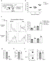
(a) Schematic of assays evaluating infection-driven NK cell proliferation and apoptosis in b-f. Equal numbers of Ly49H+ NK cells from WT (CD45.1) and IRE1NK (CD45.2) donors were labelled with cell proliferation tracing dye CTV, and then co-transferred into recipient Ly49H-deficient mice 1 day before infection with MCMV. (b) Relative percentages of transferred WT and IRE1NK Ly49H+ NK cells in the spleen of recipient Ly49H-deficient mice at specified time points PI. (c) Representative plots (left) and quantifications (right) of flow cytometric analysis showing CTV dilution of transferred WT and IRE1NK Ly49H+ (responsive) and Ly49H (bystander) NK cells in the spleen at day 4 PI. Flow cytometric analysis of EdU (d) and Annexin V (e) in co-transferred Ly49H+ NK cells at day 3.5 PI. EdU was injected intraperitoneally into mice 12 hr before measurement. (f) Representative flow cytometric plots (left) and quantifications (right) of percentage of FLICA+ cells in co-transferred Ly49H+ NK cells as in d. n = 4 mice/group for all experiments except n = 3 mice/group in c. All error bars represent mean with s.d.. **p<0.01, ***p<0.001, ****p<0.0001, ns = not significant. Data were analyzed by two-way analysis of variance (ANOVA) with the Sidak post-test (b), one-way analysis of variance (ANOVA) with the Tukey post-test (c), or two tailed unpaired Student’s t-test (d-f). Data are representative of 3 (b, c, f) or 2 (d, e) independent experiments.
Figure 4.
Figure 4.. IRE1α-XBP1 supports NK homeostatic proliferation.
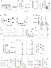
(a) Schematic of lymphopenia-induced homeostatic proliferation experiments in b, c. (b) Relative percentage of transferred WT and IRE1NK cells in the spleen of Rag2−/−Il2rg−/− recipients at specified time points after transfer. (c) Representative flow cytometric plots and quantification of CTV dilution of transferred NK cells in the spleen of recipient mice at day 4 after transfer. (d) Relative percentages and absolute number of WT and IRE1NK cells after co-incubation with IL-2 and IL-15. (e) Representative flow cytometric plots of FSC and SSC of cells in d. (f, g) IRE1NK and IRE1f/f cells from littermates were pre-labeled with CTV and cultured with IL-2 and IL-15. Flow cytomemetic assays showing (f) CTV dilution and (g) Ki-67 levels at specified time points. (h) Human primary NK cells were cultured with IL-2 and IL-15, in the presence or absence of the IRE1 inhibitor 4μ8C. (left) Quantitative real-time PCR analysis of XBP1 splicing activity after 16 hr. β-ACTIN was used as reference. (middle) Flow cytometric analysis of CTV dilution at specified time points. Plots showed representative data derived from three PBMC donors. (right) Quantification of percentages of divided cells at day 6. (i) Flow cytometric analysis of CTV dilution as in h, except CD56bright and CD56dim cells were plotted separately. (j) Representative flow cytometric plots and quantification of XBP1s protein levels in CD56bright and CD56dim NK cells before and after 16 hr of cytokine stimulation. n = 2 mice/group in b. n = 4 mice/group in c, f, g. n = 3 mice/group (and 2 wells/mice for ex vivo culture) in d, e. n = 6, 5 and 5 PBMC donors in h, i and j. All error bars represent mean with s.d.. *p<0.05, **p<0.01, ***p<0.001, ****p<0.0001. Data were analyzed by two-tailed unpaired Student’s t-test performed (c), two-way analysis of variance (ANOVA) with the Sidak post-test (d), one-way analysis of variance (ANOVA) with the Tukey post-test (f), or two-tailed paired Student’s t-test (h-j). Data are representative of 3 (b, c) or 2 (d-g) independent experiments, or pooled from 2 independent experiments (h-j).
Figure 5.
Figure 5.. IRE1 supports NK cell OXPHOS and mitochondrial function.
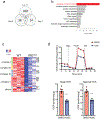
(a) Venn diagram of RNA-seq analysis showing differentially expressed genes in IRE1NK Ly49H+ NK cells compared to WT counterparts during infection with MCMV. Cells were harvested from IRE1NK (CD45.2): WT (CD45.1) mixed BM chimeras at three time points: day 0, 1.5 and 7 PI. n = 3 mice/group (b) Top 10 enriched GSEA hallmark gene sets in RNA-seq analysis at day 1.5 PI as indicated in a. n = 3 mice/group. (c) The heat map of differentially expressed OXPHOS target genes in IRE1NK versus WT NK cells at day 1.5 PI. Genes were clustered by functional annotation. c-Myc-regulated genes are shown in black text on the right. (d) Analysis of NK cell oxygen consumption rate (OCR) to assess rates of OXPHOS and maximal respiration. Primary human NK cells isolated from PBMC were incubated with IL-12 and IL-18 for 16 hr, in the presence or absence of the IRE1α inhibitor 4μ8C (5 uM). Approximately 600, 000 cells were plated per well, and the data were normalized to total protein quantification. Oligo (Oligomycin, 1 μM), FCCP (carbonyl cyanide-p-(trifluoromethoxy) phenylhydrazone, 1 μM), R (rotenone, 0.5 μM) and A (antimycin, 0.5 μM). Representative plot in d shows data from one PBMC donor with each symbol representing mean measurement of three technical replicates. Quantification in d shows the combined data from three PBMC donors and two independent experiments; two-tailed unpaired Student’s t-test was performed, with error bars representing mean with s.e.m. and ****p<0.0001.
Figure 6.
Figure 6.. XBP1 promotes NK cell proliferation at least partially via direct regulation of c-Myc.
(a) Top 10 enriched GSEA gene clusters in RNA-seq analysis of IRE1NK versus WT Ly49H+ splenic NK cells harvested from IRE1NK (CD45.2): WT (CD45.1) mixed BM chimera mice day 1.5 PI. n = 3 BM chimera mice. (b) The heat map of differentially expressed canonical c-Myc target genes in IRE1NK versus WT NK cells. Genes were clustered by functional annotation. Each column is a different mouse in group. (c) Quantitative real-time PCR analysis of c-Myc expression in transferred WT and IRE1NK Ly49H+ NK cells in the spleen of recipient Ly49H-deficient mice at day 1 PI. Equal numbers of Ly49H+ NK cells from WT and KO donors were co-transferred into recipient Ly49H-deficient mice 1 day before infection. n = 4 recipient mice. (d) Representative immunoblot and quantification of c-Myc protein in IRE1NK and WT NK cells sorted from the same mixed BM chimera mice at day 1.5 PI; quantification shows cumulative data from 4 mixed BM chimeras. (e) As in c, flow cytometric analysis of c-Myc expression in transferred WT and IRE1NK Ly49H+ NK cells in the spleen of recipient Ly49H-deficient mice at day 1 PI. n = 3 recipient mice. (f) Chromatin immunoprecipitation assays using NK cell lines MNK-1 (mouse), NKL and KHYG-1 (human) to assess XBP1 binding to the c-Myc locus. Anti-XBP1s Ab was used and IgG was used as mock control. n = 3 technical replicates/group. (g) Representative flow cytometric plots and quantification of c-Myc reporter expression. Before assessment, splenic NK cells from c-Myc reporter mice (MycGFP) were treated with indicated cytokines for 16 hr, either in the presence or absence of the IRE1 inhibitor 4μ8C (5uM). n=3 wells/treatment in ex vivo culture. (h) (left) Percentages of transferred WT (CD45.1) and MycNK (CD45.2) Ly49H+ NK cells in peripheral blood (except the endpoint using spleen) at specified time points after infection with MCMV. Equal numbers of Ly49H+ NK cells from WT and MycNK donors were co-transferred into recipient Ly49H-deficient mice 1 day before infection. (right) The relative percentages within the transferred Ly49H+ NK cells. n = 5 recipient mice. All error bars represent mean with s.d.. **p<0.01, ****p<0.0001. Data were analyzed by two-tailed one sample t-test (c, d), two-tailed unpaired Student’s t-test (e), or two-way analysis of variance (ANOVA) with the Sidak post-test (h). Data are representative of 3 (c, e, g) or 2 (d, f, g) independent experiments, or pooled from 2 experiments (h).
Figure 7.
Figure 7.. Restoration of c-Myc in the absence of IRE1 rescues the NK cell proliferation defect.
(a) Real-time PCR analysis of c-Myc mRNA in naïve NK cells from MycOE (Mycfsf/+ Ncr1Cre+) and littermate control (Mycfsf/+Ncr1Cre-) mice. (b) NK cells as in a, representative flow cytometric plots of CTV dilution after ex vivo culture with IL-2 and IL-15 for 3 days, in the presence or absence of IRE1α inhibitor 4μ8C (5 μM). (c-e) WT (Mycfsf/+ IRE1f/f Ncr1Cre-), IRE1NK (Myc+/+ IRE1f/f Ncr1Cre+), MycOE (Mycfsf/+ IRE1+/+ Ncr1Cre+) and MycOE IRE1NK (Mycfsf/+ IRE1f/f Ncr1Cre+) NK cells were pre-labeled with CTV and cultured ex vivo with IL-2 and IL-15. (c) representative flow cytometric plots and quantification of CTV dilution at day 3, and (d) representative flow cytometric plots of Ki-67 levels and quantification of percentage of proliferating cells (defined as Ki-67+ CTVlo) at day 3. (e) Absolute numbers of NK cells at day 6. n=5 mice/group in a, n = 2 mice/group in b and n= 4 mice/group in c-e. Technical duplicates in culture per mouse in b-e. All error bars represent mean with s.e.m.. ***p<0.001, ****p<0.0001. Data were analyzed by two-tailed one sample t-test (a), or one-way analysis of variance (ANOVA) with the Tukey post-test performed (c-e). Data are representative of 3 (b-e) independent experiments or pooled from 2 experiments (a).
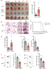
Figure 8.
Figure 8.. Intrinsic requirement of IRE1α/XBP1 for NK cell-mediated antitumor immunity.
(a) Gross morphology of lungs and individual lung lobes from IRE1f/f and IRE1NK littermates at day 10 following intravenous injection of B16F10 melanoma. Quantification of total extrapulmonary metastatic nodules is shown on the right. n = 10 mice/group. (b) H&E microscopic analysis of lungs from IRE1f/f and IRE1NK mice described in a. (c) Survival Curve of B16F10-innoculated IRE1f/f (n=11) and IRE1NK (n=14) mice described in a. (d) Similar as in a, quantification of total extrapulmonary metastatic nodules from XBP1f/f and XBP1NK littermates at day 20 following intravenous injection of B16F10 melanoma. n = 8 mice/group. (e-g) Flow cytometry analysis of lungs from B16F10 tumor-inoculated mice described in a. Graphs shown are (e) percentage of NK cells in total lymphocyte population and the absolute numbers (n = 8 for IRE1f/f, n = 9 for IRE1NK), (f) Ki-67 (n=6 mice/group) and (g) c-Myc expression (n = 6 mice/group) in lung-infiltrated NK cells. Each symbol is a different mouse (a, d-g). All error bars represent mean with s.e.m.. **p<0.01, ***p<0.001, ****p<0.0001. Data were analyzed by two-tailed Mann Whitney test (a, d, e-g), or two-tailed Log-rank test (c). Data are representative of 2 independnet experiments (b, f, g), or pooled from 3 (a) or 2 experiments (c-e).

References

References (for main text only)

    1. Caligiuri MA Human natural killer cells. Blood 112, 461–469 (2008). - PMC - PubMed
    1. Madera S et al. Type I IFN promotes NK cell expansion during viral infection by protecting NK cells against fratricide. J Exp Med 213, 225–233 (2016). - PMC - PubMed
    1. Zawislak CL et al. Stage-specific regulation of natural killer cell homeostasis and response against viral infection by microRNA-155. Proceedings of the National Academy of Sciences of the United States of America 110, 6967–6972 (2013). - PMC - PubMed
    1. Sun JC et al. Proinflammatory cytokine signaling required for the generation of natural killer cell memory. J Exp Med 209, 947–954 (2012). - VSports app下载 - PMC - PubMed
    1. Beaulieu AM, Zawislak CL, Nakayama T & Sun JC The transcription factor Zbtb32 controls the proliferative burst of virus-specific natural killer cells responding to infection. Nature immunology 15, 546–553 (2014). - PMC - PubMed

References (Methods-only)

    1. Fodil-Cornu N et al. Ly49h-deficient C57BL/6 mice: a new mouse cytomegalovirus-susceptible model remains resistant to unrelated pathogens controlled by the NK gene complex. Journal of immunology (Baltimore, Md. : 1950) 181, 6394–6405 (2008). - PMC - PubMed
    1. Iwawaki T, Akai R, Yamanaka S & Kohno K Function of IRE1 alpha in the placenta is essential for placental development and embryonic viability. Proceedings of the National Academy of Sciences of the United States of America 106, 16657–16662 (2009). - PMC - PubMed
    1. Trapnell C et al. Differential gene and transcript expression analysis of RNA-seq experiments with TopHat and Cufflinks. Nat Protoc 7, 562–578 (2012). - PMC - PubMed
    1. Anders S, Pyl PT & Huber W HTSeq--a Python framework to work with high-throughput sequencing data. Bioinformatics 31, 166–169 (2015). - PMC - PubMed
    1. Anders S & Huber W Differential expression analysis for sequence count data. Genome Biol 11, R106 (2010). - V体育2025版 - PMC - PubMed

Publication types

MeSH terms