Skip to main page content
U.S. flag

An official website of the United States government

Dot gov

The . gov means it’s official. Federal government websites often end in . gov or . mil. Before sharing sensitive information, make sure you’re on a federal government site. VSports app下载.

Https

The site is secure V体育官网. The https:// ensures that you are connecting to the official website and that any information you provide is encrypted and transmitted securely. .

. 2019 Jul 12;27(7):827-834.
doi: 10.3727/096504018X15462920753012. Epub 2019 Mar 25.

"V体育官网入口" E2-Induced Activation of the NLRP3 Inflammasome Triggers Pyroptosis and Inhibits Autophagy in HCC Cells

Affiliations

E2-Induced Activation of the NLRP3 Inflammasome Triggers Pyroptosis and Inhibits Autophagy in HCC Cells (VSports app下载)

Qing Wei et al. Oncol Res. .

Abstract

Emerging evidence suggests that 17β-estradiol (E2) and estrogen receptor (ER) signaling are protective against hepatocellular carcinoma (HCC). In our previous study, we showed that E2 suppressed the carcinogenesis and progression of HCC by targeting NLRP3 inflammasome activation, whereas the molecular mechanism by which the NLRP3 inflammasome initiated cancer cell death was not elucidated. The present study aimed to investigate the effect of NLRP3 inflammasome activation on cell death pathways and autophagy of HCC cells VSports手机版. First, we observed an increasing mortality in E2-treated HCC cells, and then apoptotic and pyroptotic cell death were both detected. The mortality of HCC cells was largely reversed by the caspase 1 antagonist, YVAD-cmk, suggesting that E2-induced cell death was associated with caspase 1-dependent pyroptosis. Second, the key role of the NLRP3 inflammasome in autophagy of HCC cells was assessed by E2-induced activation of the NLRP3 inflammasome, and we demonstrated that autophagy was inhibited by the NLRP3 inflammasome via the E2/ERβ/AMPK/mTOR pathway. Last, the interaction of pyroptosis and autophagy was confirmed by flow cytometry methods. We observed that E2-induced pyroptosis was dramatically increased by 3-methyladenine (3-MA) treatment, which was abolished by YVAD-cmk treatment, suggesting that caspase 1-dependent pyroptosis was negatively regulated by autophagy. In conclusion, E2-induced activation of the NLRP3 inflammasome may serve as a suppressor in HCC progression, as it triggers pyroptotic cell death and inhibits protective autophagy. .


"VSports app下载" Conflict of interest statement

The authors declare no conflicts of interest.

Figures
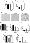
Figure 1
Figure 1
17β-Estradiol (E2)-initiated NLRP3 inflammasome activation induces hepatocellular carcinoma (HCC) cell death. (A, B) HepG2 cells were treated with E2 at concentrations of 50 nM and 100 nM for 24 h, and then caspase 1 and IL-1β expressions were detected by ELISA. (C) HepG2 cells were treated with 100 nM of E2, and the morphology of the cells was observed with a phase-contrast microscope. Scale bars: 100 μm. (D–F) HepG2 cells were treated with E2 at concentrations of 50 nM and 100 nM for 24 h, and then cell viability was detected by trypan blue stain assay, CCK-8 test, and lactate dehydrogenase (LDH) release assay. (G, H) HepG2 cells were pretreated with YVAD-cmk (50 μM) before E2 (100 nM) treatment, and cell viability and mortality were detected by trypan blue and LDH release assays. *p < 0.05, **p < 0.01, ***p < 0.001.
Figure 2
Figure 2
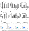
NLRP3 inflammasome activation results in caspase 1-dependent pyroptosis. (A) HepG2 cells were pretreated with YVAD-cmk (50 μM) before E2 (100 nM) treatment, and expressions of caspases 3, 8, and 9 were detected by Western blot. (B) HepG2 cells were pretreated with YVAD-cmk (50 μM) before E2 (100 nM) treatment, and caspase 1 expression was tested by ELISA. (C) Detected double positivity of caspase 1 fluorescent inhibitor probe (FAM-YVAD-FMK) and PI (Q3) using flow cytometry after 24 h. The proportions of Q3 were then presented with a histogram. (D) Representative flow cytometry scatter plots. Bars represent mean ± SEM. **p < 0.01, ***p < 0.001.
Figure 3
Figure 3
NLRP3 inflammasome activation inhibits autophagy in HCC cells. HepG2 cells were treated with different concentrations of E2 (0, 50 nM, and 100 nM) for 24 h. mRNA expressions of beclin 1 and LC3-b were detected by real-time (RT)-PCR (A), and protein levels of beclin 1 and LC3 were tested by Western blot (B). HepG2 cells were pretreated with PHTPP (1 μM) or YVAD-cmk (50 μM) before E2 (100 nM) treatment. mRNA expressions of beclin1 and LC3-b were detected by real-time PCR (C, D), and protein levels of beclin 1, LC3, and P62 were tested by Western blot (E, F). (G) Cytoplasmic expression of LC3-b in HepG2 cells. Cells were treated with 100 nM of E2 alone or in combination with 1 μM of PHTPP or 50 μM of YVAD-cmk and incubated for 24 h. The level of autophagy was evaluated using a Cyto-ID® detection agent on an inverted fluorescence microscope. Scale bar: 50 μm. *p < 0.05, **p < 0.01, ***p < 0.001.
Figure 4
Figure 4
AMP-activated protein kinase (AMPK)/mTOR pathway is required for E2-induced autophagy inhibition. (A) HepG2 cells were treated with different concentrations of E2 (0, 50 nM, and 100 nM) for 24 h, and phosphorylation of key enzymes in AMPK/mTOR pathway were tested by Western blot. (B) HepG2 cells were pretreated with pEGFP-C1-AMPK or rapamycin (100 nM) before E2 (100 nM) treatment, and phosphorylation of key enzymes in AMPK/mTOR pathway were tested by Western blot. (C) The intensity of the LC3 band was quantified by LC3 II/I ratio using the densitometry analysis. *p < 0.05, **p < 0.01.
Figure 5
Figure 5
Autophagy deficiency promotes pyroptosis in HCC cells. (A–C) HepG2 cells were pretreated with 3-MA (50 μM) or YVAD-cmk (50 μM) before E2 (100 nM) treatment, and cell viability and mortality were detected by trypan blue, CCK-8, and LDH release assays. (D, E) HepG2 cells were pretreated with 3-MA (50 μM) or YVAD-cmk (50 μM) before E2 (100 nM) treatment. Caspase 1 and IL-1β expressions were tested by ELISA. (F) Detected double positivity of caspase 1 fluorescent inhibitor probe (FAM-YVAD-FMK) and PI (Q3) using flow cytometry after 24 h. The proportions of Q3 were then presented with a histogram. (G) Representative flow cytometry scatter plots. Bars represent mean ± SEM. *p < 0.05, **p < 0.01, ***p < 0.001.

References

    1. El-Serag HB. Epidemiology of viral hepatitis and hepatocellular carcinoma. Gastroenterology 2012;142(6):1264–73.e1. - PMC - PubMed
    1. Yeh SH, Chen PJ. Gender disparity of hepatocellular carcinoma: The roles of sex hormones. Oncology 2010;78(Suppl 1):172–9. - "V体育平台登录" PubMed
    1. Naugler WE, Sakurai T, Kim S, Maeda S, Kim K, Elsharkawy AM, Karin M. Gender disparity in liver cancer due to sex differences in MyD88-dependent IL-6 production. Science 2007;317(5834):121–4. - PubMed
    1. Wei Q, Mu K, Li T, Zhang Y, Yang Z, Jia X, Zhao W, Huai W, Guo P, Han L. Deregulation of the NLRP3 inflammasome in hepatic parenchymal cells during liver cancer progression. Lab Invest. 2014;94(1):52–62. - PubMed
    1. Wei Q, Guo P, Mu K, Zhang Y, Zhao W, Huai W, Qiu Y, Li T, Ma X, Liu Y, Han L. Estrogen suppresses hepatocellular carcinoma cells through ERbeta-mediated upregulation of the NLRP3 inflammasome. Lab Invest. 2015;95(7):804–16. - PubMed

MeSH terms

LinkOut - more resources