Skip to main page content (V体育2025版)
U.S. flag

An official website of the United States government

Dot gov

The . gov means it’s official. Federal government websites often end in . gov or . mil. Before sharing sensitive information, make sure you’re on a federal government site VSports app下载. .

Https

The site is secure. The https:// ensures that you are connecting to the official website and that any information you provide is encrypted and transmitted securely. V体育官网.

. 2019 Dec 19;11(12):1069-1082.
doi: 10.1093/jmcb/mjz020.

Mitochondrial ROS promote macrophage pyroptosis by inducing GSDMD oxidation

Affiliations

"V体育官网入口" Mitochondrial ROS promote macrophage pyroptosis by inducing GSDMD oxidation

Yufang Wang et al. J Mol Cell Biol. .

V体育官网 - Abstract

Disrupted mitochondrial membrane potential (MMP) and reactive oxygen species (ROS) generation are often associated with macrophage pyroptosis. It remains unclear how these forms of mitochondrial dysfunction relate to inflammasome activation and gasdermin-D (Gsdmd) cleavage, two central steps of the pyroptotic process. Here, we also found MMP collapse and ROS generation induced by Nlrp3 inflammasome activation as previous studies reported. The elimination of ROS alleviated the cleavage of Gsdmd, suggesting that Gsdmd cleavage occurs downstream of ROS release. Consistent with this result, hydrogen peroxide treatment augmented the cleavage of Gsdmd by caspase-1. Indeed, four amino acid residues of Gsdmd were oxidized under oxidative stress in macrophages. The efficiency of Gsdmd cleavage by inflammatory caspase-1 was dramatically reduced when oxidative modification was blocked by mutation of these amino acid residues. These results demonstrate that Gsdmd oxidation serves as a de novo mechanism by which mitochondrial ROS promote Nlrp3 inflammasome-dependent pyroptotic cell death VSports手机版. .

Keywords: Nlrp3; ROS; gasdermin-D; mitochondria; oxidation V体育安卓版. .


Figures

Figure 1
Figure 1
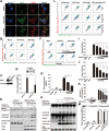
Gsdmd is not required for the mitochondrial dysfunction in pyroptosis. (A) Immunostaining of the subcellular location of Gsdmd and the mitochondrial protein COX IV in fixed BMDMs in response to inducers of the Nlrp3 inflammasome. Scale bar, 5 μm. (B–D) Primed WT and Gsdmd-deficient BMDMs were treated with 10 μM nigericin for 30 min and then labeled with MitoTracker Deep Red and MitoTracker Green before flow cytometric analysis. (E) Detection of the subcellular location of ROS by labeling with the probe DCFH-DA and MitoTracker CMXRos in live Gsdmd-deficient BMDMs. Scale bar, 5 μm. Data represent the average of three measurements. Mean ± SD of three experiments is shown. NS, P > 0.05, not significant.
Figure 2
Figure 2
Multiple inflammasome activation mediates mitochondrial dysfunction. (A) Immunostaining of the subcellular location of Nlrp3 and the mitochondria outer membrane protein Hsp60 in fixed BMDMs in response to pyroptosis. Scale bar, 5 μm. (B–D) Flow cytometric analysis of primed WT and Nlrp3-knockdown J774A.1 cells stimulated for 30 min with nigericin and stained with MitoTracker Red and MitoTracker Green. (E) Pre-treatment of 15 μM colchicine 25 min before stimulating primed macrophages with 10 μM nigericin. Immunoblot analysis of Nlrp3 translocation, Gsdmd cleavage, and caspase-1 activation in J774A.1 cells following fractionation (cytosolic and mitochondrial fractions). (F and G) Flow cytometric analysis of the MMP of primed J774A.1 cells pretreated with colchicine (10 and 15 μM) 25 min before stimulation with nigericin. (H) Immunoblot analysis of Gsdmd cleavage and caspase-1 activation in J774A.1 cells given gradient colchicine (1, 5, 10, and 15 μM) pretreatment before stimulation with nigericin. (I and J) The greyscale analysis of Gsdmd-N domain and caspase-1 p20 subunit with loading control (β-actin). (K) The LDH release assay was used to quantify cell death. (L and M) Flow cytometric analysis of the MMP of primed WT, Casp1KO, and GsdmdKO BMDMs stimulated with nigericin (10 μM) and poly(dA:dT) (2 μg/ml). Input, whole-cell lysate; SN, supernatant; WT, wild-type. Data shown in this figure represent the average of three measurements. Mean ± SD of three experiments is shown. **P < 0.01 and ***P < 0.001.
Figure 3
Figure 3
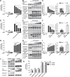
Redox status affects the cleavage of Gsdmd. (A, D, and G) Quantification of cell death via LDH release assay. (B) Immunoblot analysis of Gsdmd cleavage, caspase-1 activation, and IL-1β in cell lysates and supernatants of J774A.1 cells treated with gradient NAC (5, 10, 15, and 20 mM) and then stimulated with nigericin (10 μM). (C) The greyscale analysis of Gsdmd-N terminal in cell lysate (Input) and caspase-1 p20 subunit in both cell lysate (Input, black columns) and supernatant (SN, white columns) with loading control (β-actin), consisted with Figure 3B. (E) Primed J774A.1 cells were given gradient NAC (10, 15 and 20 mM) preincubation. Cell lysates and supernatants were collected for immunoblot analysis of Gsdmd cleavage, caspase-1 activation, and IL-1β at 1 h after poly (dA:dT) (2 μg/ml) transfection. (F) The greyscale analysis of Gsdmd-N terminal in cell lysate (Input) and caspase-1 p20 subunit in both cell lysate (Input, black columns) and supernatant (SN, white columns) with loading control (α-tubulin), consisted with Figure 3E. (H) LPS-primed J774A.1 cells were prestimulated with gradient 3-MA (1, 2.5, 5, and 10 mM) before the end of the priming process. Gsdmd cleavage, caspase-1 activation, and IL-1β were analyzed 1 h after nigericin stimulation via western blot. (I) The greyscale analysis of Gsdmd-N terminal in cell lysate (Input) and caspase-1 p20 subunit in both cell lysate (Input, black columns) and supernatant (SN, white columns) with loading control (β-actin), consisted with Figure 3H. (J) Immunoblot analysis of the cleavage of human GSDMD and mouse Gsdmd by active CASP1 subunits after gradient H2O2 (100 and 200 μM) incubation. (K) LDH assay of cell death in 293T cells expressing GSDMD/Gsdmd and active CASP1 subunits treated with gradient H2O2 or left untreated. SN, supernatant; Input, whole-cell lysate; OE, overexpression; clea., cleavage. Data represent the average of three measurements. Mean ± SD of three experiments is shown. *P < 0.05, **P < 0.01, ***P < 0.001.
Figure 4
Figure 4
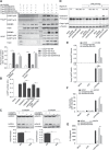
Four cysteine residues are required for human GSDMD to sense oxidative stress. (A) Model graph of four oxidized cysteine residues on GSDMD. (B) Cys38, Cys56, Cys268, and Cys467 were oxidized after incubation with H2O2. (C) Immunoblot analysis of the cleavage of WT, 3CS, and 4CS GSDMD mutants by artificially activated CASP1 treated with H2O2 or left untreated. (D) The LDH release assay was used to quantify cell death in 293T-acCASP1 cells overexpressing WT, 3CS, and 4CS GSDMD mutants treated with 100 μM H2O2 for 6 h or left untreated. (E and F) Immunoblot analysis of single CS and 2CS mutants of GSDMD cleavage by artificially activated CASP1 treated with H2O2 or left untreated. (G) Cell culture supernatants were collected for the LDH release assay of single CS and 2CS groups. Data from western blots and LDH assays represent the average of three measurements. The Arabic numerals listed below the western blot images representing the greyscale analysis of GSDMD-N domain with loading control (α-tubulin). Mean ± SD of three experiments is shown. *P < 0.05, **P < 0.01, ***P < 0.001.
Figure 5
Figure 5
The 4CS mutant of GSDMD blocks the protease activity of CASP1. (A) Co-immunoprecipitation analysis of the interaction of acCASP1 and GSDMD (WT, 4CS, D275A, and 4CS/D275A). (B) Exogenous CASP1 activity was analyzed following co-transfection with GSDMD (WT, 4CS, D275A, and 4CS/D275A). (C) Immunoblot analysis of the cleavage of human GSDMD and mouse Gsdmd (WT and 4CS) by active CASP1. (D–G) Overexpression of exogenous Gsdmd (WT and 4CS) in J774A.1 cells for 24 h that endogenous Gsdmd expression was downregulated by siRNA for 5 days. Then, Gsdmd cleavage and caspase-1 activation (D and E), cell death (F), and IL-1β release (G) were measured 1 h after Nlrp3 inflammasome activation. WT, wild-type; hGD, human GSDMD; mGd, mouse Gsdmd; simGd, mouse Gsdmd siRNA interference. Data represent the average of three measurements. Mean ± SD of three experiments is shown. *P < 0.05, **P < 0.01, ***P < 0.001.
Figure 6
Figure 6
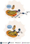
Model graph proposing how oxidative stress affects Gsdmd cleavage. In dying cells, mtROS oxidize four cysteine residues in Gsdmd and promote Gsdmd cleavage by proinflammatory caspase-1, and thus enhance Gsdmd-N domain release and cell death (above). When ROS production is inhibited, Gsdmd without oxidative modification is less cleaved by caspase-1 and blocks the enzyme activity of inflammasome-activated caspase-1, resulting in less cytokine release and cell death (below).

V体育官网 - References

    1. Abais J.M., Xia M., Zhang Y., et al. (2015). Redox regulation of NLRP3 inflammasomes: ROS as trigger or effector? Antioxid. Redox Signal. 22, 1111–1129. - PMC - PubMed
    1. Agostini L., Martinon F., Burns K., et al. (2004). NALP3 forms an IL-1β processing inflammasome with increased activity in Muckle–Wells autoinflammatory disorder. Immunity 20, 319–325. - PubMed (V体育官网入口)
    1. Ahmad S., Khan H., Shahab U. et al. (2017). Protein oxidation: an overview of metabolism of sulphur containing amino acid, cysteine. Front. Biosci. 9, 71–87. - PubMed
    1. Alfonso-Loeches S., Urena-Peralta J.R., Morillo-Bargues M.J., et al. (2014). Role of mitochondria ROS generation in ethanol-induced NLRP3 inflammasome activation and cell death in astroglial cells. Front. Cell. Neurosci. 8, 216. - PMC - PubMed
    1. Bauernfeind F., Bartok E., Rieger A., et al. (2011). Cutting edge: reactive oxygen species inhibitors block priming, but not activation, of the NLRP3 inflammasome. J. Immunol. 187, 613–617. - V体育ios版 - PMC - PubMed

Publication types

VSports注册入口 - MeSH terms