Skip to main page content
U.S. flag

An official website of the United States government

Dot gov

The VSports app下载. gov means it’s official. Federal government websites often end in . gov or . mil. Before sharing sensitive information, make sure you’re on a federal government site. .

Https

The site is secure. The https:// ensures that you are connecting to the official website and that any information you provide is encrypted and transmitted securely V体育官网. .

. 2019 Mar 4;216(3):556-570.
doi: 10.1084/jem.20181776. Epub 2019 Feb 20.

A major role for ferroptosis in VSports手机版 - Mycobacterium tuberculosis-induced cell death and tissue necrosis

Affiliations

A major role for ferroptosis in Mycobacterium tuberculosis-induced cell death and tissue necrosis

Eduardo P Amaral et al. J Exp Med. .

Abstract

Necrotic cell death during Mycobacterium tuberculosis (Mtb) infection is considered host detrimental since it facilitates mycobacterial spread. Ferroptosis is a type of regulated necrosis induced by accumulation of free iron and toxic lipid peroxides. We observed that Mtb-induced macrophage necrosis is associated with reduced levels of glutathione and glutathione peroxidase-4 (Gpx4), along with increased free iron, mitochondrial superoxide, and lipid peroxidation, all of which are important hallmarks of ferroptosis. Moreover, necrotic cell death in Mtb-infected macrophage cultures was suppressed by ferrostatin-1 (Fer-1), a well-characterized ferroptosis inhibitor, as well as by iron chelation. Additional experiments in vivo revealed that pulmonary necrosis in acutely infected mice is associated with reduced Gpx4 expression as well as increased lipid peroxidation and is likewise suppressed by Fer-1 treatment. Importantly, Fer-1-treated infected animals also exhibited marked reductions in bacterial load. Together, these findings implicate ferroptosis as a major mechanism of necrosis in Mtb infection and as a target for host-directed therapy of tuberculosis. VSports手机版.


Figures

None
Graphical abstract
Figure 1.
Figure 1.
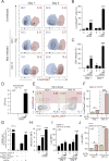
Mtb triggers necrotic cell death in BMDMs by a mechanism associated with increased intracellular labile iron, mitochondrial superoxide, and lipid peroxidation. C57BL/6 BMDMs were infected with H37Rv Mtb at different MOIs as indicated. (A and B) Sample FACS plots demonstrating Mtb-induced macrophage necrosis in vitro as measured by Live/Dead staining at day 1 versus day 4 p.i. and at different MOI. (B) Summary graph of data shown in A presenting the means ± SEM of triplicate samples analyzed. (C) Necrotic cell death measured by LDH released in the supernatants from the macrophage cultures shown in A and B. The data reported in A–C are representative results from at least six independent experiments performed. (D) The release of live mycobacteria from necrotic cells was examined by CFU quantification in macrophage culture supernatants on day 4 p.i. Results are representative of three separate experiments. (E and F) Necrosis of Mtb-infected macrophages was specifically assessed by using H37Rv-RFP infection and Live/Dead staining. (F) Summary graph of data shown in E presenting the means ± SEM of triplicate samples analyzed. (G) Intracellular free iron levels were assessed by calcein AM staining of macrophages at 24 h p.i. Representative data of two separate experiments are shown. (H) Mitochondrial superoxide was evaluated by MitoSOX staining and analyzed by flow cytometry at 24 h p.i. Results are representative of two separate experiments performed. (I) The kinetics of lipid peroxidation in CD11b+/live cells was examined by LAA staining and analyzed by flow cytometry. Representative data from one of four independent experiments are shown. (J) Lipid peroxidation in H37Rv-RFPpos or H37Rv-RFPneg CD11b+ cells was measured by flow cytometry at 24 h p.i. Results are representative of three separate experiments performed. The data shown in A–J represent the means ± SEM of triplicate samples. Statistical significance was assessed by one-way ANOVA analysis for the indicated experimental conditions (*, P < 0.05; **, P < 0.01; ***, P < 0.001). FSC, forward scatter.
Figure 2.
Figure 2.
Necrotic cell death of Mtb-infected macrophages is associated with reduced GSH levels and Gpx4 expression. C57BL/6 BMDMs were infected with H37Rv Mtb at the different MOIs indicated and the following measurements performed at 24 h p.i. (A) Intracellular GSH levels measured in whole-cell lysates. (B) Intracellular Gpx4 protein levels detected by Western blotting and quantitated by densitometry. (C) Gpx4 expression assessed by MFI of Gpx4 staining in CD11b+/live cells by flow cytometry. (D) Gpx4 expression (y axis) versus H37Rv-RFP (x axis; left) in infected cells was analyzed by flow cytometry. Necrotic cell death was simultaneously evaluated in the indicated gates (Q1–Q4; small insets) by Live/Dead staining (middle). Summary graph (right) of data shown in Q3 and Q4 (small insets) indicating the means ± SEM of triplicate samples analyzed. (E) Gpx4 expression, bacterial infection level (as measured by Mtb-RFP MFI) and lipid peroxidation in Live/Deadlow and Live/Deadhigh subsets from gate Q4 (small inset) as analyzed by flow cytometry. The data shown represent the means ± SEM of triplicate samples. Significant differences are indicated with asterisks (*, P < 0.05; **, P < 0.01; ***, P < 0.001). Results are representative of three separate experiments performed for each analysis.
Figure 3.
Figure 3.
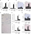
Necrotic cell death of Mtb-infected macrophages is inhibited by both iron chelation and Fer-1, a well-known ferroptosis inhibitor. C57BL/6 BMDMs were infected with H37Rv Mtb at different MOIs as indicated. (A–C) Macrophages were treated with the iron chelator PIH (1 µM). Cell death and lipid peroxidation were assessed on day 1 p.i. (A) Necrotic cell death measured by Live/Dead staining. (B) LDH release measured in supernatants from macrophage cultures. (C) Lipid peroxidation measured by LAA staining and analyzed by flow cytometry in live (left) and dead cells (right). (D) Representative images of infected macrophage cultures untreated (upper) or treated (bottom) with Fer-1 (10 µm) on day 4 p.i. Dead cells were evaluated by trypan blue staining (bars, 50 µm). (E–G) Macrophage cultures were treated with Fer-1 at the different concentrations indicated. Necrotic cell death was evaluated on day 4 p.i. (E) Necrosis assessed by Live/Dead staining and analyzed by flow cytometry. (F) LDH release measured in supernatants from macrophage cultures. (G) Lipid peroxidation measured by LAA staining and analyzed by flow cytometry in live (upper) and dead cells (bottom). Each data point represents the means ± SEMs of triplicate samples. Significant differences are indicated with asterisks (*, P < 0.05; **, P < 0.01; ***, P < 0.001). Results are representative of three independent experiments performed.
Figure 4.
Figure 4.
Gpx4, a key regulator of ferroptosis, is down-regulated in the lung following Mtb-infection. C57BL/6 mice were infected by intrapharyngeal inoculation with ∼103 bacilli of Mtb (H37Rv) as a model of severe TB (n = 4–5). (A and B) Kinetics of gpx4 (A) and slc7a11 (B) mRNA expression following Mtb infection. (C) Gpx4 expression was measured by flow cytometric analysis in CD11b+Ly6G myeloid cells in the lungs of mice at 28 d p.i. (D) Lipid peroxidation in CD11b+Ly6G myeloid subset was assessed by LAA staining and analyzed by flow cytometry. The data shown represent the means ± SEM of four to five samples per experiment. Significant differences are indicated with asterisks (*, P < 0.05; **, P < 0.01). Results are representative of two independent experiments performed.
Figure 5.
Figure 5.
Lipid peroxidation induced by Mtb in vivo is inhibited by Fer-1 treatment. (A–D) C57BL/6 mice were infected by intrapharyngeal inoculation with ∼103 bacilli of Mtb (H37Rv) as a model of severe TB and the animals sacrificed at 28 d p.i. Infected mice were treated daily by intraperitoneal injection with vehicle or Fer-1 (3 mg/kg/body weight) starting on day 15 p.i. (n = 4–5). (A) Schematic presentation of the experimental protocol. (B) Lipid peroxidation (malondialdehyde) measured in lung homogenates from uninfected and Mtb-infected mice. (C) Sample FACS plot and summary data of frequency and cell numbers of monocyte/macrophage (CD11b+Ly6G) and neutrophil subsets (CD11b+Ly6C+Ly6G+) in the lungs. (D) Lipid peroxidation in monocyte/macrophage (CD11b+Ly6G) and neutrophil subsets (CD11b+Ly6C+Ly6G+) in the lungs analyzed by flow cytometry. The data shown represent the means ± SEM of four to five samples per experiment. Significant differences are indicated with asterisks (*, P < 0.05; **, P < 0.01; ***, P < 0.001). The data shown are representative of three separate experiments performed.
Figure 6.
Figure 6.
Fer-1 treatment reduces lung pathology and CFU burden in Mtb-infected mice. (A–H) C57BL/6 mice were infected by intrapharyngeal inoculation with ∼103 bacilli of Mtb (H37Rv) as a model of severe TB and the animals sacrificed at 28 d p.i. Infected mice were treated daily by intraperitoneal injection with vehicle or Fer-1 (3 mg/kg/body weight) starting on day 15 p.i. (n = 4–5). (A–C) Lung weight (A), lung mass (B), and representative H&E image of merged lung sections from vehicle versus Fer-1–treated mice (C; bars, 250 µm). Each image is a composite of sections from three individual mice. The data are representative of three separate experiments performed. (D–F) Lung necrosis evaluated by Sytox Green DNA staining. (D) Representative bright-field versus fluorescence and merged images (bars, 500 µm) of whole-lung lobes from uninfected, infected, and infected Fer-1–treated mice either uninjected or injected with Sytox Green 20 min before euthanasia. (E and F) MFI of Sytox Green staining in whole-lung samples from five mice (E) or of individual TB lesions in these animals (F). The data are representative of two separate experiments performed. (G and H) Pulmonary (G) and splenic (H) bacterial loads in mice untreated and treated with Fer-1 at 28 d p.i. (n = 4–5). The data are representative of three separate experiments performed. The data shown represent the means ± SEM of four to five samples per experiment. Significant differences are indicated with asterisks (*, P < 0.05; **, P < 0.01; ***, P < 0.001).

Comment in

References

    1. Amaral E.P., Conceição E.L., Costa D.L., Rocha M.S., Marinho J.M., Cordeiro-Santos M., D’Império-Lima M.R., Barbosa T., Sher A., and Andrade B.B.. 2016b N-acetyl-cysteine exhibits potent anti-mycobacterial activity in addition to its known anti-oxidative functions. BMC Microbiol. 16:251 10.1186/s12866-016-0872-7 - DOI - PMC - PubMed
    1. Amaral E.P., Lasunskaia E.B., and D’Império-Lima M.R.. 2016a Innate immunity in tuberculosis: how the sensing of mycobacteria and tissue damage modulates macrophage death. Microbes Infect. 18:11–20. 10.1016/j.micinf.2015.09.005 - DOI - PubMed
    1. Andrade B.B., Pavan Kumar N., Mayer-Barber K.D., Barber D.L., Sridhar R., Rekha V.V., Jawahar M.S., Nutman T.B., Sher A., and Babu S.. 2013. Plasma heme oxygenase-1 levels distinguish latent or successfully treated human tuberculosis from active disease. PLoS One. 8:e62618 10.1371/journal.pone.0062618 - DOI - PMC - PubMed
    1. Behar S.M., Divangahi M., and Remold H.G.. 2010. Evasion of innate immunity by Mycobacterium tuberculosis: is death an exit strategy? Nat. Rev. Microbiol. 8:668–674. 10.1038/nrmicro2387 - DOI - PMC - PubMed
    1. Boelaert J.R., Vandecasteele S.J., Appelberg R., and Gordeuk V.R.. 2007. The effect of the host’s iron status on tuberculosis. J. Infect. Dis. 195:1745–1753. 10.1086/518040 - DOI - PubMed

Publication types

MeSH terms

Substances