Skip to main page content
U.S. flag

An official website of the United States government

Dot gov

The VSports app下载. gov means it’s official. Federal government websites often end in . gov or . mil. Before sharing sensitive information, make sure you’re on a federal government site. .

Https

The site is secure V体育官网. The https:// ensures that you are connecting to the official website and that any information you provide is encrypted and transmitted securely. .

. 2019 Feb 13;10(1):57.
doi: 10.1186/s13287-019-1167-3.

Inhibition of PTGS1 promotes osteogenic differentiation of adipose-derived stem cells by suppressing NF-kB signaling

Affiliations

Inhibition of PTGS1 promotes osteogenic differentiation of adipose-derived stem cells by suppressing NF-kB signaling

Yuejun Wang et al. Stem Cell Res Ther. .

Abstract

Background: Tissue inflammation is an important problem in the field of human adipose-derived stem cell (ASC)-based therapeutic bone regeneration. Many studies indicate that inflammatory cytokines are disadvantageous for osteogenic differentiation and bone formation. Therefore, overcoming inflammation would be greatly beneficial in promoting ASC-mediated bone regeneration. The present study aims to investigate the potential anti-inflammatory role of Prostaglandin G/H synthase 1 (PTGS1) during the osteogenic differentiation of ASCs. VSports手机版.

Methods: We performed TNFα treatment to investigate the response of PTGS1 to inflammation V体育安卓版. Loss- and gain-of-function experiments were applied to investigate the function of PTGS1 in the osteogenic differentiation of ASCs ex vivo and in vivo. Western blot and confocal analyses were used to determine the molecular mechanism of PTGS1-regulated osteogenic differentiation. .

Results: Our work demonstrates that PTGS1 expression is significantly increased upon inflammatory cytokine treatment. Both ex vivo and in vivo studies indicate that PTGS1 is required for the osteogenic differentiation of ASCs. Mechanistically, we show that PTGS1 regulates osteogenesis of ASCs via modulating the NF-κB signaling pathway V体育ios版. .

Conclusions: Collectively, this work confirms that the PTGS1-NF-κB signaling pathway is a novel molecular target for ASC-mediated regenerative medicine. VSports最新版本.

Keywords: ASCs; NF-κB; Osteogenic differentiation; PTGS1. V体育平台登录.


Conflict of interest statement

Ethics approval and consent to participate

This study was carried out in strict accordance with the recommendations of the Guide for the Care and Use of Laboratory Animals of the National Institutes of Health VSports注册入口. The protocol was approved by the Institutional Animal Care and Use Committee of the Peking University Health Science Center (approval no. LA2014233). All surgeries were performed under anesthesia, and all efforts were made to minimize animal suffering.

Consent for publication

Not applicable.

Competing interests

The authors declare that they have no competing interests.

Publisher’s Note

Springer Nature remains neutral with regard to jurisdictional claims in published maps and institutional affiliations.

Figures

Fig. 1
Fig. 1
PTGS1 expression is stimulated by TNFα treatment. a, b PTGS1 expression was significantly induced when treated with TNFα for 0.5, 1, and 2 h as determined by real-time RT-qPCR and Western blot analysis. GAPDH was used as an internal control. All data are shown as the mean ± SD, n = 3. **P < 0.01. h hour
Fig. 2
Fig. 2
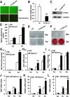
Knockdown of PTGS1 enhances the osteogenic differentiation of ASCs in vitro. a GFP-positive ASCs under light and fluorescence microscopy. Scale bar, 500 μm. b, c Knockdown of PTGS1 was verified by real-time RT-qPCR and Western blot. d, e PTGS1 silencing increased the ALP activity and ALP staining in ASCs. f, g PTGS1 knockdown increased mineralization, as shown by Alizarin red staining and quantitative calcium analysis. hl The mRNA expressions of ALP, OCN, BSP, RUNX2, and OSX were determined by RT-qPCR. GAPDH was used as an internal control. All data are shown as the mean ± SD, n = 3. **P < 0.01. NC negative control cells, PTGS1sh PTGS1 knockdown cells, d day, w week
Fig. 3
Fig. 3
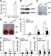
PTGS1 overexpression inhibits the osteogenic differentiation of ASCs in vitro. a, b EGFP-PTGS1-infected ASCs showed overexpression of PTGS1, as shown by real-time RT-qPCR and Western blot. c PTGS1 overexpression reduced ALP staining. d PTGS1 overexpression decreased mineralization, as shown by Alizarin red staining. eh Real-time RT-qPCR analysis of the expression of ALP, OCN, RUNX2, and OSX. GAPDH was used as an internal control. All data are shown as the mean ± SD, n = 3. **P < 0.01. w week
Fig. 4
Fig. 4
PTGS1 affects osteogenesis of ASCs in vivo. a PTGS1 knockdown promoted ASC-mediated bone-like tissue formation in vivo, as measured by HE staining, Masson’s trichrome staining, and immunohistochemistry staining of OCN in PTGS1sh and NC groups. Scale bar represents 50 μm. b Quantitative measurements of bone-like tissues showed that the area of bone formation was notably increased in PTGS1 knockdown cells compared with NC cells. c, d PTGS1 overexpression impaired the osteogenic differentiation in ASCs, as shown by HE staining, Masson’s trichrome staining, immunohistochemistry staining of OCN, and quantitative measurements of bone-like tissue. Scale bar represents 50 μm. All data are shown as the mean ± SD, n = 3. **P < 0.01
Fig. 5
Fig. 5
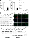
Knockdown of PTGS1 inhibits the translocation of p65 from the cytoplasm to the nucleus. ac Real-time RT-qPCR showed that expressions of p65 target genes IL6, IL8, and SELE were reduced when PTGS1 was silenced with a short hairpin RNA. GAPDH was used as an internal control. d The protein levels of p-IκBα, total IκBα, p-p65, and total p65 were measured by Western blot in the absence and presence of TNFα for 0.5 h. e The cellular localization of endogenous p65 was observed by confocal microscopy in both control and PTGS1 knockdown cells with or without TNFα treatment. Scale bars, 100 μm. f The nuclear and cytoplasmic levels of p65, tubulin, and PCAF were measured by Western blot after subcellular fractionation from PTGS1sh cells untreated or treated with TNFα for 0.5 h. GAPDH was used as an internal control. All data are shown as the mean ± SD, n = 3. **P < 0.01. NC negative control cells, PTGS1sh PTGS1 knockdown cells
Fig. 6
Fig. 6
PTGS1 promotes the translocation of p65 from the cytoplasm to the nucleus. a The protein levels of p-IκBα, total IκBα, p-p65, and total p65 were measured by Western blot in the absence and presence of TNFα for 0.5 h. b The cellular localization of endogenous p65 was observed by confocal microscopy in both control and PTGS1-overexpressing cells with or without TNFα treatment. Scale bars, 100 μm. c The nuclear and cytoplasmic protein levels of p65, tubulin, and PCAF were measured by Western blot after subcellular fractionation from EGFP-PTGS1 and vector groups untreated or treated with TNFα for 0.5 h. GAPDH was used as an internal control. All data are shown as the mean ± SD, n = 3

References

    1. Ito K, Yamada Y, Nagasaka T, Baba S, Ueda M. Osteogenic potential of injectable tissue-engineered bone: a comparison among autogenous bone, bone substitute (Bio-Oss), platelet-rich plasma, and tissue-engineered bone with respect to their mechanical properties and histological findings. J Biomed Mater Res A. 2005;73:63–67. doi: 10.1002/jbm.a.30248. - DOI (VSports注册入口) - PubMed
    1. Levi B, Longaker M. Concise review: adipose-derived stromal cells for skeletal regenerative medicine. Stem Cells. 2011;29:576–582. doi: 10.1002/stem.612. - DOI - PMC - PubMed
    1. Wilson A, Butler PE, Seifalian AM. Adipose-derived stem cells for clinical applications: a review. Cell Prolif. 2011;44:86–98. doi: 10.1111/j.1365-2184.2010.00736.x. - DOI - PMC - PubMed
    1. Zuk PA, Zhu M, Ashjian P, De Ugarte DA, Huang JI, Mizuno H, et al. Human adipose tissue is a source of multipotent stem cells. Mol Biol Cell. 2002;13:4279–4295. doi: 10.1091/mbc.e02-02-0105. - DOI - PMC - PubMed
    1. Smith WL, DeWitt DL, Garavito RM. Cyclooxygenases: structural, cellular, and molecular biology. Annu Rev Biochem. 2000;69:145–182. doi: 10.1146/annurev.biochem.69.1.145. - DOI - PubMed

Publication types

MeSH terms