"VSports手机版" Skip to main page content
U.S. flag

An official website of the United States government

Dot gov

The . gov means it’s official. Federal government websites often end in . gov or VSports app下载. mil. Before sharing sensitive information, make sure you’re on a federal government site. .

Https

The site is secure V体育官网. The https:// ensures that you are connecting to the official website and that any information you provide is encrypted and transmitted securely. .

. 2019 Nov;26(11):2284-2299.
doi: 10.1038/s41418-019-0299-4. Epub 2019 Feb 8.

Ischemia-induced ACSL4 activation contributes to ferroptosis-mediated tissue injury in intestinal ischemia/reperfusion

Affiliations

"VSports手机版" Ischemia-induced ACSL4 activation contributes to ferroptosis-mediated tissue injury in intestinal ischemia/reperfusion

"V体育2025版" Yang Li et al. Cell Death Differ. 2019 Nov.

Abstract

Ferroptosis is a recently identified form of regulated cell death defined by the iron-dependent accumulation of lipid reactive oxygen species. Ferroptosis has been studied in various diseases such as cancer, Parkinson's disease, and stroke. However, the exact function and mechanism of ferroptosis in ischemia/reperfusion (I/R) injury, especially in the intestine, remains unknown. Considering the unique conditions required for ferroptosis, we hypothesize that ischemia promotes ferroptosis immediately after intestinal reperfusion. In contrast to conventional strategies employed in I/R studies, we focused on the ischemic phase. Here we verified ferroptosis by assessing proferroptotic changes after ischemia along with protein and lipid peroxidation levels during reperfusion. The inhibition of ferroptosis by liproxstatin-1 ameliorated I/R-induced intestinal injury. Acyl-CoA synthetase long-chain family member 4 (ACSL4), which is a key enzyme that regulates lipid composition, has been shown to contribute to the execution of ferroptosis, but its role in I/R needs clarification VSports手机版. In the present study, we used rosiglitazone (ROSI) and siRNA to inhibit ischemia/hypoxia-induced ACSL4 in vivo and in vitro. The results demonstrated that ACSL4 inhibition before reperfusion protected against ferroptosis and cell death. Further investigation revealed that special protein 1 (Sp1) was a crucial transcription factor that increased ACSL4 transcription by binding to the ACSL4 promoter region. Collectively, this study demonstrates that ferroptosis is closely associated with intestinal I/R injury, and that ACSL4 has a critical role in this lethal process. Sp1 is an important factor in promoting ACSL4 expression. These results suggest a unique and effective mechanistic approach for intestinal I/R injury prevention and treatment. .


"V体育2025版" Conflict of interest statement

The authors declare that they have no conflict of interest.

Figures (V体育2025版)
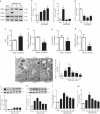
Fig. 1
Fig. 1
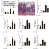
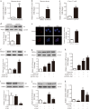
Ferroptosis is present in intestinal I/R injury. ad Mice were subjected to intestinal ischemia (I) or sham surgery (sham) and divided into four groups: sham, I30min, I45min, and I60min (n = 6). Intestines were collected after ischemia. The expressions of ACSL4, GPx4, and FTH1 were detected by western blotting (n = 3). eh Mice were subjected to 45 min of intestinal ischemia and intestines were collected to measure iron, GPx4 activity, GSH level, and the GSH/GSSG ratio after ischemia (n = 6). im Mice were subjected to 45 min of ischemia and different durations of reperfusion (R15min, R30min, R60min, R120min, and R240min). All samples were collected at the respective reperfusion times. i Representative TEM images and quantification are shown. The white arrow indicates outer mitochondrial membrane rupture and the black arrows indicate the reduction or disappearance of mitochondrial cristae. j, k GPx4 and COX2 protein levels in intestines were analyzed by western blotting (n = 3). l, m 12-HETE and 15-HETE levels were assayed by ELISA kits (n = 6). All results are expressed as the mean ± SD. *p < 0.05, **p < 0.01 vs. the sham group
Fig. 2
Fig. 2
Liproxstatin-1 ameliorates ferroptosis and intestinal injury in vivo. Mice were treated with liproxstatin-1 (10 mg/kg) by intraperitoneal injection 1 h before ischemia and then subjected to ischemia/reperfusion (I, 45 min of ischemia; R, 30 min of reperfusion) or sham surgery (sham). All samples were collected after I/R. a GPx4 and COX2 levels were assessed by western blotting (n = 3). b Representative H&E-stained intestinal slices were imaged by microscopy (scale bar = 100μm). c The intestinal permeability was detected by measuring serum FD-4 content (n = 6). df 12-HETE, 15-HETE, and LPO were assayed by corresponding kits (n = 6). gi Serum from mice was used to detect the levels of LDH, TNF-α, and IL-6 (n = 6). All results are expressed as the mean ± SD. **p < 0.01 vs. the sham group; #p < 0.05 vs. the I/R group
Fig. 3
Fig. 3
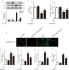
Liproxstatin-1 inhibits lipid peroxidation and cell death in vitro. Caco-2 cells were pretreated with liproxstatin-1 (200 nM) for 12 h before being subjected to H/R (H, 12 h of hypoxia; R, 2 h of reoxygenation). All samples were collected after H/R. a GPx4 and COX2 expression in cells were detected by western blotting (n = 3). b Cell survival was determined by CCK-8 kit (n = 6). c Transepithelial electrical resistance (TEER) level (n = 6). dg Cell lipid peroxidation were detected by BODIPY 581/591 C11 staining using fluorescence microscopy (scale bar = 100 μm) and 12-HETE, 15-HETE, and LPO assay kits (n = 6). h The level of LDH (n = 6). All results are expressed as the mean ± SD. **p < 0.01 vs. the control group; #p < 0.05, ##p < 0.01 vs. the H/R group
Fig. 4
Fig. 4
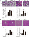
Liproxstatin-1 protects remote organs after intestinal I/R. Mice were treated with liproxstatin-1 (10 mg/kg) by intraperitoneal injection 1 h before ischemia and then subjected to ischemia/reperfusion (I, 45 min of ischemia; R, 30 min of reperfusion) or sham surgery (sham). All samples were collected after I/R. a,d Slices of remote organs (liver and lung) were stained by H&E and representative images were acquired by microscopy (scale bar = 100 μm). b,c Lung injury was measured by the wet/dry ratio and MPO assay kit (n = 6). e,f Liver injury was assessed by the Eckhoff’s score and MPO assay kit (n = 6). All results are expressed as the mean ± SD. **p < 0.01 vs. the sham group; #p < 0.05, ##p < 0.01 vs. the I/R group
Fig. 5
Fig. 5
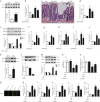
ACSL4 regulates ferroptosis in vivo and in vitro. a Western blotting was used to determine ACSL4 expression in normal and ischemic human intestines (n = 3). **p < 0.01 vs. the normal group. bj ROSI (0.4 mg/kg, intravenous injection, 1 h before ischemia) was administered to mice. Then mice were subjected to ischemia, I/R, or sham surgery (I, 45 min of ischemia; R, 30 min of reperfusion). b ACSL4 activity in 45 min ischemic intestines (n = 6). *p < 0.05, **p < 0.01 vs. the sham group; #p < 0.05 vs. the ischemia group. c Representative H&E-stained images were acquired by microscopy after I/R (scale bar = 100 μm). d The intestinal permeability was detected by measuring serum FD-4 content after I/R (n = 6). e GPx4 and COX2 protein levels were assessed by western blotting after I/R (n = 3). fi Lipid peroxidation was analyzed by 12-HETE, 15-HETE, 5-HETE, and LPO kits after I/R (n = 6). j Serum from mice was used to detect the level of LDH after I/R (n = 6). Results are expressed as the mean ± SD. *p < 0.05, **p < 0.01 vs. the sham group; #p < 0.05, ##p < 0.01 vs. the I/R group. ku Caco-2 cells were transfected with si-NC or si-ACSL4 for 2 days before hypoxia or H/R (H, 12 h of hypoxia; R, 2 h of reoxygenation). k,l ACSL4 expression under hypoxia for 12 h or after siRNA transfection was assessed by western blotting (n = 3). m GPx4 and COX2 protein levels were determined by western blotting after H/R (n = 3). n Cell survival was measured by CCK-8 kit after H/R (n = 6). o Transepithelial electrical resistance (TEER) after H/R (n = 6). pt Cell lipid peroxidation were detected by BODIPY 581/591 C11 staining using fluorescence microscopy (scale bar = 100 μm) and 12-HETE, 15-HETE, 5-HETE, and LPO assay kits after H/R (n = 6). u The level of released LDH after H/R (n = 6). Results are expressed as the mean ± SD. **p < 0.01 vs. the control group; #p < 0.05 vs. the H/R group
Fig. 6
Fig. 6
Sp1 regulates ACSL4 transcription and expression. ac Ischemic human intestines, 45 min ischemic mouse intestines and 12 h hypoxic Caco-2 cells were used for the determination of ACSL4 mRNA levels by qPCR (n = 3). **p < 0.01 vs. the normal/sham/normoxia groups. d, e Expression of nuclear Sp1 at 12 h of hypoxia was determined by western blotting (n = 3) and laser scanning confocal microscopy (n = 6). **p < 0.01 vs. the normoxia group. fk Caco-2 cells were transfected with Sp1 plasmid or siRNA for 2 days, and then subjected to 12 h of hypoxia. The Sp1 protein levels, the protein and mRNA levels of ACSL4 were determined after hypoxia (n = 3). Results are expressed as the mean ± SD. *p < 0.05, **p < 0.01 vs. the control group; #p < 0.05, ##p < 0.01 vs. the hypoxia group
Fig. 7
Fig. 7
Sp1 binds the ACSL4 promoter region. Caco-2 cells were collected for assay with or without 12 h of hypoxia. a, b Cells cotransfected with the Sp1 and WT (− 500~ + 200) plasmids were collected and luciferase activity was analyzed (n = 3). *p < 0.05 vs. the empty vector group. c ChIP assay results for three sites were presented on agarose gels, grouped as input, positive and Sp1 (n = 3). d,e Cells transfected with two Del plasmids were used in the luciferase assay and the results were normalized to those of the WT group (n = 3). **p < 0.01, ***p < 0.001 vs. the WT group

References

    1. Acosta S. Epidemiology of mesenteric vascular disease: clinical implications. Semin Vasc Surg. 2010;23:4–8. doi: 10.1053/j.semvascsurg.2009.12.001. - DOI - PubMed
    1. Mallick IH, Yang WX, Winslet MC, Seifalian AM. Ischemia-reperfusion injury of the intestine and protective strategies against injury. Dig Dis Sci. 2004;49:1359–77. doi: 10.1023/B:DDAS.0000042232.98927.91. - DOI - PubMed
    1. Stone JR, Wilkins LR. Acute mesenteric ischemia. Tech Vasc Interv Radiol. 2015;18:24–30. doi: 10.1053/j.tvir.2014.12.004. - "V体育官网入口" DOI - PubMed
    1. Cheng J, Wei Z, Liu X, Li X, Yuan Z, Zheng J, et al. The role of intestinal mucosa injury induced by intra-abdominal hypertension in the development of abdominal compartment syndrome and multiple organ dysfunction syndrome. Crit Care. 2013;17:R283. doi: 10.1186/cc13146. - VSports注册入口 - DOI - PMC - PubMed
    1. Linkermann A, Brasen JH, Himmerkus N, Liu S, Huber TB, Kunzendorf U, et al. Rip1 (receptor-interacting protein kinase 1) mediates necroptosis and contributes to renal ischemia/reperfusion injury. Kidney Int. 2012;81:751–61. doi: 10.1038/ki.2011.450. - DOI - PubMed

MeSH terms

LinkOut - more resources