Skip to main page content
U.S. flag

An official website of the United States government

Dot gov

The . gov means it’s official. Federal government websites often end in . gov or . mil VSports app下载. Before sharing sensitive information, make sure you’re on a federal government site. .

Https

The site is secure V体育官网. The https:// ensures that you are connecting to the official website and that any information you provide is encrypted and transmitted securely. .

. 2019 Apr 15;79(8):1913-1924.
doi: 10.1158/0008-5472.CAN-18-3037. Epub 2019 Feb 1.

The Deubiquitylase OTUB1 Mediates Ferroptosis via Stabilization of SLC7A11

Affiliations

The Deubiquitylase OTUB1 Mediates Ferroptosis via Stabilization of SLC7A11

Tong Liu et al. Cancer Res. .

Abstract

Although cell-cycle arrest, senescence, and apoptosis are established mechanisms of tumor suppression, accumulating evidence reveals that ferroptosis, an iron-dependent, nonapoptotic form of cell death, represents a new regulatory pathway in suppressing tumor development. Ferroptosis is triggered by lipid peroxidation and is tightly regulated by SLC7A11, a key component of the cystine-glutamate antiporter. Although many studies demonstrate the importance of transcriptional regulation of SLC7A11 in ferroptotic responses, it remains largely unknown how the stability of SLC7A11 is controlled in human cancers. In this study, we utilized biochemial purification to identify the ubiquitin hydrolase OTUB1 as a key factor in modulating SLC7A11 stability. OTUB1 directly interacted with and stabilized SLC7A11; conversely, OTUB1 knockdown diminished SLC7A11 levels in cancer cells. OTUB1 was overexpressed in human cancers, and inactivation of OTUB1 destabilized SLC7A11 and led to growth suppression of tumor xenografts in mice, which was associated with reduced activation of ferroptosis. Notably, overexpression of the cancer stem cell marker CD44 enhanced the stability of SLC7A11 by promoting the interaction between SLC7A11 and OTUB1; depletion of CD44 partially abrogated this interaction VSports手机版. CD44 expression suppressed ferroptosis in cancer cells in an OTUB1-dependent manner. Together, these results show that OTUB1 plays an essential role in controlling the stability of SLC7A11 and the CD44-mediated effects on ferroptosis in human cancers. SIGNIFICANCE: This study identifies OTUB1 as a key regulator of ferroptosis and implicates it as a potential target in cancer therapy. See related commentary by Gan, p. 1749. .


"V体育官网入口" Conflict of interest statement

There are no potential conflicts of interest disclosed.

Figures
Figure 1.
Figure 1.. OTUB1 is a bona fide binding partner of SLC7A11 both in vitro and in vivo.
A. Schematic representation of the SLC7A11 protein used for protein complex purification. B. Coomassie blue staining of affinity-purified protein complexes from Flag-HA-SLC7A11 H1299 stable cell line (lane 3) and the parental H1299 cell line (lane 2) in the upper panel. Specific SLC7A11-interacting protein bands were analyzed by LC-MS/MS and identified OTUB1 peptide sequences in the lower panel. C. Western Blot analysis for OTUB1 after immunoprecipitation (IP) of Flag-SLC7A11, with anti-Flag (M2) antibody-coupled beads, from H1299 cells transfected with Flag-SLC7A11 and OTUB1 individually or together. 1% of the sample was loaded as input. D. Western Blot analysis for SLC7A11 after immunoprecipitation of Flag-OTUB1, with anti-Flag (M2) antibody-coupled beads, from H1299 cells transfected with Flag-SLC7A11 and OTUB1 individually or together. 2.5% of the sample was loaded as input. E. Western Blot analysis for endogenous OTUB1 after immunoprecipitation of endogenous SLC7A11 from parental SK-N-BE(2)C cells. F. Western Blot analysis for endogenous SLC7A11 after immunoprecipitation of endogenous OTUB1 from parental SK-N-BE(2)C cells. G. Western Blot analysis for pulldown of purified Flag-SLC7A11 incubated with purified GST or GST-OTUB1 fusion protein. 1% of the cell lysate was loaded as input. Ponceau staining of GST and GST-OTUB1 is shown in the bottom panel.
Figure 2.
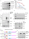
Figure 2.. OTUB1 acts as a major regulator for SLC7A11 stability in human cancer cells.
A. Western Blot analysis for SLC7A11(Long Exposure, LE and Short Exposure, SE) and Flag-OTUB1, from H1299 cells transfected with an SLC7A11-expressing plasmid and either an empty vector or increasing amounts of a Flag-OTUB1-expressing vector. B. Densitometry quantification of SLC7A11 protein levels calculated using ImageJ software (National Institutes of Health [NIH], Bethesda, MD) and plotted for half-life determination corresponding to Supplemental Fig. 2A. Error bars represent the mean ± SEM of 3 independent experiments. C. Western Blot analysis for HA-ubiquitin(Ub) after incubation of anti-Flag-coupled (M2) beads with lysates from HEK293 cells transfected with empty vector (–) or those expressing Flag-SLC7A11 either alone or together with OTUB1. D. Western Blot analysis for SLC7A11 and OTUB1 from H1299 cells transfected with control siRNA (ctrl) or a pool of OTUB1-specific siRNAs or the individual OTUB1 oligos composing the pool for 72 hours. E. Western Blot analysis for SLC7A11 and OTUB1 from U2OS cells transfected with control siRNA (ctrl) or OTUB1 siRNA and treated with DMSO/MG132 10μg/ml for 10 hours. F. A schematic structure of wild-type OTUB1 (top panel), E2-conjugating enzyme binding defect mutant, OTUB1 D88A (middle panel) and catalytic activity defect mutant, OTUB1 C91A (bottom panel). G. Western Blot analysis for SLC7A11 and Flag-OTUB1 WT, D88A and C91A, from H1299 cells transfected with an SLC7A11-expressing plasmid and either an empty vector or indicated Flag-OTUB1-expressing vectors (WT, D88A, C91A).
Figure 3.
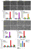
Figure 3.. OTUB1 inactivation promotes ferroptosis in human cancer cells primarily by down-regulating SLC7A11 levels.
A. Representative phase-contrast images of H1299 parental cells and OTUB1 CRISPR clones treated with TBH (40 μM) and Ferr-1 (2 μM) as indicated for 6 hours. B.C.D. H1299 control cells and OTUB1-null cells were treated with TBH (40 μM) for 6 hours, erastin (25 μM) for 16 hours and cystine starvation for 48 hours or together with Ferr-1(2 μM) as indicated respectively. Quantification of cell death from three replicates is shown; error bars represent the mean ±s.d. E. Representative phase-contrast images of SK-N-BE(2)C parental cells and OTUB1-null cells treated with erastin (35 μM) and Ferr-1 (2 μM) as indicated for 20 hours. F.G. SK-N-BE(2)C parental cells and OTUB1-null cells were treated with erastin (35 μM) for 20 hours and TBH (40 μM) for 7 hours. Quantification of cell death from three replicates is shown; error bars represent the mean ± s.d. H. SK-N-BE(2)C parental cells and OTUB1-null cells were transfected with empty vector or a SLC7A11 expressing plasmid and treated with TBH (250 μM) and Ferr-1 (2 μM) as indicated for 7 hours. Quantification of cell death from three replicates is shown; error bars represent the mean ± s.d. I. Cystine uptake levels (D.P.M., disintegrations per minutes) were measured in U2OS control cells or U2OS OTUB1-null treated with erastin (30 μM) for 6 hours as indicated.
Figure 4.
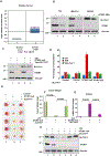
Figure 4.. OTUB1 is overexpressed in human cancers and the OTUB1-SLC7A11 interaction is critical for tumor growth.
A. Box plots derived from gene expression data in ONCOMINE (https://www.oncomine.org/resource/login.html), comparing the expression of OTUB1 mRNA in 3 normal human samples (left plots) and 39 cancer tissues (right plots) from bladder cancer samples. B. Western blot analysis for OTUB1 and SLC7A11 from T24, UM-UC-3 and SW780 cells transfected with control siRNA (ctrl) or a pool of OTUB1-specific siRNAs for 72 hours. C. Western Blot analysis for OTUB1 and SLC7A11 in T24 control cells and OTUB1-null cells that were transfected with either an empty vector or SLC7A11. D. T24 control cells and OTUB1-null cells were transfected with SLC7A11 as in Fig 4C and then treated with TBH (10 μM) and Ferr-1 (2 μM) as indicated for 16 hours. Quantification of cell death from three technical replicates is shown; error bars represent the mean ± s.d. E. Image of xenograft tumors that were inoculated into nude mice with T24 control cells, OTUB1-null cells and OTUB1-null cells stably transfected with SLC7A11 expression vector for 9 weeks. F. Weight (mg) of tumors as shown in Fig 4E was determined and compared, error bars represent the mean ±s.d. from 5 tumors per group. G. PTGS2 mRNA level from tumors was determined by qPCR; three replicates are shown; error bars represent the mean ± s.d. H. Western Blot analysis for OTUB1 and SLC7A11 from xenograft tumors obtained from the groups of T24 control cells, OTUB1-null cells and OTUB1-null cells stably transfected with SLC7A11 expression vector.
Figure 5.
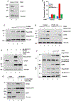
Figure 5.. The OTUB1-SLC7A11 interaction is tightly regulated by CD44 in human cancer cells.
A. Western Blot analysis for CD44 and SLC7A11 from H1299 cells transfected with control siRNA or a pool of CD44-specific siRNAs. B. H1299 cells transfected with control siRNA or a pool of CD44 siRNAs and then treated with TBH (40 μM) and Ferr-1 (2 μM) as indicated for 6 hours. Quantification of cell death from three technical replicates is shown; error bars represent the mean ±s.d. C. Western Blot analysis for SLC7A11, OTUB1 and CD44 from H1299 cells transfected with HA-SLC7A11 and either an empty vector or increasing amounts of a Flag-CD44 expressing vector. D. Western Blot analysis for SLC7A11, OTUB1 and CD44 from T24 control cells and OTUB1-null cells transfected with HA-SLC7A11 and either an empty vector or increasing amounts of a CD44-expressing vector as indicated. E. Western Blot analysis for HA-SLC7A11, OTUB1 and CD44 from H1299 control cells and OTUB1 CRISPR cells transfected with HA-SLC7A11 and either an empty vector or a CD44-expressing vector. F. Western Blot analysis for OTUB1 after immunoprecipitation of SLC7A11, with anti-HA antibody-coupled beads, from HA-SLC7A11 stable H1299 cells transfected with CD44 and Flag-OTUB1 as indicated. The amount of SLC7A11 was normalized after immunoprecipitation. 1% of the sample was loaded as input. G. Western Blot analysis for OTUB1 after immunoprecipitation of Flag-SLC7A11, with anti-Flag (M2) antibody-coupled beads, from H1299 cells transfected with control siRNA or a pool of CD44-specific siRNAs, Flag-SLC7A11 and OTUB1 individually or together as indicated. The amount of SLC7A11 was normalized after immunoprecipitation. 1% of the sample was loaded as input.
Figure 6.
Figure 6.. CD44 and OTUB1 interact with different domains of SLC7A11.
A. Western Blot analysis for CD44 after immunoprecipitation of SFB-OTUB1, with Streptavidin-coupled beads, from HEK293 cells transfected with CD44 and SFB-OTUB1 individually or together as indicated. 1% of the sample was loaded as input. B. A schematic structure of wild-type Flag-HA tagged SLC7A11 Full length (FL, top panel), 1–43 amino acids N terminal deletion (ΔNT), 471–501 amino acids C terminal deletion (ΔCT) (bottom panel). 1% of the sample was loaded as input. C. Western Blot analysis for HA-SLC7A11/ΔNT/ΔCT after immunoprecipitation (IP) of SFB-OTUB1 with Streptavidin-coupled beads, from HEK293 cells transfected with HA-SLC7A11 FL or ΔNT/ΔCT and SFB-OTUB1 individually or together. 1% of the sample was loaded as input. D. Western Blot analysis for CD44 after immunoprecipitation (IP) of HA-SLC7A11, with anti-HA antibody-coupled beads, from HEK293 cells transfected with HA-SLC7A11 FL or ΔNT/ΔCT and CD44 individually or together. 1% of the sample was loaded as input. E. T24 cells transfected with or without CD44 and then treated with erastin (15 μM) as indicated for 70 hours. Quantification of cell death from three replicates is shown; error bars represent the mean ± s.d. F. T24 OTUB1 CRISPR cells transfected with or without CD44 and then treated with erastin (15 μM) as indicated for 70 hours. Quantification of cell death from three replicates is shown; error bars represent the mean ± s.d.
Figure 7.
Figure 7.. Model of Deubiquitination of SLC7A11 by OTUB1 inhibits ferroptosis and promotes tumorigenesis.
Schematic model where OTUB1 stabilizes SLC7A11 through deuibiquitination of SLC7A11, which is enhanced by CD44. OTUB1 inhibits ferroptosis and promotes tumorigenesis.

Comment in

References

    1. Xie Y, Hou W, Song X, Yu Y, Huang J, Sun X, et al. Ferroptosis: process and function. Cell death and differentiation 2016;23:369–79 - "V体育官网" PMC - PubMed
    1. Gao M, Jiang X. To eat or not to eat-the metabolic flavor of ferroptosis. Current opinion in cell biology 2017;51:58–64 - PMC - PubMed
    1. Stockwell BR, Friedmann Angeli JP, Bayir H, Bush AI, Conrad M, Dixon SJ, et al. Ferroptosis: A Regulated Cell Death Nexus Linking Metabolism, Redox Biology, and Disease. Cell 2017;171:273–85 - PMC - PubMed
    1. Zhang Y, Shi J, Liu X, Feng L, Gong Z, Koppula P, et al. BAP1 links metabolic regulation of ferroptosis to tumour suppression. Nature cell biology 2018;20:1181–92 - PMC - PubMed
    1. Conrad M, Sato H. The oxidative stress-inducible cystine/glutamate antiporter, system × (c) (−): cystine supplier and beyond. Amino acids 2012;42:231–46 - PubMed