V体育安卓版 - Skip to main page content
U.S. flag

An official website of the United States government

Dot gov

The . gov means it’s official. Federal government websites often end in . gov or . mil. Before sharing sensitive information, make sure you’re on a federal government site. VSports app下载.

Https

The site is secure. The https:// ensures that you are connecting to the official website and that any information you provide is encrypted and transmitted securely V体育官网. .

. 2019 Mar;52(2):e12568.
doi: 10.1111/cpr.12568. Epub 2019 Jan 31.

Blockage of SLC31A1-dependent copper absorption increases pancreatic cancer cell autophagy to resist cell death

Affiliations

Blockage of SLC31A1-dependent copper absorption increases pancreatic cancer cell autophagy to resist cell death

Ze Yu et al. Cell Prolif. 2019 Mar.

Abstract (V体育平台登录)

Objectives: Clinical observations have demonstrated that copper levels elevate in several cancer types, and copper deprivation is shown to inhibit tumour angiogenesis and growth in both animal models and preclinical trials. However, the content of copper in pancreatic duct adenocarcinoma (PDAC) and whether it is a potential therapy target is still unknown VSports手机版. .

Materials and methods: The levels of copper in PDAC specimens were detected by ICP-MS assays. Copper depletion in Panc-1 or MiaPaCa-2 cells was conducted via copper transporter 1 (SLC31A1) interference and copper chelator tetrathiomolybdate (TM) treatment. The effects of copper deprivation on cancer cells were evaluated by cell proliferation, migration, invasion, colony formation and cell apoptosis V体育安卓版. The mechanism of copper deprivation-caused cancer cell quiescence was resolved through mitochondrial dysfunction tests and autophagy studies. The tumour-suppression experiments under the condition of copper block and/or autophagy inhibition were performed both in vitro and in xenografted mice. .

Results: SLC31A1-dependent copper levels are correlated with the malignant degree of pancreatic cancer. Blocking copper absorption could inhibit pancreatic cancer progression but did not increase cell death. We found that copper deprivation increased mitochondrial ROS level and decreased ATP level, which rendered cancer cells in a dormant state. Strikingly, copper deprivation caused an increase in autophagy to resist death of pancreatic cancer cells. Simultaneous treatment with TM and autophagy inhibitor CQ increased cell death of cancer cells in vitro and retarded cancer growth in vivo. V体育ios版.

Conclusions: These findings reveal that copper deprivation-caused cell dormancy and the increase in autophagy is a reason for the poor clinical outcome obtained from copper depletion therapies for cancers. Therefore, the combination of autophagy inhibition and copper depletion is potentially a novel strategy for the treatment of pancreatic cancer and other copper-dependent malignant tumours VSports最新版本. .

Keywords: SLC31A1; autophagy; copper; dormancy; pancreatic cancer V体育平台登录. .


Conflict of interest statement (VSports在线直播)

All co‐authors implicated in this research approved this article to be published. The authors declare that they have no conflict of interest V体育官网入口.

Figures (VSports在线直播)

Figure 1
Figure 1
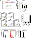
Copper content increased in pancreatic cancer tissues. A, the copper content was determined by ICPMS assay in pancreatic cancer (P1‐P10 and six mixed samples) and adjacent non‐cancer (AC) tissue specimens. Data shown are mean ± SD, n = 6, **P < 0.01 (Student's t test). B, the correlation between copper content and the survival time was analyzed in eight patients. C, GEO data analysis of Slc31a1 expression in pancreatic cancer and normal tissues. D, The SLC31A1 protein expression was examined by immunohistochemical staining. E, the correlation of Slc31a1 mRNA levels and the survival time was analyzed in 87 patients using data from the OncoLnc database
Figure 2
Figure 2
Slc31a1 knock‐down inhibited pancreatic cancer progression. A, after transfection of 50 nmol/L si‐Slc31a1 or NC for 72 h, the mRNA or protein expressions of SLC31A1 in Panc‐1 cells were measured by qPCR or Western blot, respectively. β‐actin was used as an internal control. B and C, the effect of si‐Slc31a1 (50 nmol/L) on Panc‐1 and MiaPaCa‐2 cell growth was measured by CCK‐8 assay for 5 days. D, the migration of Panc‐1 cells transfected with si‐nc or si‐Slc31a1 was tested via wound healing assay. The representative pictures were shown at 0 and 48 h after the wounds were made. E, the invasion of Panc‐1 cells transfected with NC or si‐Slc31a1 was studied via transwell assay. F, the colony formation capacities of Panc‐1 cells transfected with NC or si‐ Slc31a1 were determined. All results are presented as the means ± SD of values obtained in three independent experiments, n = 3, ***P < 0.001 (Student's t test)
Figure 3
Figure 3
Tetrathiomolybdate (TM) inhibited pancreatic cancer progression. A, the inhibition of Panc‐1 cell proliferation by the copper chelator TM was determined by CCK‐8 assay. B, CuSO 4 addition reversed the growth inhibition by TM. C, the effects of si‐Slc31a1 (50 nmol/L) and TM (50 μmol/L) on apoptosis in Panc‐1 cells were measured by flow cytometry, cisplatin was used as the positive control. D, the effects of si‐Slc31a1 on the cell cycle were measured by flow cytometry with PI staining. All results are presented as the means ± SD of values obtained in three independent experiments, n = 3, **P < 0.01, ***P < 0.001 (Student's t test)
Figure 4
Figure 4
The removal of tetrathiomolybdate (TM) restored the proliferation of pancreatic cancer cells. A, the inhibition of the growth of MiaPaCa‐2 cells by TM (50 μmol/L) was determined by CCK‐8 assay. B, Cell proliferation was tested after TM was withdrawn for 24 or 48 h. DMSO‐treated cells served as the negative control. C, the growth of MiaPaCa‐2 cell spheres was inhibited by TM and recovered after TM withdrawal. A total of 50 μmol/L TM was added on the seventh day after the cells were plated and then removed on the tenth day. All results are presented as the means ± SD of values obtained in three independent experiments, n = 3, *P < 0.05, **P < 0.01, ***P < 0.001 (Student's t test)
Figure 5
Figure 5
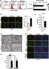
Copper deprivation by si‐Slc31a1 and tetrathiomolybdate (TM) inhibited the mitochondrial activity of pancreatic cancer cells. A, The ROS levels of Panc‐1 and MiaPaCa‐2 cells that were transfected with 50 nmol/L si‐Slc31a1 or treated with 50 μmol/L TM for 24 h were measured by flow cytometry. B, Mitochondrial membrane potential of Panc‐1 transfected with NC or si‐Slc31a1 (50 nmol/L) was tested. Rhodamine123 was used to test mitochondrial membrane potential. C, The GSH/GSSG ratio of Panc‐1 cells that was induced by Slc31a1 interference was presented via GSH/GSSG kit instructions. D, the mitochondrial morphology of Slc31a1 knocked down Panc‐1 cells was presented via transmission electron microscope. The red arrow showed mitochondrial morphology. E, The Slc31a1 knocked down Panc‐1 cells were treated with NAC (1 mmol/L) for 24 h, and ROS levels were then measured via fluorescence detection. F, The ATP levels in Panc‐1 and MiaPaCa‐2 cells were shown after the cells were transfected with si‐Slc31a1 or NC for 48 h. All results are presented as the means ± SD of values obtained in three independent experiments, n = 3, *P < 0.05, **P < 0.01, ***P < 0.001 (Student's t test)
Figure 6
Figure 6
Copper deprivation increased pancreatic cancer cell autophagy. A, the autophagy of Panc‐1 cells that was induced by Slc31a1 interference was presented via transmission electron microscope. B and C, Fluorescence detection of mCherry‐GFP‐LC3 in TM‐treated or Slc31a1 knocked down Panc‐1 and MiaPaCa‐2 cells. Red is representative of an early stage of autophagy, and green quenching indicates the increased function of lysosomes. Data shown are mean ± SD, n = 3, **P < 0.01, ***P < 0.001 (Student's t test). D, Western blot detected LC3I and LC3II in si‐Slc31a1‐transfected Panc‐1 cells
Figure 7
Figure 7
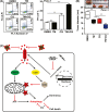
A combination of copper deprivation with autophagy inhibition repressed pancreatic cancer progression. A, Cell apoptosis treated with tetrathiomolybdate (TM) (50 μmol/L) combined with CQ (10 μmol/L) was measured by flow cytometry in Panc‐1 cells. Data shown are mean ± SD, n = 3, **P < 0.01, ***P < 0.001 (Student's t test). B, the effects of TM or CQ individually or TM combined with CQ treatment on tumour formation were tested by transplanting treated Panc‐1 cells into NPG immune‐deficient mice. For TM and CQ treatments, the NPG mice were injected subcutaneously with 2 × 106 Panc‐1 cells, and the xenografted mice were then administered TM, CQ, or a combination of TM and CQ in their water. Twenty days after transplantation, the tumours were removed and weighed. Each group included three mice. The xenografted tumours shown here were from one of three groups. The sizes of the tumours in each group were calculated and compared using Student's t test, n = 3, **P < 0.01, ***P < 0.001 (Student's t test). C, Schematic diagram of pancreatic cancer cells resisting the effect of copper shortage caused by SLC31A1 interference or TM treatment through entering a dormant state and increasing autophagy

References

    1. Zhou B, Gitschier J. hCTR1: a human gene for copper uptake identified by complementation in yeast. Proc Natl Acad Sci USA. 1997;94:7481‐7486. - VSports最新版本 - PMC - PubMed
    1. Culotta VC, Klomp LW, Strain J, et al. The copper chaperone for superoxide dismutase. J Biol Chem. 1997;272:23469‐23472. - PubMed
    1. Hamza I, Faisst A, Prohaska J, et al. The metallochaperone Atox1 plays a critical role in perinatal copper homeostasis. Proc Natl Acad Sci USA. 2001;98:6848‐6852. - PMC - PubMed
    1. Kim BE, Nevitt T, Thiele DJ. Mechanisms for copper acquisition, distribution and regulation. Nat Chem Biol. 2008;4:176‐185. - PubMed
    1. Margalioth EJ, Schenker JG, Chevion M. Copper and zinc levels in normal and malignant tissues. Cancer. 1983;52:868‐872. - PubMed

MeSH terms