Skip to main page content
U.S. flag

An official website of the United States government

Dot gov

The . gov means it’s official. Federal government websites often end in . gov or VSports app下载. mil. Before sharing sensitive information, make sure you’re on a federal government site. .

Https

The site is secure. The https:// ensures that you are connecting to the official website and that any information you provide is encrypted and transmitted securely. V体育官网.

. 2019 May;38(19):3636-3650.
doi: 10.1038/s41388-019-0684-y. Epub 2019 Jan 17.

The P2X7 receptor modulates immune cells infiltration, ectonucleotidases expression and extracellular ATP levels in the tumor microenvironment

Affiliations

The P2X7 receptor modulates immune cells infiltration, ectonucleotidases expression and extracellular ATP levels in the tumor microenvironment

Elena De Marchi et al. Oncogene. 2019 May.

Abstract

In the tumor microenvironment (TME) ATP and its receptor P2X7 exert a pivotal influence on cancer growth and tumor-host interactions. Here we analyzed the different effect of P2X7 genetic deficiency versus its antagonism on response against P2X7-expressing implanted tumors. We focused on immune cell expression of ATP degrading enzymes CD39 and CD73 and in vivo measured TME's ATP. The immune infiltrate of tumors growing in P2X7 null mice shows a decrease in CD8+ cells and an increased number of Tregs, overexpressing the fitness markers OX40, PD-1, and CD73. A similar Treg phenotype is also present in the spleen of tumor-bearing P2X7 null mice and it is paralleled by a decrease in proinflammatory cytokines and an increase in TGF-β VSports手机版. Differently, systemic administration of the P2X7 blocker A740003 in wild-type mice left unaltered the number of tumor-infiltrating CD8+ and Treg lymphocytes but increased CD4+ effector cells and decreased their expression of CD39 and CD73. P2X7 blockade did not affect spleen immune cell composition or ectonucleotidase expression but increased circulating INF-γ. Augmented CD73 in P2X7 null mice was mirrored by a decrease in TME ATP concentration and nucleotide reduced secretion from immune cells. On the contrary, TME ATP levels remained unaltered upon P2X7 antagonism, owing to release of ATP from cancerous cells and diminished ectonucleotidase expression by CD4+ and dendritic cells. These data point at P2X7 receptor as a key determinant of TME composition due to its combined action on immune cell infiltrate, ectonucleotidases, and ATP release. .


Conflict of interest statement (VSports在线直播)

Professor FDV is a member of the Scientific Advisory Board of Biosceptre Ltd, a Biotech Company involved in the development of P2X7-targeted therapies. The remaining authors declare that they have no conflict of interest V体育安卓版.

"V体育平台登录" Figures
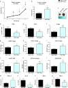
Fig. 1
Fig. 1
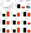
P2X7 loss enriches tumor infiltrate in Tregs overexpressing OX40, PD-1, and CD73 and decreases proinflammatory cytokines. a–c C57bl/6 mice were inoculated into the right hind flank with B16-pmeLUC cells in WT and P2X7 null mice. a Tumor volume was in vivo assessed at the indicated time points, b ex vivo tumor volume assessed by a calliper, and c representative pictures of tumors from WT and P2X7 null mice at post-inoculum day 14. Data are shown as the mean ± SEM (WT, n = 6–12; P2X7 null, n = 5–12). d–l Flow cytometric analysis of tumor masses from C57bl/6 WT and P2X7 null mice. d Percentage of CD8+ infiltrating T cells, e percentage of Teff (CD25, Foxp3, CD4+ gate), f percentage of Tregs (CD25+, Foxp3+, CD4+ gate), g–i mean fluorescence intensity (MFI) of OX40 (g), PD-1(h), CD73 (i) on Tregs, j MFI of CD73 on Teff, k MFI of CD73 on monocytes/macrophages gate (CD11b+Ly6C+ F4/80+), l MFI of CD39 on CD4+, CD25 Teff. Data are shown as the mean ± SEM (WT, n = 4–9; P2X7 null, n = 4–9). m–p Levels of plasma cytokines of tumor-bearing C57/bl6 mice inoculated with B16-pmeLUC cells. TGF-β1 (m), IL-1β (n), IFN-γ (o), and TNF-α (p) were evaluated in plasma samples obtained at post-inoculum day 14. Data are shown as the mean ± SEM (WT, n = 5–14; P2X7 null, n = 5–11). *P < 0.05, **P < 0.01, and ***P < 0.001
Fig. 2
Fig. 2
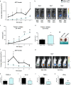
P2X7 antagonism increases tumor-infiltrating T effector lymphocytes and decreases ectonucleotidases expression on T effector and dendritic cells. a–c C57bl/6 mice were inoculated into the right hind flank with B16-pmeLUC in WT mice. A740003 (50 µg/kg) was intra-peritoneum administered to mice at post-inoculum days 5, 7, 9, 11, and 13. a Tumor volume was in vivo assessed at the indicated time points, b ex vivo tumor volume assessed by a calliper, c representative pictures of tumors from treated mice at post-inoculum day 14. Data are shown as the mean ± SEM (n = 9 per group). d–j Flow cytometric analysis of tumor masses from C57bl/6 WT mice treated with placebo or A740003 (50 µg/kg). d Percentage of CD4+-infiltrating T cells (CD45+T gate), e percentage of Teff (CD25-, Foxp3, CD4+ gate), f mean fluorescence intensity (MFI) of OX40 on Tregs, g, h MFI of CD39 (g) and CD73 (h) on Teff, i, j MFI of CD39 (i) and CD73 (j) on cDC (cDC, Cd11b+ Ly6Clow, Cd11c+ gate). Data are shown as the mean ± SEM. (placebo, n = 6–8; A740003, n = 4–8). k, l P2X7 antagonism increases circulating levels of IFN-γ while decreasing IL-1β. IFN-γ (n = 6 per group) (k) and IL-1β (l) (n = 12 per group) were evaluated in plasma samples obtained at post-inoculum day 14. Data are shown as the mean ± SEM. *P < 0.05, **P < 0.01, and ***P < 0.001
Fig. 3
Fig. 3
P2X7 ablation leads to a decrease in tumor ATP levels. a–g C57bl/6 (a, b) and BALBc/J (c–g) mice were inoculated into the right hind flank with B16-pmeLUC or WEHI-3B-pmeLUC cells, respectively in WT and P2X7 null mice. a, f Measure of ATP levels in tumor-bearing mice estimated by pmeLUC luminescence emission (p/s/cm2/sr), b, g representative pictures of pmeLUC luminescence emission in C57bl/6 (b) tumor-bearing mice at post-inoculum days 5, 7, and 9 and in BALBc/J (g) tumor-bearing mice at post-inoculum day 7, c tumor volume was in vivo assessed at the indicated time points, d ex vivo tumor volume assessed by a calliper, e representative pictures of tumors from WT and P2X7 null mice at post-inoculum day 14. Data are shown as the mean ± SEM (C57bl/6 WT, n = 6–12; C57bl/6 P2X7 null, n = 5–12; BALBc/J n = 12 per group). h–k Levels of plasma cytokines of tumor-bearing BALBc/J mice inoculated with WEHI-3B-pmeLUC cells. TGF-β1 (h) (n = 9 per group), IL-1β (i) (n = 17 per group), TNF-α (j) (n = 10 per group), and IFN-γ (k) (WT, n = 4; P2X7 null, n = 5) were evaluated in plasma samples obtained at post-inoculum day 14. Data are shown as the mean ± SEM. *P < 0.05, **P < 0.01, and ***P < 0.001
Fig. 4
Fig. 4
P2X7 null immune cells, alone or in co-colture with cancer cells, show lowered ATP release. a Supernatant ATP assessed in B16 and peritoneal macrophages coltures from WT and P2X7 null mice treated with either vehicle (PBS + DMSO 0,1%) or A740003 20 µM. Data are shown as the mean ± SEM (Vehicle, n = 7; A740003, n = 8). b, c Peri-cellular ATP levels in B16-pmeLUC cells (b) or WEHI-3B-pmeLUC cells (c) alone and in co-culture with peritoneal macrophages derived from C57bl/6 (B16-pmeLUC n = 17; Co-culture WT n = 34; Co-culture P2X7 null n = 25) or BALBc/J (WEHI-3B-pmeLUC n = 6; Co-culture WT n = 6; Co-culture P2X7 null n = 9) WT and P2X7 null mice, respectively. Data are shown as the mean ± SEM. d Peri-cellular ATP levels in B16-pmeLUC cells alone or in co-culture with splenocytes derived from C57bl/6 WT and P2X7 null mice. Data are shown as the mean ± SEM (B16-pmeLUC n = 8; Co-culture WT n = 13; Co-culture P2X7 null n = 15). Luminescence data were expressed as total photons acquired in a 5 min session. *P < 0.05, **P < 0.01, and ***P < 0.001
Fig. 5
Fig. 5
P2X7 antagonist systemic administration does not influence ATP levels in vivo. C57bl/6 (a, b) and BALBc/J (c–g) mice were inoculated into the right hind flank with B16-pmeLUC or WEHI-3B-pmeLUC cells, respectively, in WT mice. A740003 (50 µg/kg) was intra-peritoneum administered to mice at post-inoculum days 5, 7, 9, 11, and 13. a, f Measure of ATP levels in treated mice estimated by pmeLUC luminescence emission (p/s/cm2/sr), b, g representative picture of pmeLUC luminescence emission in mice at post-inoculum day 7, c tumor volume was in vivo assessed at the indicated time points, d ex vivo tumor volume assessed by a calliper, e representative pictures of tumors from treated mice at post-inoculum day 14. Data are shown as the mean ± SEM (n = 9 per group). *P < 0.05 and **P < 0.01
Fig. 6
Fig. 6
A740003 treatment leads to B16-pmeLUC cell growth arrest accompanied by ATP release. a Proliferation at 0, 24, and 48 h of B16-pmeLUC cells treated with vehicle (PBS + 0.1% DMSO) or A740003 20 µM (n = 6 per group). b ATP levels of B16-pmeLUC cells treated as above described. Luminescence data are normalized on cell number and expressed as total photons measured in a 5 min acquisition (n = 6 per group). c, d P2X7 antagonism in co-culture of tumor cells and macrophages or splenocytes results in unaltered ATP levels. c ATP levels in B16-pmeLUC cells alone and in co-culture with peritoneal macrophages derived from C57bl/6 WT mice treated with vehicle (PBS + 0.1% DMSO) or A740003 20 µM (Vehicle, n = 4; A740003, n = 6), d ATP levels in B16-pmeLUC cells alone and in co-culture with splenocytes derived from C57bl/6 WT and P2X7 null mice treated with vehicle (PBS + 0.1% DMSO) or A740003 20 µM (Vehicle, n = 6–10; A740003, n = 9–12). e Tregs reduce ATP secretion from cancer cells. ATP levels in B16-pmeLUC cells alone and in co-culture with Treg cells derived from C57bl/6 WT and P2X7 null mice treated with vehicle (PBS + 0.1% DMSO) or A740003 20 µM (n = 9 per group). c–e Luminescence data are expressed as total photons measured in a 5 min acquisition. Data are shown as the mean ± SEM. *P < 0.05, **P < 0.01, and ***P < 0.001
Fig. 7
Fig. 7
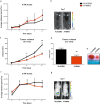
P2X7 blockade reduces tumor growth in P2X7 null mice. a–e C57bl/6 mice were inoculated into the right hind flank with B16-pmeLUC in P2X7 null mice. A740003 (50 µg/kg) was intra-peritoneum administered to mice at post-inoculum days 5, 7, 9, 11, and 13. a Tumor volume was in vivo assessed at the indicated time points, b ex vivo tumor volume assessed by a calliper, c representative pictures of tumors from treated mice at post-inoculum day 14, d kinetics of ATP levels in treated mice estimated by pmeLUC luminescence emission (p/s/cm2/sr), e representative picture of pmeLUC luminescence emission in mice at post-inoculum day 9 (placebo, n = 7; A740003, n = 8). f–h Ex vivo characterization of T cells by cytometric analysis in tumor masses derived from C57bl/6 P2X7 null mice treated with placebo or A740003 (50 µg/kg). f Percentage of CD4+ infiltrating T cells (n = 5 per group), g, h MFI of CD73 on Tregs (g) (placebo, n = 5; A740003, n = 7) and Teff (h) (placebo, n = 5; A740003, n = 6). Data are shown as the mean ± SEM. *P < 0.05, **P < 0.01, and ***P < 0.001
Fig. 8
Fig. 8
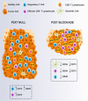
Schematic representation of the main findings of the study. Tumors growing in P2X7 null host show an increase of volume accompanied by a generation of an immunosuppressive microenvironment, characterized by a decrease of ATP levels and an augmented number of regulatory T cells overexpressing OX40 and CD73. P2X7 pharmacological blockade leads to a reduction of tumor volume and an increase of tumor-infiltrating T lymphocytes that express low levels of CD39 and CD73. Tregs and conventional dendritic cells while remaining unaltered in numbers down-modulate, respectively, OX40, CD39 and CD73

References

    1. Anderson KG, Stromnes IM, Greenberg PD. Obstacles posed by the tumor microenvironment to T cell activity: a case for synergistic therapies. Cancer Cell. 2017;31:311–25. doi: 10.1016/j.ccell.2017.02.008. - DOI - PMC - PubMed
    1. Vijayan D, Young A, Teng MWL, Smyth MJ. Targeting immunosuppressive adenosine in cancer. Nat Rev Cancer. 2017;17:765. doi: 10.1038/nrc.2017.110. - DOI - PubMed
    1. Di Virgilio F, Adinolfi E. Extracellular purines, purinergic receptors and tumor growth. Oncogene. 2017;36:293–303. doi: 10.1038/onc.2016.206. - DOI - PMC - PubMed
    1. Di Virgilio F, Sarti AC, Falzoni S, De Marchi E, Adinolfi E. Extracellular ATP and P2 purinergic signalling in the tumour microenvironment. Nat Rev Cancer. 2018;18:601–18. doi: 10.1038/s41568-018-0037-0. - DOI - PubMed
    1. Adinolfi E, Giuliani AL, De Marchi E, Pegoraro A, Orioli E, Di Virgilio F. The P2X7 receptor: a main player in inflammation. Biochem Pharmacol. 2018;151:234–44. doi: 10.1016/j.bcp.2017.12.021. - "VSports手机版" DOI - PubMed

Publication types

MeSH terms

Substances