Skip to main page content
U.S. flag

An official website of the United States government

Dot gov

The . gov means it’s official VSports app下载. Federal government websites often end in . gov or . mil. Before sharing sensitive information, make sure you’re on a federal government site. .

Https

The site is secure. The https:// ensures that you are connecting to the official website and that any information you provide is encrypted and transmitted securely V体育官网. .

. 2019 Jan;565(7737):101-105.
doi: 10.1038/s41586-018-0806-7. Epub 2018 Dec 19.

Metabolic heterogeneity underlies reciprocal fates of TH17 cell stemness and plasticity

Affiliations

Metabolic heterogeneity underlies reciprocal fates of TH17 cell stemness and plasticity

Peer W F Karmaus et al. Nature. 2019 Jan.

Abstract

A defining feature of adaptive immunity is the development of long-lived memory T cells to curtail infection. Recent studies have identified a unique stem-like T-cell subset amongst exhausted CD8-positive T cells in chronic infection1-3, but it remains unclear whether CD4-positive T-cell subsets with similar features exist in chronic inflammatory conditions VSports手机版. Amongst helper T cells, TH17 cells have prominent roles in autoimmunity and tissue inflammation and are characterized by inherent plasticity4-7, although how such plasticity is regulated is poorly understood. Here we demonstrate that TH17 cells in a mouse model of autoimmune disease are functionally and metabolically heterogeneous; they contain a subset with stemness-associated features but lower anabolic metabolism, and a reciprocal subset with higher metabolic activity that supports transdifferentiation into TH1-like cells. These two TH17-cell subsets are defined by selective expression of the transcription factors TCF-1 and T-bet, and by discrete levels of CD27 expression. We also identify signalling via the kinase complex mTORC1 as a central regulator of TH17-cell fate decisions by coordinating metabolic and transcriptional programmes. TH17 cells with disrupted mTORC1 signalling or anabolic metabolism fail to induce autoimmune neuroinflammation or to develop into TH1-like cells, but instead upregulate TCF-1 expression and acquire stemness-associated features. Single-cell RNA sequencing and experimental validation reveal heterogeneity in fate-mapped TH17 cells, and a developmental arrest in the TH1 transdifferentiation trajectory upon loss of mTORC1 activity or metabolic perturbation. Our results establish that the dichotomy of stemness and effector function underlies the heterogeneous TH17 responses and autoimmune pathogenesis, and point to previously unappreciated metabolic control of plasticity in helper T cells. .


"VSports最新版本" Figures

Extended Data Figure 1.
Extended Data Figure 1.. CD27 expression on TH17 cells during autoimmunity and cellular homeostasis of CD27+ and CD27 TH17 subsets.
a, Analysis of publically available single cell transcriptomics data of IL-17-GFP+ cells from draining lymph nodes (dLN) compared to central nervous system (CNS) (Gaublomme et al.). b, Flow cytometry analysis of putative memory surface markers on CD4+TCRβ+YFP+ cells from dLN and spleen of mice at day 9 post MOG-immunization. c, Summary of CD27 expression on CD4+TCRβ+YFP+ cells in dLN (red; n = 5, day 5; n = 3, day 7; n = 7, day 9; n = 8, day 16) overlaid with clinical EAE score (black, n = 5). d, Ki-67 expression in CD27+ and CD27 cell populations. e, Flow cytometry analysis (left) and summary (right) of CD27 expression on transferred CD27+ or CD27 YFP+ cells, at day 14 after transfer into CD45.1+ hosts (n = 14, CD27+; n = 15, CD27). f, Flow cytometry analysis of BCL-2 expression in CD27+ and CD27 populations. Data are means ± s.e.m and representative of three (b–d, f) or at least five (e) independent experiments. Numbers in plots represent frequencies of cells in gates; numbers within histogram represent mean fluorescence intensities. Mann-Whitney U test (two-sided; non-parametric) was used in e to determine statistical significance.
Extended Data Figure 2.
Extended Data Figure 2.. Gene expression profiles associated with CD27+ and CD27 TH17 subsets.
a, Volcano plot of transcriptomics data between CD27+ and CD27 CD4+TCRβ+YFP+ cells. b, GSEA plots comparing CD27+ and CD27 populations using gene sets of antigen-specific CXCR5+ and CXCR5 exhausted CD8+ T cells from chronic infection. Gene expression heat maps normalized by row (z-score) for the top 30 leading-edge genes between microarray samples from CD27+ compared to CD27 CD4+TCRβ+YFP+ cells, using gene sets derived from CXCR5+ and CXCR5 exhausted CD8 T cells (Im et al.). c, Gene expression heat maps normalized by row (z-score) for the top 30 leading-edge genes between CD27+ and CD27 CD4+TCRβ+YFP+ cells, using the “HALLMARK_APOPTOSIS” gene set. d, GSEA plots comparing CD27+ and CD27 populations using “Hallmark” gene sets, showing the enrichment of mTORC1 signaling, Myc targets, and selective metabolic pathways in CD27 TH17 cells. Data are from one experiment (a–d).
Extended Data Figure 3.
Extended Data Figure 3.. Cell-intrinsic requirement of Raptor in TH17 cells.
a, Naïve CD4+ T cells were differentiated under TH17-polarizing conditions and analyzed for cytokine expression after PMA/ionomycin stimulation in vitro (left) or for proliferation (CellTrace) and RORγt expression (right). b, Flow cytometry analysis (left) and total number of CD4+ T cells (right) from spinal cord at day 16 post-immunization (n = 10, WT; n = 8, Rptorll17aCre). c, Experimental design for the generation (left) and clinical score (right) of WT and RptorIl17aCre (R26ReYFP) bone marrow (BM) chimeras for restriction of Raptor deficiency specifically to TCRα-expressing IL-17+ T cells (n = 5 per genotype). Specifically, 5:1 ratio of Tcra–/– and Rptorll17aCre (or WT control) BM cells were transferred into sublethally irradiated Tcra –/– recipients, followed by reconstitution. In this system, all T cells in the chimeras were derived from Rptorll17aCre or WT BM cells, whereas the majority of non-T cell compartments were derived from WT cells. d, Experimental design for the generation (left) and clinical score (right) of WT and RptorIl17aCre (R26ReYFP) BM chimeras for equal inflammatory conditions. Specifically, mixed BM chimeras were generated using 1:1 ratio of congenically marked CD45.2+ RptorIl17aCre (or WT control) and CD45.1+ WT BM-derived cells, which mediated CNS inflammation in EAE (n = 3, WT; n = 4, Rptorll17aCre). e–k, Equi-inflammatory chimeric mice generated in (d) were analyzed at day 18 after EAE immunization, for frequencies of CD4+ T cells positive for CD45.2 or YFP (e) (n = 12, WT; n =14, Rptorll17aCre), and YFP+CD4+ T cells expressing IL-17 or IFNγ within the spinal cord (f) (n = 6, WT; n = 7, Rptorll17aCre), expression of Ki-67 (g) (n = 8, WT; n = 10, Rptorll17aCre), T-bet (as fold change in mean fluorescence intensity after normalization to WT cells; h) (n = 10 per genotype), and active caspase-3 in splenic YFP+CD4+ T cells (i) (n = 6 per genotype), and expression of CCR6 (j) (n = 7, WT; n = 6, Rptorll17aCre) and CXCR3 (k) (n = 8, WT; n = 10, Rptorll17aCre) in YFP+ cells from different organs. Data are means ± s.e.m and representative of three (a, c, i–k) or four (b, d–h) independent experiments. Numbers in plots represent frequencies of cells in gates or quadrants. Student’s t-test (two-sided) was used in h, k, or Mann-Whitney U test (two-sided) was used in b, e, f, g to determine statistical significance.
Extended Data Figure 4.
Extended Data Figure 4.. Raptor deficiency induces selective phenotypic changes in fate-mapped TH17 cells.
a–g, WT and RptorIl17aCre (R26ReYFP) mice were immunized with MOG, followed by analysis of YFP+ cells from draining lymph nodes (dLN) at day 9 post-immunization. a, Frequency of YFP+ cells (n = 7 per genotype). b, Efficiency of Rptor deletion (left) (n = 7 per genotype) and flow cytometry analysis of phosphorylated S6 (S235/236) and 4E-BP1 (T37/46) (right) in YFP+ cells. c, Flow cytometry of active caspase-3 and 7-AAD staining in YFP+ cells. d, Flow cytometry analysis of CXCR3 and CCR6 expression on YFP+ cells. e, Cytokine production by YFP+ cells from draining lymph nodes (dLN) (n = 7 per genotype). f, Real-time PCR analysis of Tbx21 (n = 8 per genotype), Rorc (n = 6 per genotype), Il12rb2 (n = 7 per genotype), and Il23r (n = 7 per genotype) expression in YFP+ cells. g, Flow cytometry analysis of Foxp3 expression. h, i, Cytokine production (h, i) and proliferation (h) of YFP+ cells from dLN of the indicated mice after 4 days of stimulation with MOG alone (h) or MOG+IL-23 (i) (n = 7 per genotype). j, Sorted YFP+ cells were stimulated with IL-23 or IL-12 for 30 min in vitro and stained for specific phospho-antibodies to STAT3 (left), STAT4 (right), or isotype controls. Data are means ± s.e.m and representative of seven (a), three (b–f, j), two (g), or five (h, i) independent experiments. Numbers in plots represent frequencies of cells in quadrants; numbers within histograms represent mean fluorescence intensities. Student’s t-test (two-sided) was used in b, or Mann-Whitney U test (two-sided) was used in a, e, i to determine statistical significance.
Extended Data Figure 5.
Extended Data Figure 5.. Altered gene expression profiles in Raptor-deficient cells and shared functional pathways with CD27+ TH17 cells.
a–d, WT and RptorIl17aCre (R26ReYFP) mice were immunized with MOG, and sorted YFP+ cells from draining lymph nodes (dLN) were analyzed by microarray. a, Expression of individual genes with vertical lines indicating a 1.5 fold cutoff and a horizontal line indicating the 0.05 P-value cutoff, with gene ontology (GO) gene sets for acetylation and cholesterol biosynthesis colorized and Hmgcr indicated. b, GSEA reveals significant Hallmark gene sets downregulated in RptorIl17aCre compared to WT cells. c, d, Comparison of functional enrichment of co-regulated gene sets between RptorIl17aCre and CD27+ YFP+ cells. The downregulated genes in CD27+ compared with CD27 TH17 cells, and RptorIl17aCre compared with WT (R26ReYFP) TH17 cells (FDR < 0.05 and top 200 genes based on fold change) were used for functional enrichment using (c) Hallmark and (d) KEGG pathway gene sets. Legends indicate FDR values and enrichment scores. e, Comparison of microarray analyses of CD27+ vs CD27 and RptorIl17aCre (R26ReYFP) vs WT samples from mice at day 9 post MOG-immunization. Shown is empirical cumulative distribution function for the changes in expression (Log2 values) of all genes expressed in RptorIl17aCre (R26ReYFP) TH17 cells (red line, change relative to that in WT TH17 cells) and for subsets of genes downregulated (green line) or upregulated (blue line) by CD27+ vs CD27 (< 5% FDR) in TH17 cells. P-value is calculated by using Kolmogorov–Smirnov test. f, Real-time PCR analysis of Cd27 expression in WT and RptorIl17aCre YFP+ cells (n = 5 per genotype). Data are means ± s.e.m and from one experiment (a–e), or representative of two independent experiments (f). Student’s t-test (two-sided) was used in f to determine statistical significance.
Extended Data Figure 6.
Extended Data Figure 6.. Anabolic metabolism promotes TH17 transdifferentiation into TH1-like IFNγ-producing cells.
a, Ingenuity Pathway Analysis (IPA) of upstream transcriptional regulators between WT and RptorIl17aCre samples. b, WT and RptorIl17aCre (R26ReYFP) mice were immunized with MOG, and YFP+ cells from draining lymph nodes (dLN) were analyzed by flow cytometry for intracellular expression of Myc. c, d, WT and MycIl17aCre (R26ReYFP) mice were immunized with MOG, and YFP+ cells from dLN were analyzed by real-time PCR at day 9 (c) (n = 4, Tbx21; n = 2, Rorc; n = 4, Il12rb2; n = 4, Il23r), or dLN cells were cultured with MOG, MOG+IL-12, or MOG+IL-23 for 4 days for cytokine expression analysis within YFP+ cells (d). e, f, WT and HmgcrIl17aCre (R26ReYFP) mice were immunized with MOG and YFP+ cells were isolated from dLN and analyzed by real-time PCR (e) (n = 4, Tbx21; n = 2, Rorc; n = 4, Il12rb2; n = 4, Il23r), or dLN cells were cultured with MOG, MOG+IL-12, or MOG+IL-23 for 4 days for cytokine expression analysis within YFP+ cells (f). g, Flow cytometry analysis of CD27 expression on YFP+ cells from WT, RptorIl17aCre, MycIl17aCre and HmgcrIl17aCre mice at day 9 post MOG-immunization. h, i, WT (R26ReYFP) mice were immunized with MOG, and dLN cells were stimulated with MOG and IL-12 in the presence of vehicle, PF-4708671 or Cbz-B3A at indicated concentrations for 4 days for cytokine expression analysis within YFP+ cells (h; right, summary plots) (n = 9, vehicle; n = 7, PF-4708671 (5 μM); n = 9, PF-4708671 (10 μM); n = 7, Cbz-B3A (5 μM); n = 9, Cbz-B3A (10 μM)) and CellTrace dilution (i). Data are means ± s.e.m and from one experiment (a), or representative of four (b–f, h, i) or three (g) independent experiments. Numbers in plots represent frequencies of cells in gates or quadrants; numbers within histograms represent mean fluorescence intensities. Student’s t-test (two-sided; parametric) was used to determine statistical significance in h.
Extended Data Figure 7.
Extended Data Figure 7.. Analysis of histone acetylation, and ATAC-Seq overview and specific gene loci.
a, ChIP qRT-PCR of pan-acetyl histone bound to the Ifng promoter of WT or RptorIl17aCre (R26ReYFP) YFP+ cells from draining lymph nodes (dLN) (n = 4 per genotype). b, c, WT and RptorIl17aCre (R26ReYFP) mice were immunized with MOG, and YFP+ cells from dLN and spleen at day 9 post-immunization were analyzed by ATAC-Seq. b, Density plot and heat maps of a representative individual ATAC-Seq sample, demonstrating separation into different fragment lengths implicating nucleosome-free, mono-nucleosome, di-nucleosome, tri-nucleosome patterns, consistent with Buenrostro et al. c, Correlation plot of nucleosome-free fragments. d, Nucleosome-free ATAC-Seq tracks at the Tbx21 and Il12rb2 gene loci, with immediate promoter regions indicated by red boxes. e, Summary of ATAC-Seq motif enrichment data showing Log2 (odds ratio) and Log10 (Fisher P-value) of cells from dLN. f, Tn5 insert sites from ATAC-Seq analysis from dLN were aligned to motifs for transcription factors from the TRANSFAC database, and the binding profiles of selected TCF-LEF family transcription factors are shown. Data are means ± s.e.m and representative of three independent experiments (a), or from one experiment (b–f). Student’s t-test (two-sided; parametric) was used in a to determine statistical significance.
Extended Data Figure 8.
Extended Data Figure 8.. ATAC-Seq in-depth analyses, TCF-1 binding activity, and effects of 2-DG on cytokine expression.
a, Analysis of common regions between ATAC-Seq and ChIP-Seq peaks for TCF-LEF family and T-bet transcription factor binding motifs from spleen samples. Number of motif matches and associated Fisher’s exact test P-values and log2 (odds ratio) (positive log2 (odds ratio) values indicate that the chance of occurrence of the motif is higher in RptorIl17aCre compared to WT samples, and negative values indicate that the chance of occurrence is lower in the RptorIl17aCre group). b, Nucleosome-free ATAC-Seq tracks at the Il6ra and Lrig1 gene loci, with TCF-1 binding sites indicated by red boxes, based on the alignment with TCF-1 binding sites from published data (GEO accession numbers shown). c, ChIP assay to measure TCF-1 binding to Il6ra and Lrig1 gene loci (Il6ra, n = 2 per genotype; Lrig1, n = 6 for WT, n = 5 for RptorIl17aCre). d, Cells from dLN of the indicated mice at day 9 post MOG-immunization were cultured for 4 days with MOG and IL-12 and sorted on the YFP+ population prior to intracellular staining. Flow cytometry analysis of T-bet and TCF-1 expression in YFP+ cells from WT and MycIl17aCre (R26ReYFP) mice. e, Tn5 insert sites from ATAC-Seq analysis of YFP+ cells from PBS- or 2-DG-treated mice were aligned to motifs for transcription factors from the TRANSFAC database, and the binding profiles of selected TCF-LEF family transcription factors are shown. f, Cytokine expression in dLN YFP+ cells from MOG-immunized Il17aCre (R26ReYFP) mice after culture with MOG and IL-12 for 4 days in the presence of vehicle (PBS) or 2-DG (1 mM). g, Cytokine expression in splenic YFP+ cells from MOG-immunized Il17aCre (R26ReYFP) mice after treatment with 2-DG (2 g/kg body weight) or PBS. Numbers in plots represent frequencies of cells in gates or quadrants. Data are means ± s.e.m and from one experiment (a, b, e), or representative of three independent experiments (c, d, f, g). Student’s t-test (two-sided) was used to determine statistical significance in c.
Extended Data Figure 9.
Extended Data Figure 9.. Single cell transcriptomics analysis.
WT and RptorIl17aCre (R26ReYFP) mice were immunized with MOG, and YFP+ cells were analyzed by single cell transcriptomics analysis at day 9 post-immunization. a, Individual cell cluster membership in two dimensional tSNE projections from scRNA-Seq data. b, tSNE visualization of nine clusters partitioned by unsupervised clustering. c, Frequencies of WT and RptorIl17aCre cells in different clusters (n = 3 per genotype). d, Top three enriched gene sets for each of the clusters using Hallmark, Canonical, and GO gene sets. For example, genes enriched in cluster 1 compared to other clusters were associated with proliferative events. e, Summary of cluster-specific functional enrichment analysis via Fisher’s exact test using signatures of “T-bet targets”, “late memory”, “memory TFH overlap DOWN”, “HALLMARK_GLYCOLYSIS”, “memory TFH overlap UP”, and “early memory” as described in methods. f, tSNE visualization of signature scores of “T-bet targets” and “HALLMARK_GLYCOLYSIS” expressed in individual cells. g, Violin plots of Bcl2, Cd27, and Tcf7 gene expression among the nine clusters. A violin plot combines the box plot and the local density estimation into a single display. The black bars and thin lines within the violin plots indicate the interquartile range (1st quantile – 3rd quantile) and the entire range of the data (up to 1.5 fold of interquartile range from 1st/3rd quantile), respectively, and the white dots in the center indicate the median values. Data are from one experiment (a–g) (n = 3 per genotype).
Extended Data Figure 10.
Extended Data Figure 10.. Pseudotime analysis and experimental validation.
a–h, WT and RptorIl17aCre (R26ReYFP) mice were immunized with MOG, and YFP+ cells were analyzed by single cell transcriptomics analysis at day 9 post-immunization. a, Empirical dispersion and mean expression from Monocle 2, indicating genes used for temporal ordering in black; each dot represents one gene. b, Pseudotime densities for each individual cluster. For example, cluster 1 associated with the proliferative signature was in the center of the pseudotime spectrum, while clusters 2, 3 and 8 (early in pseudotime; predominantly Raptor-deficient cells) and clusters 7, and to a lesser extent, 4 and 5 (late in pseudotime; predominantly WT cells) were on the opposite end of the spectrum. c, Projection of “early memory”, “late memory”, and “T-bet targets” signature scores onto pseudotime trajectory; legend indicates relative score per cell. d, tSNE visualization of Tbx21 and Ifng gene expression. e, Tbx21 and Ifng gene expression during pseudotime; cells without gene expression for Tbx21 or Ifng were filtered out in their respective graphs. f, Pseudotime assignment for WT and RptorIl17aCre cells colorized by genotype: WT (black) and RptorIl17aCre (red); each dot represents one cell. g, Cd27 and Tcf7 gene expression across pseudotime, colorized by genotypes: WT (black) and RptorIl17aCre (red). h, tSNE visualization of Cd27 and Tcf7 expression. i, Flow cytometry analysis of T-bet expression in freshly-isolated CD27+ and CD27 cells from dLN of Il17aCre (R26ReYFP) mice at day 9 post MOG-immunization. j, Fold change of the IL-17IFNγ+ cell percentage in MOG+IL-12-stimulated CD27+ or CD27 YFP+ cells as compared to freshly-isolated cells (n = 12). k, CD27+YFP+ cells from MOG-immunized WT and RptorIl17aCre mice were sorted and transferred into CD45.1+ hosts. The following day, CD45.1+ host mice were immunized with MOG, and four days later, YFP+ cells were analyzed by flow cytometry for surface CD27 expression (k; right, summary plots) (n = 6, WT; n = 5, RptorIl17aCre). l, CD27+YFP+ cells from MOG-immunized WT and RptorIl17aCre mice were stimulated with MOG and IL-12 for four days, followed by analysis of CD27 expression. m, TH17 cells are functionally and metabolically heterogeneous and are comprised of a subset with stemness features but lower anabolic metabolism and a reciprocal subset with higher metabolic activity that supports the transdifferentiation into TH1 cells. These two subsets are further distinguished by selective expression of transcription factors TCF-1 and T-bet, respectively, and discrete levels of CD27 expression. mTORC1 activation drives anabolic metabolism to support metabolic reprogramming favoring transcription mediated by T-bet over TCF-1, and consequently, TH17 transdifferentiation into TH1-like TH17 cells occurs. Memory/stem-like TH17 cells can become reactivated and have the potential to undergo terminal differentiation and acquire TH1-like phenotypes. Data are means ± s.e.m and from one experiment (a–h), or representative of three (i) or five (j–l) independent experiments. Numbers in plots represent frequencies of cells in gates; numbers within histograms represent mean fluorescence intensities. Mann-Whitney U test (two-sided) was used in j to determine statistical significance.
Figure 1.
Figure 1.. CD27+ TH17 cells have memory-like features and low metabolic activity.
a, Summary of CD27 expression on CD4+TCRβ+YFP+ cells at day 16 post MOG-immunization in draining lymph nodes (dLN), spleen, and spinal cord of Il17aCre (R26ReYFP) mice (n = 8, dLN; n = 12, spleen and spinal cord). b–i, Analysis of CD27+ and CD27 YFP+ populations (b, left) from Il17aCre (R26ReYFP) mice at day 9 post MOG-immunization. b, IL-17 and IFNγ expression (n = 6, CD27+/CD27 IL-17; n = 8, CD27+ IFNγ; n = 9, CD27 IFNγ). c, In vitro culture with MOG for analyses of proliferation (CellTrace) and CD27 expression. d, TCF-1 expression (left) and fold change (right, expression in CD27+ population was set to 1) (n = 9). e, CD27+ or CD27 YFP+ cells were transferred into Rag1−/− mice, and analyzed at day 15 for donor cell percentages (left) and numbers (right, normalized against cell numbers at day 1) (n = 3, CD27+; n = 4, CD27). f, GSEA using gene sets related to T cell memory from acute (top 4 panels) and chronic (bottom 4 panels) infection. g, h, Flow cytometry of phosphorylated S6 and 4E-BP1 (g) and Myc (h). i, CD27 expression on CD4+TCRβ+YFP+ cells stimulated with MOG and vehicle or 2-deoxyglucose (2-DG). Numbers within histograms represent mean fluorescence intensities. Data are means ± s.e.m; Mann-Whitney U test (two-sided) in b, Student’s t-test (two-sided) in d, e. Data are representative of three (a, e, g, i), four (b–d), one (f), or two (h) independent experiments. FDR, false discovery rate.
Figure 2.
Figure 2.. Deletion of Raptor in TH17 cells diminishes autoimmune pathogenesis and transdifferentiation into TH1-like cells.
WT and RptorIl17aCre (R26ReYFP) mice were immunized with MOG. a, Clinical scores (n = 15, WT; n = 12, RptorIl17aCre). b, Histopathology of spinal cord sections at day 16 post-immunization. Bar = 1 mm; arrows indicate lesions. c, Flow cytometry analysis (left) and summary (right) of cytokine expression within YFP+ cells from spinal cord at day 16 post-immunization (n = 5 per genotype). d, T-bet and RORγt expression in YFP+ cells from draining lymph nodes (dLN) at day 9 post-immunization. e, f, Cytokine production by YFP+ cells from dLN after 4 days of stimulation with MOG (e) (n = 7 per genotype) or MOG+IL-12 (f) (n = 5 per genotype). Numbers within histograms represent mean fluorescence intensities. Data are means ± s.e.m; two-way ANOVA in a; Mann-Whitney U test (two-sided) in c, e, f. Data are pooled from three experiments (a), or representative of three (b–d), seven (e), or five (f) independent experiments.
Figure 3.
Figure 3.. TCF-1 expression is regulated by mTORC1 and metabolic activity in TH17 cells.
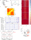
a–d, ATAC-Seq analysis of YFP+ cells isolated from draining lymph nodes (dLN) and spleen of WT and RptorIl17aCre (R26ReYFP) mice or spleen of PBS- or 2-DG-treated R26ReYFP mice at day 9 post MOG-immunization. a, Principal component analysis (PCA) plot of nucleosome-free fragments. b, Accessibility of the Ifng locus, aligned with T-bet binding sites (red box, promoter regions). c, Motif enrichment analysis of ATAC-Seq from the spleen. d, Binding profiles of selected TCF-LEF family transcription factors. e, f, Flow cytometry of T-bet and TCF-1 expression in freshly-isolated YFP+ cells from dLN of mice at day 9 post MOG-immunization (e), or after in vitro stimulation with MOG+IL-12 for 4 days (f). g, Odds ratio vs odds ratio plot of motif enrichment data from RptorIl17aCre vs WT and 2-DG vs PBS samples. h, Flow cytometry of T-bet and TCF-1 expression in YFP+ cells from Il17aCre (R26ReYFP) mice cultured with MOG+IL-12, and vehicle or 2-DG (1 mM). Data are representative of one (a–d, g), four (e, f), or three (h) independent experiments.
Figure 4.
Figure 4.. Single cell transcriptomics of TH17 cells and mTORC1-dependent licensing for TH1 transdifferentiation.
a, Two-dimensional tSNE projection of single cell transcriptomics data from WT (black) and RptorIl17aCre (red) YFP+ cells (n = 3 per genotype; 27,619 total cells). b, tSNE visualization of gene signatures of upregulated and downregulated genes shared between memory CD8+ and TFH cells (Choi et al.). c, tSNE visualization of “early memory” and “late memory” gene signatures (Wirth et al. and Muranski et al.). d, Pseudotime trajectory of single cell transcriptomics data colorized by pseudotime (arbitrary units; blue and red indicate early and late time, respectively). e, Pseudotime trajectory of cellular density: WT (gray), RptorIl17aCre (red). f, g, Cytokine production (f) and T-bet and TCF-1 expression (g) by CD27+ and CD27 YFP+ cells cultured with MOG or MOG+IL-12 for 4 days. Data are representative of one (a–e) or three (f, g) independent experiments.

Comment in (V体育官网入口)

"VSports" References

    1. Im SJ et al. Defining CD8+ T cells that provide the proliferative burst after PD-1 therapy. Nature 537, 417–421, doi:10.1038/nature19330 (2016). - DOI - PMC - PubMed
    1. He R et al. Follicular CXCR5- expressing CD8(+) T cells curtail chronic viral infection. Nature 537, 412–428, doi:10.1038/nature19317 (2016). - DOI - PubMed
    1. Leong YA et al. CXCR5(+) follicular cytotoxic T cells control viral infection in B cell follicles. Nat. Immunol. 17, 1187–1196, doi:10.1038/ni.3543 (2016). - DOI (VSports app下载) - PubMed
    1. Gagliani N et al. Th17 cells transdifferentiate into regulatory T cells during resolution of inflammation. Nature 523, 221–225, doi:10.1038/nature14452 (2015). - DOI - PMC - PubMed
    1. Lee YK et al. Late developmental plasticity in the T helper 17 lineage. Immunity 30, 92–107, doi:10.1016/j.immuni.2008.11.005 (2009). - DOI - PMC - PubMed

MeSH terms

VSports - Substances