"V体育平台登录" Skip to main page content
U.S. flag

An official website of the United States government

Dot gov

The . gov means it’s official. Federal government websites often end in VSports app下载. gov or . mil. Before sharing sensitive information, make sure you’re on a federal government site. .

Https

The site is secure. The https:// ensures that you are connecting to the official website and that any information you provide is encrypted and transmitted securely. V体育官网.

. 2018 Aug 23;7(11):e1470735.
doi: 10.1080/2162402X.2018.1470735. eCollection 2018.

Human CD4- invariant NKT lymphocytes regulate graft versus host disease

Affiliations

Human CD4- invariant NKT lymphocytes regulate graft versus host disease (V体育2025版)

Tereza Coman et al. Oncoimmunology. .

V体育2025版 - Abstract

Despite increasing evidence for a protective role of invariant (i) NKT cells in the control of graft-versus-host disease (GVHD), the mechanisms underpinning regulation of the allogeneic immune response in humans are not known. In this study, we evaluated the distinct effects of human in vitro expanded and flow-sorted human CD4+ and CD4- iNKT subsets on human T cell activation in a pre-clinical humanized NSG mouse model of xenogeneic GVHD. We demonstrate that human CD4- but not CD4+ iNKT cells could control xenogeneic GVHD, allowing significantly prolonged overall survival and reduced pathological GVHD scores without impairing human T cell engraftment. Human CD4- iNKT cells reduced the activation of human T cells and their Th1 and Th17 differentiation in vivo. CD4- and CD4+ iNKT cells had distinct effects upon DC maturation and survival. Compared to their CD4+ counterparts, in co-culture experiments in vitro, human CD4- iNKT cells had a higher ability to make contacts and degranulate in the presence of mouse bone marrow-derived DCs, inducing their apoptosis. In vivo we observed that infusion of PBMC and CD4- iNKT cells was associated with decreased numbers of splenic mouse CD11c+ DCs. Similar differential effects of the iNKT cell subsets were observed on the maturation and in the induction of apoptosis of human monocyte-derived dendritic cells in vitro VSports手机版. These results highlight the increased immunosuppressive functions of CD4-versus CD4+ human iNKT cells in the context of alloreactivity, and provide a rationale for CD4- iNKT selective expansion or transfer to prevent GVHD in clinical trials. .

Keywords: Allogeneic stem cell transplantation; NSG mice; dendritic cells; graft-versus-host disease; human invariant NKT cells; immunoregulation. V体育安卓版.


Figures (VSports最新版本)

Figure 1.
Figure 1.
Human iNKT4, but not iNKT4+ cells can control the development of xeno-GVHD in irradiated NSG mice. NSG mice were irradiated (200c Gy) and injected intravenously with 3 × 106 human PBMCs alone or enriched with 2.5 × 105 human CD4 iNKT (PBMC + iNKT4) or CD4+ iNKT (PBMC+iNKT4+) cells from the same donor. (A) Median survival was prolonged in mice transplanted with PBMCs + CD4 iNKT cells (52 days) compared to control group with PBMCs alone (31 days, p = 0.0005) or those transplanted with PBMCs and iNKT CD4+ iNKT (24.5 days, p = 0.0007). Mice transplanted with PBMCs + CD4 iNKT cells had (B) reduced weight loss and (C) lower GVHD clinical scores in comparison to mice transplanted with PBMCs alone or with PBMCs and CD4+ iNKT cells in the first 30 days post-transplant. (D) Reduced pathological GVHD score in the liver compared to mice receiving PBMCs alone (p = 0.0277) or PBMC + CD4+ iNKT cells (p = 0.0138) (left panel and 5–6 of the right panel) at 4 weeks after graft infusion in mice with documented human T cell engraftment. (E) Kinetics and rates of engraftment of CD45+ human T cells were similar between all groups. (F) Percentages of human circulating CD3+ T cells among alive human CD45+ blood cells at day 21 post-transplantation (left panel) and human CD4/CD8 T cell ratios at day 21 days post-transplantation (right panel) were similar between all groups. Data are pooled from 6 different experiments containing 6 mice in the PBMC alone, 14 mice in the PBMC +NKT4neg and 12 mice in the PBMC + iNKT4pos groups (ns = non-significant, *: p ≤ 0.05, **: p ≤ 0.01, ***: p ≤ 0.001).
Figure 2.
Figure 2.
In vivo assessment of mouse serum levels of human cytokine and T cell polarization after human PBMC + iNKT CD4 or CD4+ injection. (A) BrdU staining, performed on splenic cells and assessed on human CD3+ (left panel) and CD4+ (right panel) T cells, showed similar levels of proliferating T cells between all groups on day 28 post-transplantation (p > 0.05 in all comparisons). (B) Expression of CD25 (upper panel) and CD69 (lower panel) on circulating human CD4+ T cells were reduced in mice transplanted with human PBMCs + CD4 iNKT cells (n = 15) as compared to those receiving PBMCs alone (n = 6) (p = 0.0069 and p = 0.0375, respectively) and PBMCs + CD4+ iNKT cells (n = 12) (0.0017 and 0.0038, respectively). (C) Expression of CD25 (upper panel) and CD69 (lower panel) on circulating human CD8+ T cells were reduced in mice transplanted with human PBMCs + CD4 iNKT cells (n = 15) as compared to those receiving PBMCs alone (n = 6) (p = 0.2065 and p = 0.0020, respectively) and PBMCs + CD4+ iNKT cells (n = 12) (0.0006 and 0.00158, respectively). (D) Shows comparable FoxP3 expression on human CD4+ T cells from blood, spleen and bone marrow of NSG mice transplanted with human PBMCs alone or with co-administration of CD4+ or CD4 iNKT cell. (E) Human cytokine measurement in plasma of NSG mice at day 21 after transplantation with human cells showed reduced levels of IL-17, TNF-α, INF-γ but increased concentrations of IL-4 in the group of mice transplanted with human PBMC and CD4 iNKT cells (n = 18) in comparison of those having received human PBMCs alone group (n = 8) (p ≤ 0.05 for all cytokines). Mice transplanted with human PBMCs and CD4+ iNKT cells (n = 22) had reduced plasma levels of INF-γ (p ≤ 0.05) and increased IL-4 (p ≤ 0.01) but similar IL-17 and TNF-α levels in comparison to control mice transplanted with human PBMCs alone. (F) Ratio of Th1+Th17/Th2 cytokines was significantly decreased in sera of mice injected with PBMC+CD4 iNKT compared to PBMC alone (p = 0,0397) (lower panel). (G) Intracellular staining of IL-17 and ROR-γt in peripheral blood T cells of NSG mice at day 21 after transplantation with human cells showed reduced levels of IL-17 and ROR-γt in the group of mice transplanted with human PBMC and CD4 iNKT cells (n = 6) in comparison of those having received human PBMCs alone group (n = 4) (p = 0.0293 and p = 0.0290 respectively). Data are pooled from 3 (A,B,F), 4 (C) or 7 (D,E) different experiments. (ns = non-significant; *: p ≤ 0.05, **: p ≤ 0.01, ***: p ≤ 0.001).
Figure 3.
Figure 3.
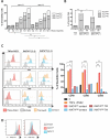
In vivo and in vitro assessment of human iNKT CD4 or CD4+ cell effects on the survival and maturation mouse bone marrow derived dendritic cells (moBMDC). (A) Mice transplanted with human PBMCs + CD4 iNKT cells (n = 10) showed lower percentage of CD11c+ DCs compared to mice transplanted with human PBMCs + CD4+ iNKT (n = 10) (p = 0.0110) or with control mice injected with human PBMCs alone (n = 13) (p = 0.0450) on day 28 after transplantation. (B) DCs from mice injected with human PBMCs + CD4+ iNKT cells (n = 9) expressed higher levels of CD40 (p = 0.0407), CD86 (p = 0.0259) and CD80 (p = 0.0004) compared to mice injected with human PBMCs alone (n = 8), whereas DCs from transplanted with human PBMCs + CD4 iNKT cells (n = 4) had similar levels of expression of CD40 and CD86 than DCs from mice injected with PBMCs alone. (C) Flow sorted human iNKT CD4+ and CD4 were cultured in a 50%/50% mixture, alone or with mouse BMDC (moBMDC) at a 2:2:1 final ratio. The image obtained by ImageStream (AMNIS) is representative of human CD107a staining of iNKT CD4 (upper panel) or CD4+ iNKT (lower panel) within the synapse gate with the moBMDC visualized. (D) At 4 hours of culture, synapses between CD11c+ moBMDC and human CD3+ iNKT cells were analyzed by Image Stream (left panel). The percentage of synapses between iNKT subsets and moBMDCs was higher with CD4 iNKT compared to CD4+ iNKT cells (76,3 vs 23,7%, p < 0,0001) (right panel). (E) CD107a expression on iNKT cells analyzed by flow cytometry showed higher expression on human CD4 iNKT cells compared to CD4+ iNKT cells (71% vs 48%, p < 0,0001) Results are representative of three separate experiments performed in duplicates. (ns = non-significant; *: p ≤ 0.05, **: p ≤ 0.01, ***: p ≤ 0.001).
Figure 4.
Figure 4.
In vitro effects of human CD4+ and CD4 iNKT cell subsets on the maturation and survival of human monocyte-derived DCs. (A) Both CD4+ and CD4 iNKT cells induce of the apoptosis of mature MoDCs in a dose dependent manner. Annexin V+ (sum of early apoptosis (Annexin V+ PI) and late apoptosis (Annexin V+ PI+)) MoDCs were significantly higher with iNKT CD4 at the following iNKT/DC ratios (2:1 p = 0,001), 5:1 (p = 0,0228), 10:1 (p = 0,0287). (B) Apoptosis of MoDCs was contact dependent in both CD4 and CD4+ iNKT conditions (2:1 ratio) and significantly higher percentages of apoptotic Annexin V+ (sum of early apoptotic (Annexin V+ PI) and late apoptotic (Annexin V+ PI+)) mature MoDCs were observed in contact with CD4 iNKT compared to CD4+ iNKT cells (p = 0.002). (C) In contact with CD4+ iNKT cells alone, monocyte-derived DCs (MoDCs) expressed higher levels of (CD80 (p = 0,001), CD86 (p = 0,0106) and CD40 (p = 0,0449)) compared to DC maturation with PGE2 and TNF-α without iNKT cells or to DC maturation in the presence of CD4 iNKT cells (CD80 (p = 0,0051), CD86 (p = 0,0004) and CD40 (p = 0,0003)). (D) CD4+ iNKT cells had higher expression of intra (p = 0,0116) and extracellular (p = 0,0343) CD40-L compared to their CD4 counterparts. Results are representative of 3 experiments performed in duplicates. (ns = non-significant; *: p ≤ 0.05, **: p ≤ 0.01, ***: p ≤ 0.001).

References (V体育平台登录)

    1. Ferrara JLM, Levine JE, Reddy P, Holler E.. Graft-versus-host disease. Lancet Lond Engl. 2009;373(9674):1550–1561. doi:10.1016/S0140-6736(09)60237-3. - DOI - PMC - PubMed
    1. Ferrara JLM, Reddy P. Pathophysiology of graft-versus-host disease. Semin Hematol. 2006;43(1):3–10. doi:10.1053/j.seminhematol.2005.09.001. - V体育2025版 - DOI - PubMed
    1. Henden AS, Hill GR. Cytokines in graft-versus-host disease. J Immunol Baltim Md 1950. 2015;194(10):4604–4612. - "VSports手机版" PubMed
    1. Di Ianni M, Falzetti F, Carotti A, Terenzi A, Castellino F, Bonifacio E, Del Papa B, Zei T, Ostini RI, Cecchini D, et al. Tregs prevent GVHD and promote immune reconstitution in HLA-haploidentical transplantation. Blood. 2011;117(14):3921–3928. doi:10.1182/blood-2010-10-311894. - DOI (VSports注册入口) - PubMed
    1. Hicheri Y, Bouchekioua A, Hamel Y, Henry A, Rouard H, Pautas C, Beaumont JL, Kuentz M, Cordonnier C, Cohen JL, et al. Donor regulatory T cells identified by FoxP3 expression but also by the membranous CD4+CD127low/neg phenotype influence graft-versus-tumor effect after donor lymphocyte infusion. J Immunother Hagerstown Md 1997. 2008;31(9):806–811. doi:10.1097/CJI.0b013e318184908d - DOI - PubMed

Publication types