Skip to main page content
U.S. flag

An official website of the United States government

Dot gov

The . gov means it’s official VSports app下载. Federal government websites often end in . gov or . mil. Before sharing sensitive information, make sure you’re on a federal government site. .

Https

The site is secure. The https:// ensures that you are connecting to the official website and that any information you provide is encrypted and transmitted securely V体育官网. .

. 2018 Nov 5;215(11):2833-2849.
doi: 10.1084/jem.20180439. Epub 2018 Sep 28.

KDM4B-regulated unfolded protein response as a therapeutic vulnerability in PTEN-deficient breast cancer (V体育平台登录)

Affiliations

KDM4B-regulated unfolded protein response as a therapeutic vulnerability in PTEN-deficient breast cancer (VSports在线直播)

Wenyu Wang et al. J Exp Med. .

Abstract (V体育ios版)

PTEN deficiency in breast cancer leads to resistance to PI3K-AKT inhibitor treatment despite aberrant activation of this signaling pathway. Here, we report that genetic depletion or small molecule inhibition of KDM4B histone demethylase activates the unfolded protein response (UPR) pathway and results in preferential apoptosis in PTEN-deficient triple-negative breast cancers (TNBCs). Intriguingly, this function of KDM4B on UPR requires its demethylase activity but is independent of its canonical role in histone modification, and acts through its cytoplasmic interaction with eIF2α, a crucial component of UPR signaling, resulting in reduced phosphorylation of this component VSports手机版. Targeting KDM4B in combination with PI3K inhibition induces further activation of UPR, leading to robust synergy in apoptosis. These findings identify KDM4B as a therapeutic vulnerability in PTEN-deficient TNBC that otherwise would be resistant to PI3K inhibition. .


"V体育官网入口" Figures

None
Graphical abstract
Figure 1.
Figure 1.
Drug screening identifies KDM inhibitor Methylstat selectively impairing PTEN-deficient breast cancer cells. (A) MCF10A parental and PTEN-KO cells treated for 4 d with a compound library consisting of 140 small molecule inhibitors. Cell viability was assessed using a CellTiter-Glo Luminescent Cell Viability Assay. Shown is the scatter plot of z-scores normalized to DMSO control in MCF10A parental (green) and PTEN-KO cells (red). Blue dashed line indicates a z-score of –3 as the significance threshold. (B) IC50s of Methylstat or GDC-0941 in indicated isogenic MCF10A cells as determined by a CellTiter-Glo Luminescent Cell Viability Assay. (C) Colony formation assay was performed in indicated isogenic MCF10A cells in 0.5% methylcellulose containing the indicated concentration of Methylstat or GDC-0941 for 1 wk. Representative data of two independent experiments are shown. (D) IC50s of Methylstat in indicated breast cell lines with different PTEN and PIK3CA status. Top: Cells were treated with Methylstat for 3 d, and viability was assessed using a CellTiter-Glo Luminescent Cell Viability Assay. Bottom: Western blot analysis of PTEN in indicated breast cell lines. MW, molecular weight. See also Fig. S1. All data are representative of three independent experiments unless stated otherwise. Data are expressed as means ± SD. P values were determined by two-tailed unpaired Student’s t test; *** P ≤ 0.001, **** P ≤ 0.0001.
Figure 2.
Figure 2.
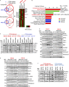
Methylstat activates the UPR pathway in PTEN-deficient TNBC cells. (A) Venn diagram showing up- and down-regulated genes (>1.5 fold, P ≤ 0.05) by Methylstat (2.5 µM) for 24 h in PTEN-deficient MB468 and HCC70 and PTEN wild-type MB231 cells (left panel). Heat map is showing common Methylstat-responsive genes in PTEN-deficient MB468 and HCC70 (right panel). (B) IPA showing nine hallmark pathways exhibiting enrichment of Methylstat-responsive genes in MB468 and HCC70. Graph displays category scores as –log10 (P value) from Fisher’s exact test. TOB, transducer of ErbB2. (C) Western blot analysis of the UPR pathway in indicated TNBC cell lines treated with 2.5 µM Methylstat for 24 h. MW, molecular weight. (D) Western blot analysis of the UPR pathway in response to indicated concentrations of Methylstat for 24 h. (E) Western blot analysis of UPR in cells treated with 2.5 µM Methylstat for indicated hours. (F) Western blot analysis of UPR in indicated TNBC cells treated with 5 µM ML324 for 24 h. See also Fig. S1 and Tables S1 and S2. All data are representative of three independent experiments.
Figure 3.
Figure 3.
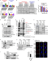
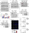
KDM4B represses UPR activity through cytoplasmic interaction with eIF2α. (A) Cell death determined by the percentage of a sub-G1 flow cytometry assay in indicated cell lines treated with indicated siRNAs for 48 h. (B) Western blot analysis of the UPR pathway in indicated cell lines treated with indicated siRNAs. MW, molecular weight. (C) Response to Methylstat in MB436 cell lines expressing empty vector, KDM4B wild-type, or KDM4B mutant plasmids. (D) Western blot analysis of MB436 cells transfected with indicated siRNAs and followed by plasmid transfection 24 h later. (E) Immunoaffinity purification of the KDM4B-containing protein complex. Cell extracts from MB436 cells stably expressing FLAG and FLAG-KDM4B were immunoprecipitated (IP) with anti-FLAG beads. All the immunoprecipitated proteins were resolved by SDS-PAGE and Coomassie blue staining. The protein bands were retrieved and analyzed by mass spectrometry. The interacting proteins were further analyzed by IPA analysis. (F) Coimmunoprecipitation study of endogenous KDM4B and eIF2α in MB436 cells treated with vehicle and 2.5 µM Methylstat for 24 h. (G) Coimmunoprecipitation study using eIF2α antibody in MB436 cells following cytoplasmic and nuclear fractionation. (H) Coimmunoprecipitation study of KDM4B and eIF2α in MB436 cells treated with vehicle and 2.5 µM Methylstat for 24 h following cytoplasmic and nuclear fractionation. (I) DMSO- and Methylstat-treated MDA-MB-436 cells were stained with specific antibodies against eIF2α (mouse) and KDM4B (rabbit) before PLA. The PLA signals between endogenous eIF2α and KDM4B are shown in the red channel; DAPI was used to stain the nuclei (blue); and the merged images show the overlay of the red and blue channels. Dots per cells were analyzed from two independent experiments. Scale bars, 10 µm. (J) Coimmunoprecipitation study using KDM4B antibody in MB436 cells following cytoplasmic and nuclear fractionation. See also Fig. S2. All data are representative of at least three independent experiments unless stated otherwise. Data are expressed as mean ± SD. P values were determined by two-tailed unpaired Student’s t test; **** P ≤ 0.0001.
Figure 4.
Figure 4.
PTEN loss results in down-regulation of KDM4B and activation of UPR. (A) Western blot analysis of the UPR pathway in indicated isogenic MCF10A cell lines. MW, molecular weight. (B) Western blot analysis of UPR in PTEN-KO–MCF10A cells with ectopic expression of wild-type PTEN and PTEN-C124S proteins. (C) Western blot analysis of PTEN wild-type MB231- and HCC1806-expressing PTEN shRNAs. (D) Western blot analysis of the UPR pathway in MB231 cells expressing PTEN shRNAs treated with vehicle or Methylstat (2.5 µM) for 24 h. (E) Representative IHC staining of PTEN, KDM4B, and P-eIF2α in a TNBC tumor specimen. Scale bars, 100 µm. (F) Correlation analysis of IHC staining in tissue microarray between PTEN, KDM4B, and P-eIF2α in TNBC patients (n = 59). Pearson correlation coefficient and two-tailed P values are shown. (G) GSEA was performed with UPR gene signatures (which was implicated in protein folding or translational control and ER-associated protein degradation; see Table S3) on TCGA dataset where PTEN deficiency–related genes were ordered from largest to smallest in 98 basal patients. Basal patients with deep deletion, mutation, mRNA down-regulation, and/or protein down-regulation of PTEN were defined as PTEN deficient; the rest were defined as PTEN wild-type (PTEN deficiency, n = 27; PTEN wild-type, n = 71). NES, normalized enrichment score; FDR, false discovery rate. See also Fig. S3 and Table S3. All Western blot data are representative of three independent experiments.
Figure 5.
Figure 5.
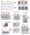
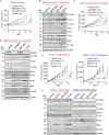
Methylstat and GDC-0941 synergize to boost UPR and cell death in PTEN-deficient TNBC cells. (A) Cellular response to Methylstat (2.5 µM), GDC-0941 (1 µM), or both in indicated cell lines as determined by a CellTiter-Glo Luminescent Cell Viability Assay. Representative data are expressed as log2-fold change normalized to day 0 of three technical replicates. (B) Colony formation assay in cells treated as indicated. Cells were stained with crystal violet when DMSO-treated cells were confluent. (C) Western blot analysis of UPR in PTEN-deficient and PTEN wild-type TNBC cells treated with Methylstat and indicated drugs for 48 h. MW, molecular weight. (D) Western blot analysis of UPR in PTEN-deficient TNBC cells treated with ML324 and indicated drugs for 48 h. (E) Western blot analysis of UPR in MB436 cells transfected with indicated KDM4B siRNAs followed by treatment of GDC-0941 for 48 h. (F) Cell death determined by sub-G1 analysis in MB436 cells in E. (G) Colony formation assay in PTEN-deficient TNBC cells expressing inducible shRNAs. Cells growing in medium with doxycycline were treated with DMSO and GDC-0941 until control cells were confluent, and then colonies were stained with crystal violet. (H) Western blot analysis of MB436 cells expressing inducible shRNAs grown in medium containing doxycycline for 48 h followed by drug treatment for another 48 h. (I) Cell death presented as sub-G1 in MB436 cells, as in H. Drug treatment: 2.5 µM Methylstat, 1 µM GDC-0941, or in combination. See also Fig. S4. All data are representative of three independent experiments unless stated otherwise. Data are expressed as mean ± SD. P values were determined by two-tailed unpaired Student’s t test; * P ≤ 0.05, ** P ≤ 0.01, *** P ≤ 0.001, **** P ≤ 0.0001.
Figure 6.
Figure 6.
FoxO1 and ATF4 cooperate to directly activate UPR pro-apoptotic gene expression. (A) Western blot analysis of MB436 treated with Methylstat, GDC-0941, or in combination for the indicated hours. MW, molecular weight. (B) Subcellular fractionation and Western blot analysis of MB436 treated with the indicated drugs for 8 and 16 h. (C) Western blot analysis of MB436 cells with induced knockdown of FoxO1 treated with Methylstat, GDC-0941, and in combination for 48 h. (D) Cell death presented as sub-G1 was assessed by flow cytometry in MB436 cells from C. (E) Western blot analysis of MB436 cells with induced knockdown of FoxO3a treated with Methylstat, GDC-0941, and in combination for 48 h. (F and G) Coimmunoprecipitation study of FoxO1 and ATF4 in MB436 treated with the indicated drugs for 24 h using FoxO1 (F) and ATF4 (G) antibody, respectively. IB, immunoblotting; WCL, whole cell lysate; IP, immunoprecipitation. (H) Indicated drug–treated MB-436 cells were stained with specific antibodies against ATF4 (mouse) and FoxO1 (rabbit) before PLA. The PLA signals between endogenous ATF4 and FoxO1 are shown in the red channel; DAPI was used to stain the nuclei (blue); and the merged images show the overlay of the red and blue channels. Dots per cells were analyzed from two independent experiments. Scale bars, 10 µm. (I) ChIP-PCR analysis of ATF4 and FoxO1 enrichments on ATF3, BIM, and CHOP promoters in MB436 cells treated with Methylstat (2.5 µM), GDC-0941 (1 µM), or in combination for 24 h. All data are representative of at least three independent experiments unless stated otherwise. P values were determined by two-tailed unpaired Student’s t test; ns P > 0.05, * P ≤ 0.05, ** P ≤ 0.01, *** P ≤ 0.001, **** P ≤ 0.0001.
Figure 7.
Figure 7.
The effect of Methylstat and GDC-0941 combination in vivo. (A) NOD-SCID mice bearing MB436 xenografts were treated with vehicle (n = 13) and Methylstat (30 mg/kg; n = 12) by intraperitoneal injection twice a week, GDC-0941 (100 mg/kg; n = 15) by oral gavage daily, or both drugs (n = 6) for 2 wk. Tumor volumes were measured twice a week. (B) Western blot analysis of representative tumors from each experimental group in A. MW, molecular weight. (C) NOD-SCID mice bearing MB436 (TetOn-shKDM4B) xenografts were fed with diets of doxycycline or treated with GDC-0941 (100 mg/kg) by oral gavage daily or in combination as indicted for 1 mo. Tumor volumes were measured twice a week. Vehicle (n = 9), doxycycline (n = 9), GDC-0941 (n = 10), combination (n = 10). (D) Western blot analysis of representative tumors from each experimental group in C. (E) NOD-SCID mice bearing PTEN-deficient PDX#41 were treated with vehicle (n = 6) and ML324 (60 mg/kg; n = 7) by intraperitoneal injection daily, GDC-0941 (100 mg/kg; n = 7) by oral gavage daily, or both drugs (n = 6) for 2 wk. NOD-SCID mice bearing PTEN wild-type PDX#EL12-58 were treated with vehicle (n = 6), ML324 (60 mg/kg; n = 6), GDC-0941 (100 mg/kg; n = 6), or both drugs (n = 6) for 2 wk. Tumor volumes were measured twice a week. (F) Western blot analysis of representative tumors from each experimental group in E. See also Fig. S5. Data are presented as mean ± SEM. P values were determined by two-tailed unpaired Student’s t test; ns P > 0.05, * P ≤ 0.05, ** P ≤ 0.01, *** P ≤ 0.001, **** P ≤ 0.0001.
Figure 8.
Figure 8.
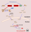
Schematic representation of the proposed mechanisms of over-activation of UPR to trigger apoptosis. Stepwise activation of the UPR pathway by PTEN deficiency, KDM4B inhibition, and PI3K/AKT inhibition leads to cell death in breast cancer cells.

References

    1. Benitez J.A., Ma J., D’Antonio M., Boyer A., Camargo M.F., Zanca C., Kelly S., Khodadadi-Jamayran A., Jameson N.M., Andersen M., et al. . 2017. PTEN regulates glioblastoma oncogenesis through chromatin-associated complexes of DAXX and histone H3.3. Nat. Commun. 8:15223 10.1038/ncomms15223 - DOI - PMC - PubMed
    1. Berry W.L., Kim T.D., and Janknecht R.. 2014. Stimulation of β-catenin and colon cancer cell growth by the KDM4B histone demethylase. Int. J. Oncol. 44:1341–1348. 10.3892/ijo.2014.2279 - V体育官网 - DOI - PubMed
    1. Beyer S., Kristensen M.M., Jensen K.S., Johansen J.V., and Staller P.. 2008. The histone demethylases JMJD1A and JMJD2B are transcriptional targets of hypoxia-inducible factor HIF. J. Biol. Chem. 283:36542–36552. 10.1074/jbc.M804578200 - DOI (VSports) - PMC - PubMed
    1. Brachmann S.M., Hofmann I., Schnell C., Fritsch C., Wee S., Lane H., Wang S., Garcia-Echeverria C., and Maira S.M.. 2009. Specific apoptosis induction by the dual PI3K/mTor inhibitor NVP-BEZ235 in HER2 amplified and PIK3CA mutant breast cancer cells. Proc. Natl. Acad. Sci. USA. 106:22299–22304. 10.1073/pnas.0905152106 - "VSports在线直播" DOI - PMC - PubMed
    1. Cancer Genome Atlas Network 2012. Comprehensive molecular portraits of human breast tumours. Nature. 490:61–70. 10.1038/nature11412 - "VSports手机版" DOI - PMC - PubMed

Publication types

MeSH terms