Skip to main page content
U.S. flag

An official website of the United States government

Dot gov

The . gov means it’s official. Federal government websites often end in . gov or . mil. Before sharing sensitive information, make sure you’re on a federal government site VSports app下载. .

Https

The site is secure. The https:// ensures that you are connecting to the official website and that any information you provide is encrypted and transmitted securely V体育官网. .

Review
. 2018 Sep 14;8(4):1433-1461.
doi: 10.1002/cphy.c170045.

Intersection of Iron and Copper Metabolism in the Mammalian Intestine and Liver

Affiliations
Review

"V体育官网" Intersection of Iron and Copper Metabolism in the Mammalian Intestine and Liver

Caglar Doguer et al. Compr Physiol. .

Abstract

Iron and copper have similar physiochemical properties; thus, physiologically relevant interactions seem likely. Indeed, points of intersection between these two essential trace minerals have been recognized for many decades, but mechanistic details have been lacking. Investigations in recent years have revealed that copper may positively influence iron homeostasis, and also that iron may antagonize copper metabolism. For example, when body iron stores are low, copper is apparently redistributed to tissues important for regulating iron balance, including enterocytes of upper small bowel, the liver, and blood. Copper in enterocytes may positively influence iron transport, and hepatic copper may enhance biosynthesis of a circulating ferroxidase, ceruloplasmin, which potentiates iron release from stores. Moreover, many intestinal genes related to iron absorption are transactivated by a hypoxia-inducible transcription factor, hypoxia-inducible factor-2α (HIF2α), during iron deficiency. Interestingly, copper influences the DNA-binding activity of the HIF factors, thus further exemplifying how copper may modulate intestinal iron homeostasis VSports手机版. Copper may also alter the activity of the iron-regulatory hormone hepcidin. Furthermore, copper depletion has been noted in iron-loading disorders, such as hereditary hemochromatosis. Copper depletion may also be caused by high-dose iron supplementation, raising concerns particularly in pregnancy when iron supplementation is widely recommended. This review will cover the basic physiology of intestinal iron and copper absorption as well as the metabolism of these minerals in the liver. Also considered in detail will be current experimental work in this field, with a focus on molecular aspects of intestinal and hepatic iron-copper interplay and how this relates to various disease states. © 2018 American Physiological Society. Compr Physiol 8:1433-1461, 2018. .


Figures

Figure 1
Figure 1
Iron and copper metabolism in mammals, highlighting points of intersection between these two essential trace minerals. Iron and copper homeostasis during physiological conditions is displayed with points of iron-copper intersection demarcated by yellow stars. Copper movement is indicated with green lines and iron flux in a rust color. Both minerals are absorbed in the duodenum. The inset shows points of iron-copper intersection in a duodenal enterocyte; more details are provided in Figure 2. Copper is mainly incorporated into ceruloplasmin (CP) in hepatocytes, which is secreted into the blood where it functions predominantly in iron metabolism, facilitating iron release from some tissues. A membrane-anchored form of CP, GPI-CP, has a similar function in some tissues. Excess body copper is excreted in bile. Ferric iron binds transferrin (TF) in the portal blood, and after reduction and import into the liver, it is utilized for metabolic purposes or stored in hepatocytes within ferritin. Ferrous iron is then exported into the serum by FPN1, where it is oxidized by CP and then binds to TF for distribution in the blood. Most diferric-TF is taken up by immature red blood cells in the bone marrow and utilized predominantly for hemoglobin synthesis. Iron utilization by developing erythrocytes is copper dependent, although the mechanism by which this occurs is unclear. Iron is also taken up into other tissues, including the brain, where iron release requires GPI-CP. The FOX zyklopen, a copper-dependent protein, may be required for proper iron flux in the placenta. Iron within hemoglobin of senescent red blood cells is recovered and stored by RE macrophages in spleen, bone marrow, and liver (i.e., Kupffer cells). Iron release from these macrophages requires CP or possibly GPI-CP. Iron homeostasis is regulated by hepcidin, which modulates iron flux by inhibiting intestinal iron absorption and iron release from stores in RE macrophages and hepatocytes. Hepcidin may be stabilized by copper, exemplifying another point of iron-copper intersection. Iron is lost from the body predominantly by desquamation of skin cells and exfoliation of enterocytes, and by blood loss, since no active, regulatory excretory system for iron has evolved in humans.
Figure 2
Figure 2
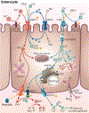
Iron-copper metabolism in a duodenal enterocyte, highlighting points of intersection between these two essential trace minerals. A duodenal enterocyte is depicted along with the proteins which mediate iron and copper absorption. Points where iron and copper metabolism intersect are demarcated by yellow stars. Both metals require reduction prior to absorption, which may be mediated by DCYTB and/or other reductases. Subsequently, iron is transported along with protons across the BBM by DMT1. The electrochemical proton gradient across the BBM that provides the driving force for ferrous iron transport is maintained via the action of a sodium-hydrogen antiporter (NHE3) and the Na+/K+ ATPase on the BLM. DMT1 may also transport copper during iron deficiency (FeD). High-iron (HFe) intake may block copper transport by DMT1 and/or CTR1, eventually leading to copper depletion. Cytosolic iron may be transported into mitochondria for metabolic use, stored in ferritin, or exported across the BLM by FPN1. FPN1 activity may be impacted by copper. Ferrous iron must then be oxidized by HEPH, CP, or other FOXs (not shown) to enable binding to TF in the interstitial fluids. After reduction, dietary copper is transported into enterocytes by CTR1 and is then distributed to various cellular locations by intracellular copper-binding proteins (i.e. chaperones). Excess copper may be stored in the cell by MT. Copper is pumped into the TGN by ATP7A, supporting cuproenzyme synthesis, or exported from the cell by ATP7A, which moves to the BLM when copper is in excess. ATP7A expression is strongly upregulated by iron depletion, suggesting that it (or copper) may positively influence iron metabolism in enterocytes. Copper is spontaneously oxidized by dissolved oxygen in the blood and then bound to mainly albumin and α2-macrogloubuoin in the portal blood and delivered to the liver.
Figure 3
Figure 3
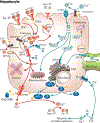
Iron-copper metabolism in a hepatocyte, highlighting points of intersection between these two essential trace minerals. Iron-copper interactions within hepatocytes are indicated by yellow stars. Hepatocytes produce and secrete the iron-regulatory, peptide hormone hepcidin (not shown), which alters intestinal iron absorption (thus justifying the consideration of liver iron homeostasis in this review). Hepcidin also acts in an autocrine fashion to block iron release from hepatocytes (bottom left). Copper may stabilize hepcidin, and thus influence its activity. Hepatocytes also play a principal role in copper metabolism by mediating the excretion of excess copper in bile. These cells assimilate iron via receptor-mediated endocytosis of diferric-TF via transferrin receptors (TFR1/2). Iron is subsequently released from TF by the action of an H+-ATPase in endosomes, reduced (perhaps by STEAP3), and is then transported into the cytosol by DMT1 (or ZIP14). Under pathological conditions of iron overload, nontransferrin bound (ferric) iron in the blood may be reduced and taken up into hepatocytes by ZIP14. This reductase may also reduce copper. Iron is used in cells for metabolic purposes, stored in ferritin, or exported by FPN1 (which may be influenced by copper levels). After reduction, cuprous copper is taken up into hepatocytes via CTR1 and distributed by chaperones. ATOX1 delivers copper to ATP7B, which transports copper into the TGN for incorporation into cuproenzymes, including the FOXs CP and GPI-CP. These FOXs mediate the oxidation of ferrous iron (in an autocrine manner) after release by hepatocytes or other cells (by paracrine of endocrine actions) to permit ferric iron binding to TF in the interstitial fluids. ATP7B also transports excess copper across the canalicular membrane into bile for excretion. ATP7B activity is modulated by COMMD1, and XIAP, a ubiquitin ligase which mediates proteasomal degradation of COMMD1.

References

    1. Abboud S, Haile DJ. A novel mammalian iron-regulated protein involved in intracellular iron metabolism. J Biol Chem 275: 19906–19912, 2000. - PubMed
    1. Aigner E, Theurl I, Haufe H, Seifert M, Hohla F, Scharinger L, Stickel F, Mourlane F, Weiss G, Datz C. Copper availability contributes to iron perturbations in human nonalcoholic fatty liver disease. Gastroenterology 135: 680–688, 2008. - PubMed
    1. Alexander RT, Rievaj J, Dimke H. Paracellular calcium transport across renal and intestinal epithelia. Biochem Cell Biol 92: 467–480, 2014. - PubMed
    1. Anderson GJ, Frazer DM, McKie AT, Vulpe CD. The ceruloplasmin homolog hephaestin and the control of intestinal iron absorption. Blood Cells Mol Dis 29: 367–375, 2002. - PubMed
    1. Anderson GJ, Frazer DM, McLaren GD. Iron absorption and metabolism. Curr Opin Gastroenterol 25: 129–135, 2009. - PubMed

VSports最新版本 - Publication types

"VSports手机版" LinkOut - more resources