<kbd date-time="xeeW5"></kbd><del id="wxzcfps"></del> Skip to main page content
U.S. flag

An official website of the United States government

Dot gov

The . gov means it’s official. Federal government websites often end in VSports app下载. gov or . mil. Before sharing sensitive information, make sure you’re on a federal government site. .

Https

The site is secure V体育官网. The https:// ensures that you are connecting to the official website and that any information you provide is encrypted and transmitted securely. .

. 2018 Sep 14;361(6407):eaao2933.
doi: 10.1126/science.aao2933.

Differential IL-2 expression defines developmental fates of follicular versus nonfollicular helper T cells (V体育2025版)

Affiliations

Differential IL-2 expression defines developmental fates of follicular versus nonfollicular helper T cells (VSports手机版)

Daniel DiToro et al. Science. .

"VSports在线直播" Abstract

In response to infection, naïve CD4+ T cells differentiate into two subpopulations: T follicular helper (TFH) cells, which support B cell antibody production, and non-TFH cells, which enhance innate immune cell functions. Interleukin-2 (IL-2), the major cytokine produced by naïve T cells, plays an important role in the developmental divergence of these populations. However, the relationship between IL-2 production and fate determination remains unclear. Using reporter mice, we found that differential production of IL-2 by naïve CD4+ T cells defined precursors fated for different immune functions. IL-2 producers, which were fated to become TFH cells, delivered IL-2 to nonproducers destined to become non-TFH cells VSports手机版. Because IL-2 production was limited to cells receiving the strongest T cell receptor (TCR) signals, a direct link between TCR-signal strength, IL-2 production, and T cell fate determination has been established. .


"VSports在线直播" Figures

Fig. 1.
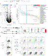
Fig. 1.. Differential expression of Bcl6 and Blimp1 by IL-2+ and IL-2 T cells.
(A) Gene targeting strategy for the generation of IL-2.eGFP knock-in reporter mice. The loxP-flanked neomycin resistance cassette was deleted by crossing founders to EIIa-Cre transgenic mice. (B) Sorted naïve (GFPCD44CD62L+) IL-2.eGFP CD4+ T cells were labeled with CellTraceViolet, stimulated in vitro with soluble anti-CD3 (5μg/mL) and irradiated CD4-depleted feeder cells, then examined for expression of CD69 and IL-2.eGFP by flow cytometry at the indicated time points. Data are representative of four experiments with at least three replicates per condition. CTV staining performed in two of four experiments. (C) Total RNA isolated from naïve IL-2.eGFP CD4+ T cells stimulated for 18–24 h as in B and FACS-purified into CD69+GFP (IL-2) or CD69+GFP+ (IL-2+) fractions was analyzed by comparative expression profiling using RNA-seq. Data depict two biological replicates per condition. (D) RNA isolated from IL-2.eGFP CD4+ T cells stimulated and FACS-purified as in C was analyzed by qPCR for expression of Il2, Bcl6 and Prdm1 at the indicated time points. Error bars represent SEM of three technical replicates per sample. Data are representative of four experiments. (E) Validation of selected transcript expression using RNA isolated from IL-2.eGFP CD4+ T cells stimulated and FACS-purified as in C. Three technical replicates per sample shown. Data were analyzed using Student’s t-tests and are representative of two experiments. For all experiments: ns, p>0.05; *, p<0.05; **, p<0.01; ***, p<0.001; ****, p<0.0001. Error bars depict SEM.
Fig. 2.
Fig. 2.. Differential chromatin accessibility and transcription factor binding at the IL-2 locus in IL-2+ and IL-2 T cells.
(A) ATAC-seq was performed on nuclei isolated from naïve (GFPCD44CD62L+) IL-2.eGFP CD4+ T cells and FACS-purified CD69+GFP+ (IL-2+) and CD69+GFP (IL-2) fractions treated as in Fig. 1C. Chromatin accessibility peaks were visualized using IGB browser and are shown aligned against a VISTA plot of syntenic regions of human and mouse chromosomes corresponding to Il2-Il21/IL2-IL21 gene loci. Data are representative of two experiments. (B) Naïve IL-2.eGFP CD4+ T cells were treated as in Fig. 1C, and the IL-2 promoter region (Il2p) and conserved non-coding sequence 35kb upstream of the Il2 transcription start site (CNS-35kb) of CD69+GFP+ (IL-2+) and CD69+GFP (IL-2) fractions were analyzed by quantitative ChIP-PCR for the presence of Bcl6, Blimp1, and Foxo1 binding, or H3K4me3 and H3K427me3 histone modifications, normalized to total DNA input. Three technical replicates per group. Data for each region analyzed separately by one-way ANOVA. ns, p>0.05; *, p<0.05; **, p<0.01; ***, p<0.001; ****, p<0.0001. Error bars depict SEM.
Fig. 3.
Fig. 3.. Bcl6 and IL-2 co-segregate early in each T effector cell developmental program.
(A) Naïve (GFPCD44CD62L+CD69CD25) IL-2.eGFP CD4+ T cells were stimulated in vitro under Th0, Th1, Th2 and Th17 conditions for 20 h and examined by flow cytometry for CD69 and IL-2.eGFP expression. Data are representative of 2 experiments. Flow plots depict cell number-controlled concatenated averages of three samples per group. Error bars depict SD. (B) Experiment performed as in A, with CD69+ IL-2.eGFP+ and IL-2.eGFP CD4+ T cells sorted 20 h after activation. RNA was isolated and analyzed by qPCR for expression of Il2, Bcl6 and Prdm1. Three technical replicates per condition are shown. Error bars depict SEM. Data for A and B are representative of two experiments each.
Fig. 4.
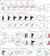
Fig. 4.. IL-2+ T cells activate IL-2 T cells via paracrine IL-2 signaling to drive differential gene expression in vivo.
(A) Sorted naïve (GFPCD44CD62L+) IL-2.eGFP CD45.2+ SMARTA CD4+ T cells were transferred into CD45.1+ WT mice infected with ActA-Lm-gp66 24 h prior to transfer. Total RNA was isolated by FACS-purified CD45.2+ CD69+GFP+ (IL-2+) and CD69+GFP (IL-2) CD4+ T cells 20–24 h after transfer and analyzed by RNA-seq. Data depict three biological replicates per condition from three separate experiments. (B) Hallmark gene set enrichment analysis of IL-2+ and IL-2 T cells from A. For each pathway, mean and 95% confidence intervals are plotted then color-coded to indicate false discovery rate corrected p-values. (C) Schematic of targeting strategy to generate IL-2.Thy1.1 Bac-In (2BiT) transgenic reporter mice. (D) Sorted naïve (Thy1.1CD44CD62L+) 2BiT CD4+ T cells were stimulated in vitro with soluble anti-CD3 (5μg/mL) and irradiated CD4-depleted feeder cells for 24 h then examined by flow cytometry for expression of CD69 and Thy1.1. RNA isolated from CD69+Thy1.1+ (IL-2+) and CD69+Thy1.1 (IL-2) CD4+ T cells was analyzed by qPCR for expression of Il2 mRNA. Error bars represent SEM of three technical replicates per sample. Data are representative of two experiments. (E) 2BiT mice were infected with ActA-Lm. After 18 h mice were sacrificed and splenic CD4+ T cells were analyzed by flow cytometry for the expression of IL-2.Thy1.1, CD25, and tyrosine phosphorylation of Stat5 (p-Stat5). Data are representative of two experiments. (F) Congenic CD45.1+ WT mice were infected with ActA-Lm-gp66. Twenty-four hours later, naïve CD45.2+ SMARTA 2BiT CD4+ T cells were transferred into infected CD45.1+ recipients. Mice were sacrificed at the indicated times, and splenic CD4+ T cells were analyzed for expression of Thy1.1, Foxp3 and p-Stat5. Data are representative of two experiments.
Fig. 5.
Fig. 5.. IL-2 producers are precursors of Tfh cells.
(A-C) 2BiT mice were injected with 250μg anti-Thy1.1 or isotype control mAb, then infected 1 day later with ActALm-gp66. Endogenous CD4+ T cells specific for IAb-gp66 were enriched from lymph nodes and spleens 3 d following infection using tetramer-based magnetic sorting and analyzed by flow cytometry for IAb-gp66 tetramer binding and expression of Ly6C, CXCR5, IL-2.Thy1.1 and PD-1 (A) or Bcl6 (B). Flow plots depict cell-number-controlled concatenated averages of all samples within a group. Data for A and B are representative of two experiments each. (C) Data from the experiments depicted in A and supplemental fig. S6 were analyzed by two-way ANOVA. A total of eight control and eight treatment animals from two separate experiments are shown. (D) 2BiT mice were injected with 250μg anti-Thy1.1 or isotype control mAb and immunized with 2×10¹⁰ CFU heat-killed Lm (HKLm). Mice were bled every 6 d for 24 days, and serum anti-LM IgG was measured by ELISA. n = 7 per group. Data are representative of two experiments. (E) Magnetically enriched WT CD45.1+ and 2BiT CD45.2+ CD4+ T cells were transferred into TCRβ-deficient mice (Tcrb–/–). Twenty-four hours later, mice were immunized with 2×10¹⁰ CFU HKLm and injected with 250μg anti-Thy1.1 or isotype control mAb. Mice were sacrificed 5 d following immunization, and splenic CD4+ T cells were analyzed by flow cytometry for expression of CD44, CD45.1, CD45.2, PD-1, and CXCR5. Results were analyzed by two-way ANOVA. n = 3 per group. Data are representative of three experiments. (F) CD4+ T cells magnetically enriched from WT CAG-eGFP (CD45.2) mice and CD45.1+ 2BiT mice were adoptively transferred into TCRβ-deficient recipients. Twenty-four hours later, the mice were immunized with 2×10¹⁰ CFU HKLm and injected with 250μg anti-Thy1.1 or isotype control mAb. Two weeks following immunization, spleens were collected and analyzed by confocal microscopy for the expression of GFP (WT), CD45.1 (2BiT), Ki67 and IgD. Quantitation of WT (GFP+), 2BiT (CD45.1+) and total T-cell numbers in germinal centers was done using computer-assisted counting. Splenic B cells were analyzed by flow cytometry for the expression of IgD, B220, GL7 and Fas in an IgDlo B-cell gate. n = 3 per group. Data are representative of three experiments. For all experiments: ns, p>0.05; *, p<0.05; **, p<0.01; ***, p<0.001; ****, p<0.0001. Error bars depict SEM.
Fig. 6.
Fig. 6.. IL-2 producers are fated to become Tfh cells in type 2 and type 3 immune responses.
(A) 2BiT mice were injected with 250μg anti-Thy1.1 or isotype control. Twenty-four hours later, they were immunized with OVA emulsified in Alum. Mice were bled and sacrificed at day 12. Splenic IAb-OVA tetramer-specific CD4+ T cells were analyzed by flow cytometry for the expression of CD44, PD-1, and CXCR5. Splenic B cells were analyzed for the expression of B220, IgD, GL7 and Fas. Serum anti-OVA IgG was measured by ELISA. n = 5 to 6 per group. (B) 2BiT mice were injected with 250μg anti-Thy1.1 or isotype control. Twenty-four hours later they were orally gavaged with 1–2×10⁹ CFU Citrobacter rodentium strain DBS100 (ATCC 51459) or the bioluminescent ICC180 derivative. Whole body bioluminescence was tracked and quantified after infection. Splenic IAb-Int884C tetramer-specific CD4+ T cells harvested on day 14 were analyzed by flow cytometry for the expression of CD44, PD-1, and CXCR5. Splenic B cells were analyzed for expression of B220, IgD, GL7 and Fas. n = 5 to 6 per group. Flow plots depict cell-number-controlled concatenated averages. Data are representative of two experiments. (C+D) Splenic (C) and MLN (D) CD4+ T cells harvested from mice treated as in (B) were isolated, re-stimulated with PMA and ionomycin then analyzed by flow cytometry for expression of CD44, Foxp3, IFNγ, and IL-17A. Flow plots and bar graphs are gated on CD4+CD44+Foxp3 cells. Flow plots depict cell-number-controlled concatenated averages. n = 5 to 6 per group. For all experiments: ns, p>0.05; *, p<0.05; **, p<0.01; ***, p<0.001; ****, p<0.0001. Data were analyzed using Student’s t-tests. Error bars depict SEM.
Fig. 7.
Fig. 7.. IL-2 production and Tfh differentiation correlate with TCR signal strength.
(A) WT CD45.1+ recipient mice were infected with ActALm-OVA-gp66 and/or ActALm-OVA-2WIS at the indicated doses. After 24 h, 106 naïve (GFPCD44CD62L+) SMARTA IL-2.eGFP CD4+ T cells were adoptively transferred into infected hosts. Splenic CD4+ T cells were harvested 15 h following transfer and analyzed for expression of CD69 and IL-2.eGFP by flow cytometry. Values in the larger boxes of flow cytometric plots represent percentages of CD69+ cells, and values in the smaller boxes represent percentages IL-2.eGFP+ cells within the CD69+ fraction. n = 4 per group. Data are representative of 2 experiments. (B) WT mice were infected with ActALm-OVA-gp66 and/or ActALm-OVA-2WIS at the indicated doses. Five days later, magnetically enriched endogenous splenic CD4+ T cells were analyzed by flow cytometry for binding of IAb-gp66 tetramer and expression of CD44, CXCR5, and PD-1. n = 3 per group. Data are representative of two separate experiments. (C) 2D affinity measurements were performed on splenic TCR transgenic CD4+ T cells via micropipette adhesion frequency assays with biotinylated pMHC IAb-gp66- and IAb-OVA3C monomers. Log-normalized data are shown. WT CD45.1+ recipient mice were infected with ActALm-OVA-gp66. After 24 h, 0.5×10⁶ naïve (GFPCD44CD62L+CD69CD25) SMARTA IL-2.eGFP and OTII IL-2.eGFP CD4+ T cells were pooled and adoptively transferred into infected hosts. Splenic CD4+ T cells were harvested 18 h following transfer and analyzed for expression of CD45.1, CD45.2, Vβ5, CD69 and IL-2.eGFP by flow cytometry. Values in the larger boxes of flow cytometric plots depicting CD69 and IL-2.eGFP represent percentages of CD69+ cells, and values in the smaller boxes represent the percentages of IL-2.eGFP+ cells within the CD69+ fraction. n = 3 per group. Data are representative of 2 experiments. (D) WT mice were infected with ActALm-OVA-gp66. Five days after infection, magnetically enriched endogenous splenic CD4+ T cells were co-stained with IAb-gp66- and IAb-OVA3C tetramers and analyzed by flow cytometry for expression of CD44, CXCR5, and PD-1. Data are representative of 4 experiments. (E) WT mice were infected with 2.5×10⁷ CFU ActALm-OVA-gp66. Enriched splenic CD4+ T cells harvested 5 d following infection were stained for IAb-gp66, CD44, TCRβ, PD-1 and CXCR5. Log-normalized 2D affinity measurements were performed on FACS-purified IAb-gp66 tetramer-positive splenic Tfh and non-Tfh cells pooled from 3–5 animals. TCR β quantifications were performed on unsorted aliquots stained separately. Data from 2 experiments are shown. (F) Naïve (GFPCD44CD62L+) SMARTA IL-2.eGFP CD4+ T cells were stimulated for 16 h with irradiated CD4-depleted feeder cells and 1μg/mL gp66 and analyzed by flow cytometry for expression of CD69, Vα2 and IL-2.eGFP. Data are representative of 2 experiments. For all experiments: ns, p>0.05; *, p<0.05; **, p<0.01; ***, p<0.001; ****, p<0.0001. Data were analyzed using Student’s t-tests. Error bars depict SEM.
Fig. 8.
Fig. 8.. IL-2 producers and Tfh exhibit enhanced cell cycle progression.
(A) Naïve (GFPCD44CD62L+CD69CD25) IL-2.eGFP CD4+ T cells were stimulated for 18 h with indicated concentrations of plate-bound anti-CD3 and 1μg/mL soluble anti-CD28 then analyzed for expression of CD69 and IL-2.eGFP by flow cytometry. The MFI of CD69 expression within the CD69+GFP and CD69+GFP+ gates was quantitated for the indicated concentrations of anti-CD3 (right). Three technical replicates per condition. Experiment performed three times. (B) Naïve (GFPCD44CD62L+CD69CD25) IL-2.eGFP CD4+ T cells were stimulated in vitro with soluble anti-CD3 (2.5μg/mL), soluble anti-CD28 (0.5μg/mL) and irradiated CD4-depleted feeder cells. qPCR was performed on CD69+ IL-2.eGFP+ and IL-2.eGFP CD4+ T cells sorted 20 h after activation. Three technical replicates per condition shown. Data are representative of two experiments and were analyzed by one-way ANOVA. (C) WT CD45.1+ recipient mice were infected with ActALm-OVA-gp66. After 24 h, 5×10⁴ CTV-labeled naïve (GFPCD44CD62L+CD69CD25) SMARTA CD4+ T cells were adoptively transferred into infected hosts. Splenic CD4+ T cells harvested 3 d following transfer were analyzed for expression of CD44, PD-1 and CXCR5 by flow cytometry. n = 4 per experiment. Experiment performed three times. For all experiments: ns, p>0.05; *, p<0.05; **, p<0.01; ***, p<0.001; ****, p<0.0001. Error bars depict SEM.

References

    1. Zhu J, Yamane H, Paul WE, Differentiation of effector CD4+ T-cell populations. Annu. Rev. Immunol 28, 445–489 (2010). - PMC - PubMed
    1. Tubo NJ et al., Single Naive CD4+ T cells from a diverse repertoire produce different effector cell types during infection. Cell. 153, 785–796 (2013). - PMC - PubMed
    1. Bucy RP et al., Single cell analysis of cytokine gene coexpression during CD4+ T-cell phenotype development. Proc. Nat.l Acad. Sci. USA 92, 7565–7569 (1995). - "V体育官网入口" PMC - PubMed
    1. Johnston RJ et al., Bcl6 and Blimp-1 are reciprocal and antagonistic regulators of T follicular helper cell differentiation. Science. 325, 1006–1010 (2009). - PMC - PubMed
    1. Nurieva RI et al., STAT5 protein negatively regulates T follicular helper (Tfh) cell generation and function. J Biol. Chem 287, 11234–11239 (2012). - VSports手机版 - PMC - PubMed

Publication types

MeSH terms