Skip to main page content
U.S. flag

An official website of the United States government

Dot gov

The . gov means it’s official. Federal government websites often end in . gov or . mil VSports app下载. Before sharing sensitive information, make sure you’re on a federal government site. .

Https

The site is secure. The https:// ensures that you are connecting to the official website and that any information you provide is encrypted and transmitted securely V体育官网. .

. 2018 Sep 4;9(5):e02290-17.
doi: 10.1128/mBio.02290-17.

Phase Transitions Drive the Formation of Vesicular Stomatitis Virus Replication Compartments

Affiliations

"VSports最新版本" Phase Transitions Drive the Formation of Vesicular Stomatitis Virus Replication Compartments

Bianca S Heinrich et al. mBio. .

Abstract

RNA viruses that replicate in the cell cytoplasm typically concentrate their replication machinery within specialized compartments. This concentration favors enzymatic reactions and shields viral RNA from detection by cytosolic pattern recognition receptors. Nonsegmented negative-strand (NNS) RNA viruses, which include some of the most significant human, animal, and plant pathogens extant, form inclusions that are sites of RNA synthesis and are not circumscribed by a membrane. These inclusions share similarities with cellular protein/RNA structures such as P granules and nucleoli, which are phase-separated liquid compartments. Here we show that replication compartments of vesicular stomatitis virus (VSV) have the properties of liquid-like compartments that form by phase separation. Expression of the individual viral components of the replication machinery in cells demonstrates that the 3 viral proteins required for replication are sufficient to drive cytoplasmic phase separation. Therefore, liquid-liquid phase separation, previously linked to organization of P granules, nucleolus homeostasis, and cell signaling, plays a key role in host-pathogen interactions. This work suggests novel therapeutic approaches to the problem of combating NNS RNA viral infections VSports手机版. IMPORTANCE RNA viruses compartmentalize their replication machinery to evade detection by host pattern recognition receptors and concentrate the machinery of RNA synthesis. For positive-strand RNA viruses, RNA replication occurs in a virus-induced membrane-associated replication organelle. For NNS RNA viruses, the replication compartment is a cytoplasmic inclusion that is not circumscribed by a cellular membrane. Such structures were first observed in the cell bodies of neurons from humans infected with rabies virus and were termed Negri bodies. How the replication machinery that forms this inclusion remains associated in the absence of a membrane has been an enduring mystery. In this article, we present evidence that the VSV replication compartments form through phase separation. Phase separation is increasingly recognized as responsible for cellular structures as diverse as processing bodies (P-bodies) and nucleoli and was recently demonstrated for rabies virus. This article further links the fields of host-pathogen interaction with that of phase separation. .

Keywords: negative-strand RNA virus; phase separation; rhabdovirus; viral replication; viroplasm; virus-host interactions. V体育安卓版.


Figures

FIG 1
FIG 1
Fluid-like properties of the non-membrane-associated VSV replication compartment. (a) Composite phase-contrast and laser micrograph of Vero cells infected with rVSV-eGFP-P 6 h postinfection (6 hpi). Scale bar, 10 µm. (b) Electron micrograph of rVSV-eGFP-P-infected Vero cells (6 hpi) showing electron-dense inclusion (I), mitochondrion (M), and cytoplasm (C). Scale bar, 0.5 µm. (c and d) Time-lapse GFP images (Movie 1) showing fusion (c) and fission (d) of viral inclusions in rVSV-eGFP-P-infected Vero cells (6 hpi). Arrow marks the clearly separated inclusion. Single-plane GFP fluorescence micrographs were captured every 30 s. Scale bars, 5 µm. (e) Phase-separated compartments form in cells from a diverse range of hosts. Shown are fluorescence micrographs of the following cell lines infected with rVSV-eGFP-P and counterstained for DNA: Homo sapiens HeLa S3 (6 hpi), Chlorocebus aethiops Vero (6 hpi), Mus musculus mouse embryonic fibroblast (8 hpi), Xenopus laevis S3 (8 hpi), Aedes albopictus C6-36 (48 hpi), and Spodoptera frugiperda Sf-9 cells (48 hpi). Scale bars, 10 µm.
FIG 2
FIG 2
Replication compartment formation and maintenance are insensitive to microtubule depolymerization. (a) GFP fluorescence micrographs of Vero cells cultured in medium alone (mock treatment) or supplemented with nocodazole and fixed at 3 or 6 hpi with rVSV-eGFP-P. Cell nuclei are outlined for reference (dotted lines). Scale bar, 10 µm. (b) Time course for the appearance of eGFP in cytosol and viroplasm in Vero cells after infection with rVSV-eGFP-P and culture in medium alone (mock treatment) or supplemented with nocodazole. (c) Time-lapse GFP images (Movie S2) of fusion between viral inclusions in nocodazole-treated Vero cells infected with rVSV-eGFP-P and imaged every minute. Scale bar, 5 µm.
FIG 3
FIG 3
Rapid exchange of the components of the replication compartments. (a) Time-lapse images and quantification of FRAP experiments in Vero cells infected with rVSV-eGFP-P (filled circles) or transfected with a cDNA encoding G250-GFP (filled squares). The photobleached region (solid line box) and regions used for quantification (dotted line boxes) are indicated. Scale bars, 5 µm. (b) Time-lapse images and quantification of whole-inclusion FRAP in Vero cells infected with rVSV-eGFP-P. Integrated fluorescence intensity over time was fit to a single-exponential recovery curve for representative inclusions from mock-treated (black; t = 0.010 s−1; R2 = 0.979) or nocodazole-treated (red; t = 006 s−1; R2 = 0.993) cells. Scale bars, 2 µm. (c) FRAP recovery half-lives for inclusions in mock-treated (5 cells; mean, 68 s; standard deviation [SD], 11 s) and nocodazole-treated (10 cells; mean, 83 s; SD, 41 s) had no significant difference (unpaired t test, P = 0.3).
FIG 4
FIG 4
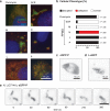
Viral replication proteins, but not replication, are required to form viroplasm in cells. (a) Representative images showing the effects on inclusion morphology in Vero cells treated with puromycin or the indicated PPMO for 3 h: VSV-L (green), VSV-N (red), and DNA (blue). Scale bar, 10 µm. (b) Classification of VSV-N and VSV-L protein staining patterns in VSV-infected Vero cells after PPMO treatment. The total cell count is indicated for each treatment. (c and d) Representative live eGFP fluorescence images of cells transfected with cDNA encoding eGFP-P (c) or L-eGFP (d). Scale bars, 10 µm. (e) Single-plane, time-lapse imaging (2 frames min−1) of inclusions in Vero cells transfected with the indicated plasmid cDNA encoding (N) eGFP-P and catalytically inactive L. Scale bar, 5 µm.

References

    1. Ahlquist P. 2006. Parallels among positive-strand RNA viruses, reverse-transcribing viruses and double-stranded RNA viruses. Nat Rev Microbiol 4:371–382. doi:10.1038/nrmicro1389. - DOI - PMC - PubMed
    1. den Boon JA, Ahlquist P. 2010. Organelle-like membrane compartmentalization of positive-strand RNA virus replication factories. Annu Rev Microbiol 64:241–256. doi:10.1146/annurev.micro.112408.134012. - DOI - PubMed
    1. Heinrich BS, Cureton DK, Rahmeh AA, Whelan SP. 2010. Protein expression redirects vesicular stomatitis virus RNA synthesis to cytoplasmic inclusions. PLoS Pathog 6:e1000958. doi:10.1371/journal.ppat.1000958. - DOI - PMC - PubMed
    1. Lahaye X, Vidy A, Pomier C, Obiang L, Harper F, Gaudin Y, Blondel D. 2009. Functional characterization of Negri bodies (NBs) in rabies virus-infected cells: evidence that NBs are sites of viral transcription and replication. J Virol 83:7948–7958. doi:10.1128/JVI.00554-09. - DOI - PMC - PubMed
    1. Nikolic J, Le Bars R, Lama Z, Scrima N, Lagaudrière-Gesbert C, Gaudin Y, Blondel D. 2017. Negri bodies are viral factories with properties of liquid organelles. Nat Commun 8:58. doi:10.1038/s41467-017-00102-9. - DOI - PMC - PubMed

V体育平台登录 - Publication types

"V体育ios版" LinkOut - more resources