Skip to main page content
U.S. flag

An official website of the United States government

Dot gov

The . gov means it’s official. Federal government websites often end in . gov or VSports app下载. mil. Before sharing sensitive information, make sure you’re on a federal government site. .

Https

The site is secure. The https:// ensures that you are connecting to the official website and that any information you provide is encrypted and transmitted securely. V体育官网.

. 2018 Oct;24(10):1550-1558.
doi: 10.1038/s41591-018-0136-1. Epub 2018 Aug 20.

Signatures of T cell dysfunction and exclusion predict cancer immunotherapy response

Affiliations

Signatures of T cell dysfunction and exclusion predict cancer immunotherapy response

Peng Jiang et al. Nat Med. 2018 Oct.

Abstract

Cancer treatment by immune checkpoint blockade (ICB) can bring long-lasting clinical benefits, but only a fraction of patients respond to treatment. To predict ICB response, we developed TIDE, a computational method to model two primary mechanisms of tumor immune evasion: the induction of T cell dysfunction in tumors with high infiltration of cytotoxic T lymphocytes (CTL) and the prevention of T cell infiltration in tumors with low CTL level. We identified signatures of T cell dysfunction from large tumor cohorts by testing how the expression of each gene in tumors interacts with the CTL infiltration level to influence patient survival. We also modeled factors that exclude T cell infiltration into tumors using expression signatures from immunosuppressive cells. Using this framework and pre-treatment RNA-Seq or NanoString tumor expression profiles, TIDE predicted the outcome of melanoma patients treated with first-line anti-PD1 or anti-CTLA4 more accurately than other biomarkers such as PD-L1 level and mutation load. TIDE also revealed new candidate ICB resistance regulators, such as SERPINB9, demonstrating utility for immunotherapy research. VSports手机版.


Figures
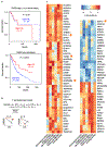
Fig. 1 |
Fig. 1 |. The interaction test identifies gene signatures of T cell dysfunction.
a, The association between the CTL level and overall patient survival for melanoma tumors with different TGFB1 levels. For each metastatic melanoma tumor in TCGA, the CTL infiltration level was estimated as the average expression level of CD8A, CD8B, GZMA, GZMB and PRF1. The association between the CTL level and overall survival was computed through the two-sided Wald test in the Cox-PH regression. Each Kaplan–Meier plot presents tumors in two groups: ‘High CTL’ (red) have above-average CTL values among all samples, while ‘Low CTL’ (blue) have values below average. Samples were split according to the TGFB1 expression level to show the association between the CTL level and survival outcome. The top panel shows tumors with high TGFB1 expression (one standard deviation above the average), while the bottom panels show the remaining samples. b, The interaction test in a Cox-PH regression to identify genes associated with the T cell dysfunction. The variable CTL represents the level of CTLs in each tumor. The variable V represents the status of a candidate gene. The coefficient d reflects the effect of interaction between the CTL and V on death hazard outcome estimated from the survival data. The graphs represent the association slopes between CTL and death hazard. The black and gold arrows represent the association slopes before and after increasing the level of V. c, Genes with significant T cell dysfunction scores in multiple cancer types. Five data sets, representing five cancer types, had more than 1% of genes passing the FDR threshold 0.1. We display the genes whose T cell dysfunction scores, defined as the z score of d/standard error (s.e.), are significantly positive or negative (two-sided Wald test P values corresponding to an FDR less than 0.1) in at least two cancer types. The orange stars indicate genes of special interest. The number of samples in each cohort is available in Supplementary Table 2b. UCEC, uterine corpus endometrial carcinoma, TNBC, triple-negative breast cancer; AML, acute myeloid leukemia; SKCM, skin cutaneous melanoma; NB, neuroblastoma.
Fig. 2 |
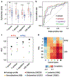
Fig. 2 |. T cell dysfunction signatures are consistent with published signatures of tumor immune evasion.
a, The consistency between T cell dysfunction signatures predicted by the interaction test and published gene signatures of tumor immune evasion. To evaluate the reliability of the T cell dysfunction gene scores, we collected four published gene signatures related to T cell dysfunction and immunotherapy resistance (Supplementary Table 4). We plotted the T cell dysfunction scores averaged across five cancer types (average profile in Fig. 1c) for the positive (red) and negative (blue) hits of each gene signature. The numbers of positive and negative hits for each signature are available in Supplementary Table 4. Within each group, the scattered dots represent all gene values, and the thick line represents the median value. The bottom and top of the boxes are the 25th and 75th percentiles (interquartile range). The whiskers encompass 1.5 times the interquartile range. The difference between positive and negative groups was compared through the two-sided Wilcoxon rank-sum test, and P values for signatures of ‘T accum’, ‘T exhaust’, ‘T regulatory’ and ‘ICB resist’ are 6.94×10⁻³, 1.20×10⁻⁸, 1.81 ×10⁻⁶ and 1.95 ×10⁻⁷, respectively. The range of P values are labeled above each boxplot with asterisks (**P<1 ×10⁻²; ***P<1 ×10⁻³). T accum: shRNA screens for regulators of T cell accumulation in tumors; T exhaust: transcriptome of exhausted T cells; T regulatory: transcriptome of CD4 regulatory T cells. ICB resist: transcriptome of murine tumors that resist anti-CTLA4 checkpoint blockade. b, The ROC curves measuring the performance of the average T cell dysfunction scores (average profile in Fig. 1c) in predicting the positive and negative gene hits in each signature in a. c, The area under the ROC curve of the average profile of all five cancer types (black squares) and each of the individual cancer types SKCM, AML, NB, UCEC and TNBC with different dot colors. d, Pearson correlations between the T cell dysfunction scores and the expression profile of exhausted T cells. The correlations were computed across 12,498 genes shared between human and mouse signatures, for all pairwise combinations between five human cancer types and different time points in a mouse model of T cell exhaustion (‘T exh fixed’ in Supplementary Table 4).
Fig. 3 |
Fig. 3 |. Immunosuppressive cell expression models gene signatures of T cell exclusion.
a, Prediction of T cell exclusion scores for tumors using immunosuppressive cell signatures. For each metastatic tumor in the TCGA melanoma data set (blue dots, n = 317), we computed the Pearson correlation between its expression profile and the expression signature of MDSCs, M2 TAMs or CAFs (Supplementary Table 4) or the average of the three expression signatures. In each graph, these values are plotted along the x axis. The y axis shows the CTL level for each tumor (average expression level of CD8A, CD8B, GZMA, GZMB and PRF1). The Pearson correlation (R) between the plotted values is shown in the upper right corner of each plot. The two-sided t-test P values for correlations in MDSCs, TAMs, CAFs and mean are 4.61 ×10−37, 1.58×10−51, 8.84×10⁻¹³ and 2.58×10−52, respectively. b, A histogram of the correlations between the CTL levels and the T cell exclusion scores across tumors. The correlations analyzed in the histogram correspond to the R value in the top right corner of a (example of TCGA melanoma) across 43 solid tumor data sets. Gliomas are excluded because of low T cell infiltration levels in most gliomas. c, Anti-correlation between T cell dysfunction scores and exclusion scores across TCGA melanoma tumors. For each metastatic melanoma tumor (colored dots, n = 317), we computed the Pearson correlation between the sample’s expression profile and the TIDE T cell dysfunction signature (y axis). The same computation was made between the tumor expression profile and the TIDE T cell exclusion signature (x axis). The Pearson correlation between the plotted values is shown in the upper right (two-sided t-test P value = 4.02 ×10⁻³⁴). The dot color indicates the CTL level in each tumor. d, Anti-correlation between T cell dysfunction scores and exclusion scores across TCGA cancer types. For each TCGA cancer type with normal control samples (n = 17), we calculated the average expression difference between tumor versus normal samples. We then computed the Pearson correlation between that value and the TIDE T cell dysfunction signature (y axis). We also made the same calculation for the TIDE T cell exclusion signature (x axis). The Pearson correlation between the plotted values is shown in the upper right (one-sided t-test P value = 0.042). The CTL level difference between tumor and normal samples is shown by the dot color.
Fig. 4 |
Fig. 4 |. TIDE signatures predict iCB immunotherapy response.
a, A waterfall plot of TIDE prediction scores across 25 melanoma tumors treated with anti-PDI. The TIDE framework divided tumors into CTL-high or -low categories based on the expression level of CTL marker genes (Supplementary Fig. 4a). Red indicates a tumor that responded to therapy. Blue indicates a non-responder. In each category, we sorted tumors in descending order according to their TIDE prediction scores. b, A waterfall plot of TIDE prediction scores across 42 melanoma tumors treated with anti-CTLA4 in the same way as in a. Besides responders or non-responders, several patients are classified as long-survival in the original study due to the long overall survival time. c, A waterfall plot of TIDE prediction scores across 33 tumors treated with anti-PD1 in the same way as in a. The gene expression profiles are measured by the NanoString platform. The 33 tumors comprise 9 melanoma, 12 lung adenocarcinoma, 9 lung squamous carcinoma and 3 head and neck tumors. d, ROC curves for the performance of the TIDE prediction score, PD-L1 expression, interferon gamma (IFNG) response and total mutation load in predicting anti-PD1 response among 25 melanoma tumors in a. e, ROC curves for the performance of several signatures in predicting anti-CTLA4 response among 42 melanoma tumors in b. f, ROC curves for the performance of several signatures in predicting anti-PD1 response among 33 tumors in c. g, The area under the ROC curve (AUC) for several signatures in predicting anti-PD1 response among 25 melanoma tumors in a. The signatures are defined in Supplementary Table 7, with TIDE in dark red and other signatures in blue. Besides the genome-wide TIDE signature, ‘TIDE.selected’ is a variation focused on 770 genes with both high expression variation across tumors and significant values in the either T cell dysfunction or exclusion signatures. The performance of a random predictor (AUC = 0.5) is represented by the dashed line. h, AUC for signatures in predicting anti-CTLA4 response among 42 melanoma tumors in b in the same way as in g. i, AUC for signatures in predicting anti-PD1 response among 33 tumors in c in the same way as in g. TIDE AUC metrics are also shown separately for nine melanoma, twelve lung adenocarcinoma (Adeno) and nine lung squamous carcinoma (Squamous) tumors. j, Kaplan–Meier plots of overall survival (OS) for 25 melanoma patients (in a) treated with anti-PD1 with the top (>1) and bottom (<1) TIDE prediction scores. The P value was calculated by testing the association between TIDE prediction scores and overall survival with the two-sided Wald test in a Cox-PH regression. k, Kaplan-Meier plots of overall survival for 42 melanoma patients (in b) treated with anti-CTLA4 in the same way as in j. l, Kaplan-Meier plots of progression-free survival (PFS) for 33 patients (in c) treated with anti-PD1 in the same way as in j.
Fig. 5 |
Fig. 5 |. Validation of SERPINB9 as a regulator of tumor immune escape.
a, The log-fold change (log[FC]) of expression between anti-CTLA4-resistant and parental B16 murine tumors for genes with significant T cell dysfunction scores in Fig. 1c. All genes are ranked increasingly with the top one labeled by name. b, The expression value of Serpinb9 between anti-CTLA4-resistant and parental B16 tumors. Within each group, the scattered dots represent Serpinb9 expression values (n = 6 samples in the resistant group, n = 4 samples in the parental group). The thick line represents the median value. The bottom and top of the boxes are the 25th and 75th percentiles (interquartile range). The whiskers encompass 1.5 times the interquartile range. The P value, testing the group difference, was calculated with the two-sided Wilcoxon rank-sum test. c, Kaplan-Meier plots of patients with top half and bottom half SERPINB9 expression levels, using the data from an anti-CTLA4 study with 42 patients profiled. Both progression-free survival and overall survival are shown. The association between SERPINB9 expression and patient survival was tested by the two-sided Wald test in a Cox-PH regression (Supplementary Table 9). d, Western blot of SERPINB9 following genetic knockout and overexpression. For knockout (KO), there are two independent CRISPR guides targeting Serpinb9 and a control non-targeting sequence. Cells were either untreated (left 3 lanes) or treated with 100 ng ml−1 IFNγ to induce Serpinb9 expression (right 3 lanes). VCL is the loading control. For overexpression, the open reading frame of Serpinb9 was cloned into the pEF1a plasmid and overexpressed in B16F10 cells. The protein level was compared between pEF1a backbone- and pEF1a-Serpinb9-transduced cells. The bands of related protein targets are cut and shown. All experiments have been repeated independently two times with similar results. e, The effect of Serpinb9 knockout on T cell-mediated tumor killing. B16F10 cancer cells were co-cultured for three days with cytotoxic T cells at three B16F10 to T cell ratios (3:1, 2:1 or 1:1). Each CRISPR gRNA-transduced GFP positive cell line (Control, KO 1, KO 2) was mixed with the parental GFP-negative cell line at a 1:1 ratio. After co-culture, the ratio of edited GFP+cells to parental cells (GFP) was determined by flow cytometry. The bar plots present the median value among three cell-culture replicates with standard deviations as the error bars. The results of the two-sided Student t-test, comparing the difference between knockout and control conditions, are available in Supplementary Table 10. f, The effect of Serpinb9 overexpression on T cell-mediated tumor killing. The effect of Serpinb9 overexpression was examined. The bar plots present the median value among two cell-culture replicates with standard deviations as the error bars. The results of the two-sided Student t-test, comparing the difference between overexpression and control conditions, are available in Supplementary Table 10.

Comment in (VSports注册入口)

References

    1. Mahoney KM, Rennert PD & Freeman GJ Combination cancer immunotherapy and new immunomodulatory targets. Nat. Rev. Drug Discov 14, 561–584 (2015). - V体育2025版 - PubMed
    1. Sharma P, Hu-Lieskovan S, Wargo JA & Ribas A Primary, adaptive, and acquired resistance to cancer immunotherapy. Cell 168, 707–723 (2017). - PMC - PubMed
    1. Van Allen EM et al. Genomic correlates of response to CTLA-4 blockade in metastatic melanoma. Science 350, 207–211 (2015). - PMC - PubMed
    1. Snyder A et al. Genetic basis for clinical response to CTLA-4 blockade in melanoma. New Engl. J. Med 371, 2189–2199 (2014). - PMC - PubMed
    1. Nishino M, Ramaiya NH, Hatabu H & Hodi FS Monitoring immune-checkpoint blockade: response evaluation and biomarker development. Nat. Rev. Clin. Oncol 14, 655–668 (2017). - PMC - PubMed

"VSports app下载" Publication types

MeSH terms