<area date-time="nffKsE"></area> "V体育2025版" Skip to main page content
U.S. flag

An official website of the United States government

Dot gov

The . gov means it’s official. Federal government websites often end in . gov or VSports app下载. mil. Before sharing sensitive information, make sure you’re on a federal government site. .

Https

The site is secure. The https:// ensures that you are connecting to the official website and that any information you provide is encrypted and transmitted securely. V体育官网.

. 2018 Oct;40(4):1971-1984.
doi: 10.3892/or.2018.6634. Epub 2018 Aug 7.

VSports app下载 - Downregulation of GSDMD attenuates tumor proliferation via the intrinsic mitochondrial apoptotic pathway and inhibition of EGFR/Akt signaling and predicts a good prognosis in non‑small cell lung cancer

Affiliations

V体育官网入口 - Downregulation of GSDMD attenuates tumor proliferation via the intrinsic mitochondrial apoptotic pathway and inhibition of EGFR/Akt signaling and predicts a good prognosis in non‑small cell lung cancer

Jianwei Gao et al. Oncol Rep. 2018 Oct.

Abstract

Gasdermin D (GSDMD) is a newly discovered pyroptosis executive protein, which can be cleaved by inflammatory caspases and is essential for secretion of IL‑1β, making it a critical mediator of inflammation. However, the precise role of GSDMD in carcinogenesis remains nearly unknown VSports手机版. Considering the vital role of inflammation in tumorigenesis, we investigated the biological function of GSDMD in non‑small cell lung cancer (NSCLC). Our study demonstrated that the GSDMD protein levels were significantly upregulated in NSCLC compared to these levels in matched adjacent tumor specimens. Higher GSDMD expression was associated with aggressive traits including larger tumor size and more advanced tumor-node-metastasis (TNM) stages. In addition, high GSDMD expression indicated a poor prognosis in lung adenocarcinoma (LUAD), but not in squamous cell carcinoma (LUSC). Knockdown of GSDMD restricted tumor growth in vitro and in vivo. Notably, intrinsic and extrinsic activation of pyroptotic (NLRP3/caspase‑1) signaling in GSDMD‑deficient tumor cells induced another type of programmed cell death (apoptosis), instead of pyroptosis. GSDMD depletion activated the cleavage of caspase‑3 and PARP, and promoted cancer cell death via intrinsic mitochondrial apoptotic pathways. In addition, co‑expression analyses indicated a correlation between GSDMD and EGFR/Akt signaling. Collectively, our results revealed a crosstalk between pyroptotic signaling and apoptosis in tumor cells. Knockdown of GSDMD attenuated tumor proliferation by promoting apoptosis and inhibiting EGFR/Akt signaling in NSCLC. In conclution, GSDMD is an independent prognostic biomarker for LUAD. .


Figures
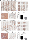
Figure 1.
Figure 1.
GSDMD protein expression levels are upregulated in NSCLC compared with adjacent tissues. (A and B) IHC staining of GSDMD in NSCLC tissue microarrays, with statistical analysis of the GSDMD IHC scores in the lower right panel. (A) IHC on 87 paired LUAD with adjacent tumor specimens plus six individual LUAD sections marked by a blue box. (B) IHC on 75 paired LUSC specimens. T, tumor; A, adjacent tumor specimen; ***P<0.001 (Student's t-test). GSDMD, gasdermin D; IHC, immunohistochemistry; NSCLC, non-small cell lung cancer; LUAD, lung adenocarcinoma.
Figure 2.
Figure 2.
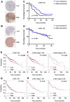
Correlation between GSDMD expression and clinical prognosis based on tissue microarrays and public database analysis. (A and B) Representative IHC images of LUAD (A) and LUSC (B) with high or low GSDMD expression levels. (C and D) Survival curves of 92 LUAD (C) and 70 LUSC (D) patients grouped according to quantitative GSDMD IHC scores. (E-H) Prognosis analysis performed using a clinical-based Kaplan-Meier plot database. (E and F) A high GSDMD expression level was correlated with shortened overall survival (OS) in LUAD patients (E), particularly in stage I and stage II patients (F). (G and H) The GSDMD expression level was not correlated with LUSC patient overall survival. GSDMD, gasdermin D; IHC, immunohistochemistry; LUAD, lung adenocarcinoma; LUSC, lung squamous cell carcinoma.
Figure 3.
Figure 3.
GSDMD knockdown attenuates proliferation of NSCLC cell lines. (A) GSDMD protein and mRNA expression level in HBE and eight NSCLC cell lines as detected by western blotting and RT-qPCR, respectively. Data are represented as the mean ± SD (n=3). (B) PC9, H1703 and H1975 cells were transfected with the indicated siRNAs (NC, si-1031 or si-1244), and the knockdown efficiency was confirmed by western blotting at 48 h post-transfection and RT-qPCR at 24 h post-transfection. Data are represented as the mean ± SD (n=3). (C) Cell proliferation was examined at the indicated time-points post-transfection using the MTT assay. Data are represented as the mean ± SD (n=3). (D) Representative images of colonies formed from transfected cells by the clonogenic assay. **P<0.01, ***P<0.001 (ANOVA test). GSDMD, gasdermin D; NSCLC, non-small cell lung cancer; NC, negative control.
Figure 4.
Figure 4.
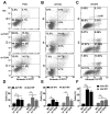
GSDMD knockdown facilitates apoptosis of NSCLC cell lines. (A-C) Cell apoptosis was analyzed by flow cytometry with Annexin V-FITC (AV)/PI double staining at 72 h post-transfection (NC, si-1031 or si-1244). (D-F) Statistical analysis of early and late apoptosis in (D) PC9, (E) H1703 and (F) H1975 cell lines. AV+/PI, AV+/PI+ indicate early apoptotic and late apoptotic cells, respectively. Data are represented as the mean ± SD (n=3). *P<0.05, **P<0.01 (ANOVA test). GSDMD, gasdermin D; NSCLC, non-small cell lung cancer; NC, negative control.
Figure 5.
Figure 5.
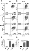
GSDMD knockdown decreases the MMP of NSCLC cell lines. (A and B) MMP was detected by the JC-1 probe at 72 h post-transfection (NC, si-1031 or si-1244). Normal MMP cells are indicated by high green fluorescence (FL1) and red fluorescence (FL2), Low MMP cells are indicated by high FL1 and low FL2. (C and D) Statistical analysis of low MMP cell percentage. Data are represented as the mean ± SD (n=3). *P<0.05, **P<0.01, ***P<0.001 (ANOVA test). MMP, mitochondrial membrane potential; GSDMD, gasdermin D; NSCLC, non-small cell lung cancer; NC, negative control.
Figure 6.
Figure 6.
NLRP3 inflammasome stimulation in GSDMD-deficient cells induce apoptosis, instead of pyroptosis. (A) Western blotting was used to examine the expression of proteins upstream of GSDMD in classical pyroptotic pathways: NLRP3, caspase-1 and its cleaved form in transfected PC9 and H1703 cells. (B) PC9 cells transfected with indicated siRNAs (NC or si-GSDMD) were primed with 1 µg/ml LPS for 5 h and subsequently incubated with 5 mM ATP for 12 h. IL-1β levels in the supernatants of cultured cell lines were detected by ELISA. Data are represented as the mean ± SD (n=3). (C) Statistical analysis of Annexin V-FITC (AV)+/PI and AV+/PI+ PC9 cells treated as described in B. (D) Cell death was analyzed by flow cytometry with Annexin V-FITC/PI double staining. Generally, AV+/PI, AV+/PI+, AV/PI and AV/PI+ indicate early apoptotic, late apoptotic, live and necrotic cells, respectively. Notably, pyroptotic cells tend to be simultaneously Annexin V/PI positive. (E) Representative light microscopy images of PC9 cells treated as described in B. PC9 cells were also treated with cisplatin at a concentration of 80 µg/ml for 24 h. *P<0.05, **P<0.01, ***P<0.001 (ANOVA test). GSDMD, gasdermin D; LPS, lipopolysaccharide; ATP, adenosine 5′-triphosphate.
Figure 7.
Figure 7.
GSDMD knockdown induces caspase-3 activation and downregulates EGFR/Akt signaling in NSCLC cell lines. (A and B) LDH release from GSDMD knockdown or control PC9 and H1703 cells was assessed at indicated time-points after transfection (NC, si-1031 or si-1244). Data are represented as the mean ± SD (n=3). (C and D) Expression of caspase-3, its substrate PARP, and their cleaved forms in cells transfected with the indicated siRNAs and detected by western blotting. (E and F) Cells were pretreated with 30 µM Z-VAD-FMK (zvad) for 24 h before transfection and cell proliferation was examined at the indicated time-points following transfection using the MTT assay. Data are represented as the mean ± SD (n=3). (G and H) Western blotting demonstrating EGFR/Akt/mTOR signaling in the transfected cells. *P<0.05 (ANOVA test). LDH, lactate dehydrogenase; GSDMD, gasdermin D; NSCLC, non-small cell lung cancer.
Figure 8.
Figure 8.
shGsd-transduced PC9 cells demonstrate decreased cell proliferation and increased cell apoptosis. (A) PC9 cells were transduced with shGsd or shNeg, and the transduction efficiency was confirmed by western blotting at 96 h post-transduction and RT-qPCR at 72 h post-transduction. (B) Cell proliferation was examined at the indicated time-points post-transduction using the MTT assay. (C) Representative images of colonies formed from transducted cells by the clonogenic assay. (D) Cell apoptosis was analyzed by flow cytometry with Annexin V-FITC (AV)/PI double staining at 96 h post-transduction. (E) Statistical analysis of early and late apoptosis in the PC9 cell line. AV+/PI, AV+/PI+ indicate early apoptotic, late apoptotic cells, respectively. Data are represented as the mean ± SD (n=3). *P<0.05, ***P<0.001 (Student's t-test).
Figure 9.
Figure 9.
GSDMD depletion suppresses tumor growth in vivo. (A) Six-week-old male Balb/c nude mice were injected subcutaneously with 2×10⁶ shGsd or shNeg transduced PC9 cells. Tumors harvested from nude mice 18 days post injection are shown. (B) Tumor volumes were determined every 3 days. Data are represented as the mean ± SD (n=5). (C) Tumor weight determined 18 days post injection. Data are represented as the mean ± SD (n=5). (D) Representative H&E, Ki-67, and TUNEL staining images of tumors from nude mice formed by shGsd or shNeg transduced PC9 cells. (E) Western blotting demonstrating EGFR/Akt/mTOR signaling in tumors harvested from nude mice injected with shGsd or shNeg transduced PC9 cells. *P<0.05, **P<0.01 (Student's t-test). GSDMD, gasdermin D.

References

    1. Chen W, Zheng R, Baade PD, Zhang S, Zeng H, Bray F, Jemal A, Yu XQ, He J. Cancer statistics in China, 2015. CA Cancer J Clin. 2016;66:115–132. doi: 10.3322/caac.21338. - DOI - PubMed
    1. Reck M, Rabe KF. Precision diagnosis and treatment for advanced non-small-cell lung cancer. N Engl J Med. 2017;377:849–861. doi: 10.1056/NEJMra1703413. - VSports app下载 - DOI - PubMed
    1. Saeki N, Usui T, Aoyagi K, Kim DH, Sato M, Mabuchi T, Yanagihara K, Ogawa K, Sakamoto H, Yoshida T, Sasaki H. Distinctive expression and function of four GSDM family genes (GSDMA-D) in normal and malignant upper gastrointestinal epithelium. Genes Chromosomes Cancer. 2009;48:261–271. doi: 10.1002/gcc.20636. - DOI - PubMed
    1. Shi J, Gao W, Shao F. Pyroptosis: Gasdermin-mediated programmed necrotic cell death. Trends Biochem Sci. 2017;42:245–254. doi: 10.1016/j.tibs.2016.10.004. - DOI - PubMed
    1. Kayagaki N, Stowe IB, Lee BL, O'Rourke K, Anderson K, Warming S, Cuellar T, Haley B, Roose-Girma M, Phung QT, et al. Caspase-11 cleaves gasdermin D for non-canonical inflammasome signalling. Nature. 2015;526:666–671. doi: 10.1038/nature15541. - V体育2025版 - DOI - PubMed

"VSports最新版本" MeSH terms

Substances