Skip to main page content
U.S. flag

An official website of the United States government

Dot gov

The . gov means it’s official. Federal government websites often end in . gov or . mil. Before sharing sensitive information, make sure you’re on a federal government site. VSports app下载.

Https

The site is secure V体育官网. The https:// ensures that you are connecting to the official website and that any information you provide is encrypted and transmitted securely. .

. 2018 Aug 3;9(1):3049.
doi: 10.1038/s41467-018-05395-y.

"VSports注册入口" The chimeric TAC receptor co-opts the T cell receptor yielding robust anti-tumor activity without toxicity

Affiliations

The chimeric TAC receptor co-opts the T cell receptor yielding robust anti-tumor activity without toxicity

"VSports注册入口" Christopher W Helsen et al. Nat Commun. .

Abstract

Engineering T cells with chimeric antigen receptors (CARs) is an effective method for directing T cells to attack tumors, but may cause adverse side effects such as the potentially lethal cytokine release syndrome. Here the authors show that the T cell antigen coupler (TAC), a chimeric receptor that co-opts the endogenous TCR, induces more efficient anti-tumor responses and reduced toxicity when compared with past-generation CARs. TAC-engineered T cells induce robust and antigen-specific cytokine production and cytotoxicity in vitro, and strong anti-tumor activity in a variety of xenograft models including solid and liquid tumors. In a solid tumor model, TAC-T cells outperform CD28-based CAR-T cells with increased anti-tumor efficacy, reduced toxicity, and faster tumor infiltration VSports手机版. Intratumoral TAC-T cells are enriched for Ki-67+ CD8+ T cells, demonstrating local expansion. These results indicate that TAC-T cells may have a superior therapeutic index relative to CAR-T cells. .


Conflict of interest statement

Christopher W V体育安卓版. Helsen, Joanne A. Hammill, Kenneth A. Mwawasi, Arya Afsahi, Galina F. Denisova, and Jonathan L. Bramson hold shares in Triumvira Immunologics. Christopher W. Helsen and Jonathan L. Bramson are founding scientists of Triumvira Immunologics. All other authors declare no competing interests.

Figures
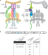
Fig. 1
Fig. 1
TAC design mimics the TCR-CD3:co-receptor complex. a Left: Naturally occurring TCR-CD3 complex interacts directly with the antigen presented by MHC. Meanwhile, the CD8/CD4 co-receptor interacts with MHC I/II in an antigen-independent manner. Together, these interactions comprise the first step in T cell activation. Right: The TAC receptor re-directs the TCR-CD3 complex towards an antigen of choice using an interchangeable antigen binding moiety (here depicted with an scFv, purple). An scFv is used to recruit the TCR-CD3 complex (blue). Co-receptor properties are incorporated by including the CD4 hinge, TM region, and cytosolic tail (green). b The TAC is incorporated into the pCCL DNA backbone containing a truncated NGFR (tNGFR), which lacks cytosolic signaling domains, as a transduction control. The vector features a bi-directional promoter system with tNGFR under control of the mCMV promoter and TAC expression being driven by the EF-1α promoter. TAC is comprised of an antigen binding domain, a CD3-binding domain, and a co-receptor domain. A variety of proteins can be used for each of these three TAC domains allowing the TAC to be modified to best respond to numerous different antigens. The specific domain combinations tested are described below
Fig. 2
Fig. 2
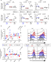
Evaluation of multiple anti-CD3 scFv domains for recruitment of TAC to the TCR-CD3 complex. a, e Schematic representation of evaluated TAC constructs. TAC receptors utilizing the (a) anti-HER2 DARPin are paired with either the UCHT1 or OKT3 anti-CD3 scFv. TAC receptors using the (e) anti-CD19 scFv are paired with either the huUCHT1, F6A, or L2K anti-CD3 scFv. b, f Relative TAC surface expression is measured by flow cytometry. Cells are stained for CD4, CD8, tNGFR and TAC, and gated on either CD4+NGFR+ or CD8+NGFR+. Representative data of three independent experiments are presented as histogram analysis of (b) HER2-TAC or (f) CD19-TAC. Surface expression of OKT3 relative to UCHT1 was significantly higher in CD4 cells (p = 0.0007) but not in CD8 cells. huUCHT1 expression is significantly higher compared to either L2K (p = 0.005 (CD4)/0.0002 (CD8)) or F6A (p < 0.0001 (CD4) p < 0.0001 (CD8)). For the gating strategy see Supplementary Fig. 13A. c, g HER2- and CD19-specific TAC-T cells are stimulated with antigen-positive (c) SK-OV-3 and (g) Raji tumor cells, respectively. Data are presented as percent of CD4 or CD8 T cells producing cytokine. Cytokine producing cells are compared from (c) TAC-T cells bearing UCHT1 (square) or OKT3 (inverted triangle), or (g) TAC-T cells bearing huUCHT1 (square), F6A (triangle), or L2K (diamond). Lines represent the mean. Multiple t-test is used to determine significance in all cases. For the gating strategy see Supplementary Fig. 13B. d, h HER2- and CD19-TAC and vector control (vector only carrying tNGFR) T cells are co-cultured with (d) SK-OV-3 and (h) NALM-6 tumor cells, respectively, to measure TAC-T cell-mediated cytotoxicity. Vector control T cells (circles) are compared against d HER2-specific TAC-T cells bearing UCHT1 (square) or OKT3 (triangle), or (h) CD19-specific TAC-T cells bearing huUCHT1 (square), F6A (triangle), or L2K (diamond). Data are from three independent experiments with three different donors, error bars are standard deviation
Fig. 3
Fig. 3
Relative expression of checkpoint receptors and memory T cell subsets in CAR- and TAC-engineered T cells. T cells were transduced with HER2-TAC, or a second-generation anti-HER2 CAR including the CD28 (28ζ CAR) or 4-1BB (BBζ CAR) costimulatory receptor domains, or a vector control (tNGFR). Engineered T cells are stained for surface marker expression and CD4+NGFR+ or CD8+NGFR+ populations are analyzed by flow cytometry for (a) expression of checkpoint receptors PD-1, LAG-3, and TIM-3. All data is normalized to TAC-engineered T cells, and lines represent the mean of four donors. Each donor is represented by a unique symbol to highlight donor-to-donor variations. Multiple t-test is used to determine significance. For the gating strategy see Supplementary Fig. 14. b Memory T cell subsets of TAC-, 28ζ CAR-, and BBζ CAR-T cells, relative to T cells engineered with a vector control (tNGFR). T cell subsets are defined as naïve (CD45RA+, CCR7+), central memory (CM) (CD45RA, CCR7+), effector memory (EM) (CD45RA, CCR7), and terminal effectors (EMRA) (CD45RA+, CCR7). Lines represent the mean of four donors. Multiple t-test is used to determine significance. c CD27 and CD28 expression; representative histograms show data from one donor, median fluorescence intensity is indicated. Data are from two independent experiments with four different donors. For both (b) and (c), the gating strategy is shown in Supplementary Fig. 15
Fig. 4
Fig. 4
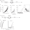
TAC-T cells demonstrate in vivo efficacy against solid and liquid tumors. a Treatment schema for OVCAR-3 tumor-bearing mice. In short, NRG mice receive 2.5 × 106 OVCAR-3 cells subcutaneously. Tumors grow for 35–42 days until an average size of ~100 mm3 is achieved. T cells are delivered over two doses, 48 h apart. b Tumor-bearing mice receive intravenous delivery of 4–6 × 106 HER2-TAC-T cells, CD19-TAC-T cells, or an equivalent dose of vector control T cells. Tumor growth is followed over time. Each curve shows data from a single treated tumor. Data are from three donors, collected over two independent experiments, n = 11 for each of HER2-TAC and vector control groups; CD19-TAC data generated from one donor, one experiment, n = 4. Using curve fitting analysis and multiple t-test HER2-TAC induced regression is significantly different from controls, while tumor growth between vector control and CD19-TAC was not significantly different. c Treatment schema for NALM-6 tumor-bearing mice. In short, 0.5 million NALM-6 cells are administered intravenously and allowed to establish for 5 days. Mice were treated with a total dose of 4 × 106 cryopreserved CD19-TAC-T cells. T cells are delivered over two doses, 7 days apart. d Mice are treated with either TAC-Δantigen-binding domain or CD19-TAC-T cells. Curves each represent a single treated tumor. Data are from one donor, one experiment, n = 5 for each of CD19-TAC and control groups. Data have been replicated in independent experiments, n = 10. Tumor progression is followed weekly via luminescence
Fig. 5
Fig. 5
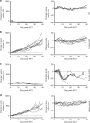
HER2-TAC-T cells demonstrate an enhanced safety profile and improved efficacy over first- and second-generation HER2-CAR-T cells in vivo. OVCAR-3 tumor-bearing mice are treated with 2.0 × 106 HER2-TAC-T cells (a), first-generation anti-HER2 CAR-T cells (b), second-generation anti-HER2 28ζ CAR-T cells (c), or a matched total number of vector control T cells (d). Mice are followed for change in body weight and tumor volume; each curve represents a single treated mouse relative to pre-treatment weight/volume. When mice reach endpoint, this is indicated via X in (c). Data has been replicated in an independent experiment. Using curve fitting analysis and multiple t-test HER2-TAC induced regression is significantly different from control, 1st, and 2nd generation CAR curves, while tumor growth between vector control and 1st gen CAR was not significantly different. The change in body weight observed in 2nd gen CAR curves is significantly different from vector control, 1st gen CAR, and TAC-T cell treated mice. However, the difference in body weight between vector control, 1st gen CAR, and TAC curves was not significantly different
Fig. 6
Fig. 6
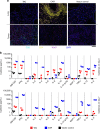
Engineered T cell distribution and cytokine release in vivo. OVCAR-3 tumor-bearing mice were treated with 6.0 × 106 anti-HER2 28ζ CAR- or HER2-TAC-T cells, or a matched total number of vector control cells. Mice are sacrificed at 1, 3, 5, and 7 days post-ACT1 for multiplex serum cytokine analysis or perfusion and fixation of tissues for subsequent histology. a Multicolor IHC is performed on tumor and lung tissue 7 days post-ACT1. Tissues are stained for CD8 (cyan), CD4 (yellow), DNA (blue) and a proliferation marker (Ki-67, purple). Data are representative of 3 mice. Scale bar indicates 100 μm. b Multiplex analysis of human cytokines in mouse serum on day 3 and 7. Measurements that fall below 0.2 pg/mL are below the calibration range and are therefore defined as 0. 0 values are depicted on the graph’s x axis. Statistical analysis is provided in Supplementary Table 1, analysis was performed using multiple t-test. Individual data points are shown, bars indicate standard deviation and center bars indicate the median

References (V体育安卓版)

    1. Ali, S. A. et al. T cells expressing an anti-B-cell-maturation-antigen chimeric antigen receptor cause remissions of multiple myeloma. Blood128, 1688–1700 (2016). - PMC - PubMed
    1. Kochenderfer JN, Yu Z, Frasheri D, Restifo NP, Rosenberg SA. Adoptive transfer of syngeneic T cells transduced with a chimeric antigen receptor that recognizes murine CD19 can eradicate lymphoma and normal B cells. Blood. 2010;116:3875–3886. doi: 10.1182/blood-2010-01-265041. - DOI - PMC - PubMed
    1. Ramos CA, Savoldo B, Dotti G. CD19-CAR trials. Cancer J. 2015;20:112–118. doi: 10.1097/PPO.0000000000000031. - "VSports app下载" DOI - PMC - PubMed
    1. Gross G, Waks T, Eshhar Z. Expression of immunoglobulin-T-cell receptor chimeric molecules as functional receptors with antibody-type specificity. Proc. Natl. Acad. Sci. U.S.A. 1989;86:10024–10028. doi: 10.1073/pnas.86.24.10024. - DOI - PMC - PubMed
    1. Finney HM, Lawson AD, Bebbington CR, Weir AN. Chimeric receptors providing both primary and costimulatory signaling in T cells from a single gene product. J. Immunol. 1998;161:2791–2797. - VSports最新版本 - PubMed

Publication types

MeSH terms