Skip to main page content
U.S. flag

An official website of the United States government

Dot gov

The . gov means it’s official. Federal government websites often end in . gov or . mil. Before sharing sensitive information, make sure you’re on a federal government site. VSports app下载.

Https

The site is secure. The https:// ensures that you are connecting to the official website and that any information you provide is encrypted and transmitted securely V体育官网. .

. 2018 Jul 26;3(14):e99208.
doi: 10.1172/jci.insight.99208.

From proteomics to discovery of first-in-class ST2 inhibitors active in vivo

Affiliations

From proteomics to discovery of first-in-class ST2 inhibitors active in vivo

VSports最新版本 - Abdulraouf M Ramadan et al. JCI Insight. .

Abstract (V体育2025版)

Soluble cytokine receptors function as decoy receptors to attenuate cytokine-mediated signaling and modulate downstream cellular responses. Dysregulated overproduction of soluble receptors can be pathological, such as soluble ST2 (sST2), a prognostic biomarker in cardiovascular diseases, ulcerative colitis, and graft-versus-host disease (GVHD) VSports手机版. Although intervention using an ST2 antibody improves survival in murine GVHD models, sST2 is a challenging target for drug development because it binds to IL-33 via an extensive interaction interface. Here, we report the discovery of small-molecule ST2 inhibitors through a combination of high-throughput screening and computational analysis. After in vitro and in vivo toxicity assessment, 3 compounds were selected for evaluation in 2 experimental GVHD models. We show that the most effective compound, iST2-1, reduces plasma sST2 levels, alleviates disease symptoms, improves survival, and maintains graft-versus-leukemia activity. Our data suggest that iST2-1 warrants further optimization to develop treatment for inflammatory diseases mediated by sST2. .

Keywords: Drug screens; Stem cell transplantation; Th2 response; Therapeutics; Transplantation V体育安卓版. .


VSports注册入口 - Conflict of interest statement

Conflict of interest: SP has a patent (US 20130115232A1, WO 2013066369A3) on “Methods of detection of graft-versus-host disease” licensed to Viracor-IBT laboratories.

Figures

Figure 1
Figure 1. Flowchart of integrated high-throughput screening and computational analysis for discovery of ST2 inhibitors.
PAINS, pan-assay interference compounds; MACCS, Molecular ACCess System; hPBMC, human peripheral blood mononuclear cell.
Figure 2
Figure 2. In vitro and in vivo toxicity evaluation of selected candidates.
(A) Human PBMC death induced by compounds at 3 concentrations after incubation for 20 hours (duplicate per compound at each concentration). IC50 values determined by the AlphaLISA are shown in parentheses. Compounds belonging to chemotypes I, II, and III and unclassified are grouped together. CB6114052, CB5107562, and NAT13-343201 are 3 initial hits. The average IC50 values of 17 compounds at 63.7 μM were used as references for setting the maximum concentration at 66 μM. (B) Titration curves for iST2-1, iST2-2, and iST2-4 measured with the AlphaLISA and the HEK-Blue IL-33 assay. (C) In vivo dose-escalation toxicity evaluation of 7 selected compounds in healthy mice. Data represent mean ± SEM (n = 3 per compound). hPBMC, human peripheral blood mononuclear cell.
Figure 3
Figure 3. IC50 values of iST2-1 and its analogs, SAXS studies of apo-ST2, ST2/iST2-1, and potential iST2-1 binding sites identified from computational simulations.
(A) Chemical structures of racemic iST2-1, (R)-iST2-1, (S)-iST2-1, analogs of iST2-1, and structural alignment of (R)-iST2-1 (purple) with (S)-iST2-1 (green). IC50 values of the inhibitors are shown in parentheses. (B) Inhibition curves and IC50 values (in parentheses) of 4 representative compounds. Each data point is an average of triplicate measurements ± SD. (C) SAXS profiles, the residual plot between the SAXS profiles of apo-ST2 and ST2/iST2-1, comparison of the Kratky plot based on the SAXS profiles of apo-ST2 and ST2/iST2-1, the pair-wise distance distribution [P(r)] of apo-ST2 and ST2/iST2-1 calculated from the SAXS profiles. Dmax values are shown in parentheses. (D) Ab initio shape reconstruction of apo-ST2 (gray) and ST2/iST2-1 (purple). (E) Mapping of the (R)-iST2-1 and (S)-iST2-1 binding sites (surface envelop) in ST2 based on 32-ns MD simulations. Maps detected from 13- and 15-Å octahedron boxes are colored in purple and green, respectively. Consensus binding sites (S1r, S2r and S1s) are circled. The D1 and D2 domains of ST2 are labeled. (F) Binding sites of (R)-iST2-1 (green mesh) and (S)-iST2-1 (red mesh) that block interaction between ST2 and IL-33 (orange) are highlighted. (G) Binding sites of (R)-, (S)-, (S,S)-iST2-1 in the glycosylated ST2 from the MD simulations. (S,S)-iST2-1 is (S)-iST2-1 in which the proton on the pyrrolidine group is in the S form and its mapped site is shown in cyan mesh. SAXS, small-angle X-ray scattering.
Figure 4
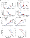
Figure 4. Dosages and treatment schedule for ST2 inhibitors and ex vivo analysis in the GVHD mouse models.
(A) The dosages were calculated using the body weight of each mouse at 20 mg and correspond to twice the IC50 values of the inhibitors determined by the HEK-Blue IL-33 assay. (B) Number of T cells infiltrating the gut in the B6C3H.SW GVHD model at day 14. Flow cytometric analysis of intestinal CD4+IFN-γ+ and FoxP3+CD4+ T cell populations on day 14 in the (C) hu-T cells → NSG model and day 21 in the (D) hu-T cells → NSG, (E) B6 → C3H.SW GVHD models treated with iST2-1, iST2-2, or iST2-3. n = 2 per group at day 14 and 21 respectively, and for each model. Data represent mean ± SEM (n = 2). TBI, total body irradiation; hPBMC, human peripheral blood mononuclear cell. *P < 0.05, **P < 0.01 by unpaired t test.
Figure 5
Figure 5. sST2 and IFN-γ levels, GVHD scores, and survival curves for GVHD disease model mice treated with ST2 inhibitors.
(A) Plasma levels of human and murine sST2 and IFN-γ in the hu-T cells → NSG and B6 → C3H.SW GVHD models from day 7 to 28. Data represent mean ± SEM (n = 3 per group for NSG and n = 6 for C3H.SW). (B) GVHD scores and survival curves for the hu-T cells → NSG and B6 → C3H.SW GVHD models. Data represent mean ± SEM for GVHD scores, and Kaplan-Meier curves for survival (n = 13 per group). P values were calculated for GVHD scores by unpaired t test and for survival by log-rank test. *P < 0.05; **P < 0.01; ***P < 0.001.
Figure 6
Figure 6. GVL activity in the C3H.SW mouse model treated with DMSO or iST2-1.
(A) GVHD scores. Data represent mean ± SEM, unpaired t test, P = 0.0001 (n = 5 per group), and (B) Kaplan-Meier survival curves of C3H.SW mice that received 2 × 10⁴ GFP+ MLL-AF9 leukemia cells with allo-HCT (B6 → C3H.SW) and were treated with DMSO control (filled circles) or iST2-1 (open circles), log-rank P = 0.013 (n = 5 per group). The treatment schedule was the same as in Figure 4A. (C) Percentage of mice that succumbed to GVHD or leukemia or remained alive with treatment of DMSO or iST2-1 at the end of the study (day 73). *P < 0.05; ***P < 0.001.

References

    1. Kotas ME, Medzhitov R. Homeostasis, inflammation, and disease susceptibility. Cell. 2015;160(5):816–827. doi: 10.1016/j.cell.2015.02.010. - DOI - PMC - PubMed
    1. Netea MG, et al. A guiding map for inflammation. Nat Immunol. 2017;18(8):826–831. doi: 10.1038/ni.3790. - DOI - PMC - PubMed
    1. Mantovani A, Locati M, Vecchi A, Sozzani S, Allavena P. Decoy receptors: a strategy to regulate inflammatory cytokines and chemokines. Trends Immunol. 2001;22(6):328–336. doi: 10.1016/S1471-4906(01)01941-X. - DOI - PubMed
    1. Levine SJ. Molecular mechanisms of soluble cytokine receptor generation. J Biol Chem. 2008;283(21):14177–14181. doi: 10.1074/jbc.R700052200. - DOI - PMC - PubMed
    1. Heaney ML, Golde DW. Soluble receptors in human disease. J Leukoc Biol. 1998;64(2):135–146. doi: 10.1002/jlb.64.2.135. - DOI - PubMed

Publication types

V体育安卓版 - MeSH terms